
Abstract
Tissue engineering may require precise patterning and monitoring of cells and bioactive factors within the scaffold. We investigated a new hybrid nanobioprinting technique that facilitates manipulation and tracking of cells and bioactive factors within a three-dimensional tissue construct. This technique combines the initial patterning capabilities of syringe-based cell deposition with the active patterning capabilities of superparamagnetic nanoparticles. Superparamagnetic iron oxide nanoparticles, either in the alginate biopolymer or loaded inside endothelial cells, were bioprinted using a solid freeform fabrication direct cell writing system. Bioprinting did not impact cell viability when nanoparticles were in the alginate. However, both control and printed samples with 0.1 or 1.0 mg/mL nanoparticles in the alginate showed a 16% or 35% viability loss at 36 h after printing, respectively. Nanoparticle loading in cells decreased cell viability to 11% and bioprinting decreased viability to an additional 29% at 36 h. No changes were observed in any samples after 36 h, suggesting that cell viability stabilized following the initial nanoparticle toxicity effect. Nanoparticles in the alginate and those loaded in cells were moved using an external magnet, depending on biopolymer viscosity, and imaged by microcomputed tomography. The hybrid nanobioprinting method can noninvasively manipulate and track bioactive factors and cells within tissue engineering structures.
Introduction
Many biofabrication techniques have been developed to incorporate living cells into functionalized scaffolds in a reproducible, 3D pattern.2,3 Rapid prototyping,4,5 inkjet-based cell printing,6–8 and microcontact printing9,10 are among the commonly used cell deposition systems for tissue engineering applications. These biofabrication methods allow initial deposition of scaffold and cells in a predefined pattern. However, the methods are often expensive, are time consuming, require chemically modified surfaces, or cause cell damage due to high temperatures and pressures used in the deposition process. We developed a direct cell writing system for the freeform construction of biopolymer-based 3D tissue scaffolds and cell-embedded tissue constructs. 11 The direct cell writing system uses micronozzles driven by pneumatic microvalves to deposit living cells, scaffold material, and bioactive components such as growth factors in controlled amounts with precise spatial positioning. The system requires no preprocessing, is computer controlled to rapidly produce sample replicates, and operates at room temperature and low pressure to maximize cell viability.
Recently, several new approaches have been proposed to actively pattern cell constructs using external forces, including dielectrophoresis, 12 an optical trap, 13 or superparamagnetic nanoparticles in a magnetic field.14,15 Superparamagnetic iron oxide nanoparticles have been of primary interest for both in vivo and in vitro applications because they exhibit magnetic behavior only in the presence of a magnetic field. 16 These nanoparticles can be conjugated with proteins or loaded inside cells, are relatively nontoxic, and can be imaged by magnetic resonance imaging (MRI) or computed tomography (CT). In vivo, superparamagnetic nanoparticles have been used to target drugs to a treatment site to increase drug efficiency and reduce systemic effects, 17 to enhance gene delivery to target cells because nanoparticles easily cross cell membranes,18,19 and to detect vascular tissues such as tumors, because iron oxide nanoparticles appear dark on MR images.20,21 In vitro, superparamagnetic nanoparticles have been used to create high resolution, two-dimensional (2D) cell patterns on nonfunctionalized surfaces. 14 More recently, Frasca et al. used magnetic fields and magnetic field gradients to achieve 3D cell patterning. 15 However, the ability of this technique to create complex 3D shapes is highly limited because the only method of shape control is with a magnetic field gradient from magnets placed under the scaffold material.
We propose a technique combining the initial patterning capabilities of the direct cell writing system with the active patterning capabilities of superparamagnetic nanoparticles. This new hybrid technique would allow biofabrication of a complex 3D tissue scaffold of magnetically labeled cells and bioactive factors, which could then be manipulated and tracked within the tissue engineering construct. In this study, superparamagnetic iron oxide nanoparticles were bioprinted either in an alginate scaffold or inside endothelial cells using the multinozzle direct cell writing system. Cell viability was assessed for various nanoparticle and alginate concentrations at a predefined printing nozzle size. The nanoparticles in both alginate and endothelial cells were manipulated using a magnetic field. Finally, the nanoparticles were patterned inside 3D biopolymer scaffolds and imaged using a microCT scanner. Bioprinting of superparamagnetic iron oxide nanoparticles could help create more versatile tissue engineering structures, as well as improve our understanding of cell behavior in 3D tissue culture.
Materials and Methods
Chemical formulation
Sodium alginate powder (FMCBioPolymer, Drammen, Norway) was dissolved in deionized water at 0.5%, 1%, 2%, and 3% (w/v) concentrations. An ionic crosslinking solution was prepared by dissolving calcium chloride (BDH Chemicals, Poole, UK) in deionized water. NanoArc magnetic iron oxide nanoparticles (Alfa Aesar, Ward Hill, MA) of 20–40 nm diameter were used in all experiments. Spherical nanoparticles of the given size were selected to obtain maximum uptake efficiency. 22 Sodium alginate–magnetic nanoparticle solutions were prepared by vigorously mixing sodium alginate with increasing concentrations of iron oxide nanoparticles to achieve a homogeneous nanoparticle distribution.
Cell culture
Porcine aortic endothelial cells (PAECs) were isolated by the collagenase dispersion method and maintained in low-glucose Dulbecco's modified Eagle's medium supplemented with 5% fetal bovine serum, 1% penicillin–streptomycin, and 2% glutamine (Invitrogen, Carlsbad, CA). Culture medium was changed every 48 h, and cells between passages 4 and 9 were used. Prior to printing, cells were gently mixed at a concentration of 1.5 × 10⁵ cells/mL in sodium alginate solution to ensure uniform cell distribution. For magnetically labeled cells, PAECs in 100-mm tissue culture dishes were loaded with different nanoparticle concentrations and incubated at 37°C in a 5% carbon dioxide incubator for 24 h. Nanoparticle uptake by cell was confirmed by transmission electron microscopy. Our transmission electron microscopy images also suggest that the majority, if not all, of the cells take up nanoparticles.
Cell-dispensing system
A proprietary solid freeform fabrication-based direct cell writing system (Fig. 1A) was developed to create 3D tissue constructs by dispensing cells and biopolymers into predefined patterns.11,23 The direct cell writing system used in this study operates at room temperature and low-pressure conditions to facilitate deposition of living cells, growth factors, or other bioactive compounds in controlled amounts with precise spatial positioning. Pneumatic microvalves (EFD, East Providence, RI) were used to apply a low printing pressure of 2 and 5 psi to minimize cell death due to the dispensing process11,23 (Fig. 1B).
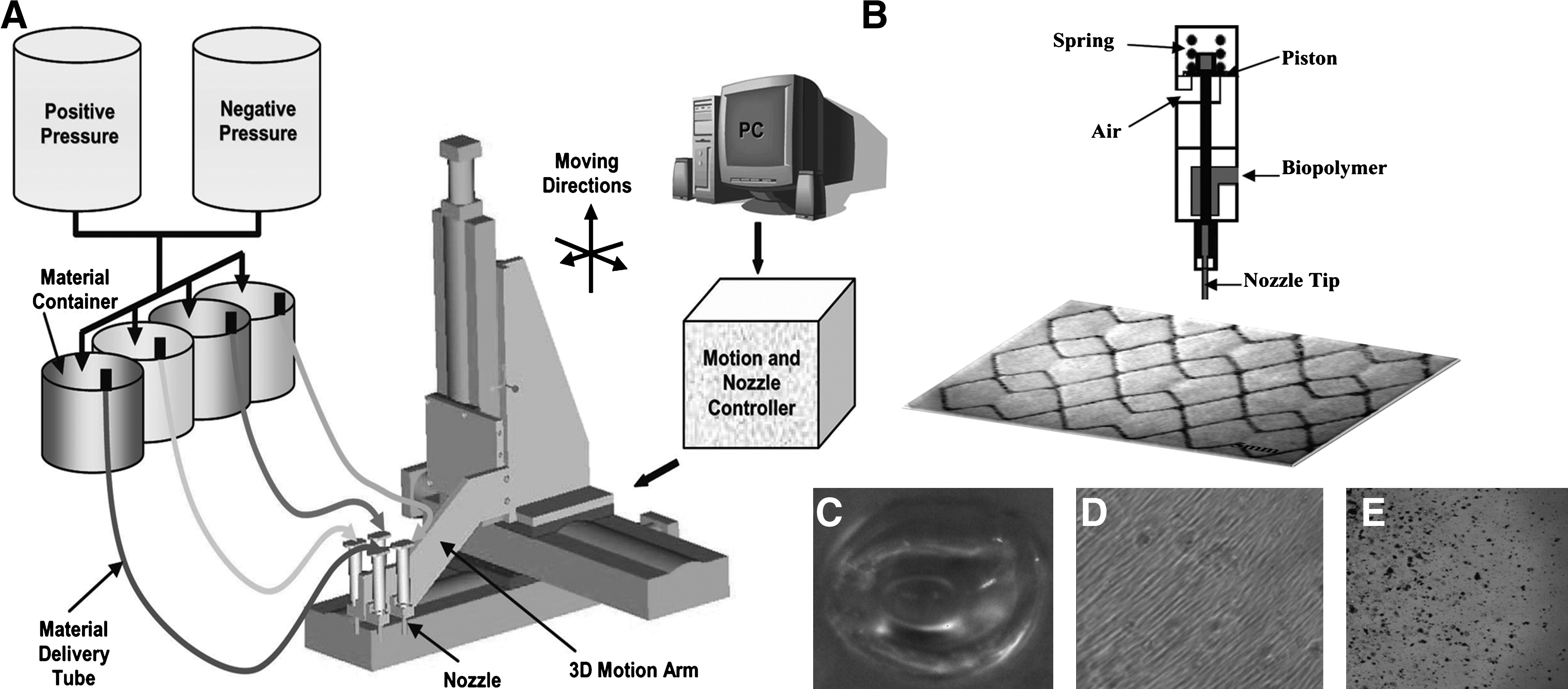
(
Sodium alginate was chosen as the scaffold biopolymer. Alginate–nanoparticle–cell mixtures with 0, 0.1, or 1.0 mg/mL nanoparticle concentration were printed using 250-μm nozzles. Control samples were dispensed in the system but without using nozzle tips. All samples were dispensed as 0.3 g of bulk material with a sample size of three, and each experiment was repeated a minimum of two times. Data presented are from one representative experiment. After dispensing, each sample was immediately submerged in a 5.0% (w/v) calcium chloride crosslinking solution for 5 min, placed in supplemented media, and returned to the incubator. Samples in the long-term study were crosslinked daily to maintain both cell immobilization and alginate structural integrity. Representative images of printed bulk samples and cell distribution in alginate bulk samples are presented in Figure 1C, D, and E.
Cell viability
Alamar blue quantitatively measures cell metabolic activity by using an oxidation–reduction indicator that fluoresces and changes color in metabolically active cells. 24 Crosslinked alginate–cell solutions in six-well plates were incubated with 2 mL supplemented media and 200 μL Alamar blue solution (AbD Serotec, Oxford, UK). After 4 h of incubation at 37°C in 5% carbon dioxide atmosphere, 100 μL of media from each well was transferred into a 96-well flat-bottomed black assay plate, and fluorescence was measured at 535/590 nm in a GENios microplate reader (Tecan, Männedorf, Switzerland). About 3 × 10⁴ cells were calibrated to a fluorescence intensity reading of 35,000. As the Alamar blue assay measures the mean metabolic activity of the cell population, cell viability was confirmed using a live/dead assay (Invitrogen) as per manufacturer's instructions.
Nanoparticle and magnetically labeled cell movement in the scaffold
Bulk samples of 1.0 mg/mL magnetic nanoparticles in 0.5%, 1.0%, and 2% (w/v) alginate were printed using the direct cell writing system. A 1-inch-diameter neodymium iron boron (NdFeB) magnet with a surface field of 6450 Gauss (K&J Magnetics, Jamison, PA) was placed under the 60-mm cell culture dishes. Specific patterns of nanoparticles and magnetically labeled cells were also printed using the cell-dispensing system. A rectangular NdFeB magnet with a surface field of 6450 Gauss (K&J Magnetics) was used to move nanoparticles to a specified location either in a new pattern or while maintaining the original printed pattern. Movement of magnetic nanoparticles and the magnetically labeled cells by the applied magnetic field was imaged using a 4-megapixel CCD camera (Alpha Innotech, San Leandro, CA).
MicroCT scan
A 1.5 mm × 1.5 mm area of 0.1 mg/mL magnetic nanoparticles was printed within a 5 mm × 5 mm × 2 mm of 2% (w/v) alginate construct and imaged using a microCT scanner (SkyScan 1172, Skyscan, Kontich, Belgium). MicroCT allows nondestructive evaluation of the internal structure and composition of the sample based on changes in X-ray absorption. Image resolution was set at 2.16 μm with a 1-mm aluminum filter. The rotation angle was 180° with a rotation step of 0.1°.
Statistical analysis
The samples were statistically compared using Student's t-test. Statistical significance was established at either p < 0.05 (#) or p < 0.01 (*). Two-way analysis of variance (ANOVA) was used to compare changes over time, with statistical significance established at p < 0.0001.
Results
Viability of cells printed with magnetic nanoparticles in the alginate
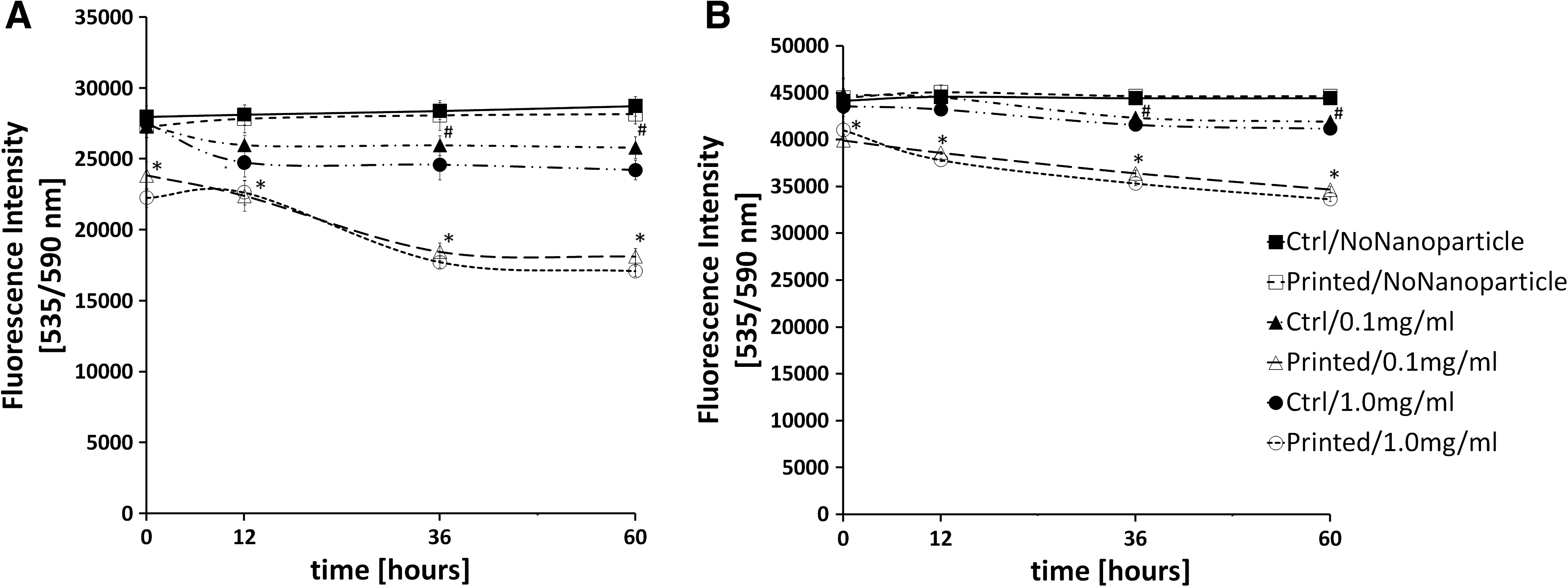
Bioprinting magnetic nanoparticles along with cells in a biopolymer scaffold may provide an effective means to track and manipulate bioactive factors in tissue-engineered structures. We now show that while nanoparticles themselves slightly decreased endothelial cell viability, bioprinting had no significant effect (Fig. 2A). At 0 and 12 h after printing, cell viability did not change significantly for unprinted or printed cells with 0 or 0.1 mg/mL nanoparticles in a 1% (w/v) alginate solution. However, at 36 h after printing, PAECs with 0.1 or 1.0 mg/mL nanoparticles were 16% or 35% less viable than cells without nanoparticles, respectively. The viability loss was independent of the printing process. Cell viability continued to decrease with time up to 60 h after cell printing (ANOVA, p < 0.0001). In a long-term assay (Fig. 2B), endothelial cell viability similarly decreased nearly 22% with 1.0 mg/mL iron oxide nanoparticles in the alginate at 72 h after printing compared with samples without nanoparticles (ANOVA, p < 0.0001). No further cell viability decrease was observed from 72 to 144 h, showing that cells maintained their viability following the initial nanoparticle toxicity effect.

Endothelial cell viability decreased in a dose-dependent manner with magnetic nanoparticles in the alginate, but printing had no effect. (
Increased nanoparticle concentration decreased cell viability, but no additional decrease was observed with printing (Fig. 2A). PAECs encapsulated in alginate with 1.0 mg/mL nanoparticles showed 20% lower viability than cells with 0.1 mg/mL nanoparticles and 36% lower than the control, suggesting a nanoparticle concentration-dependent effect on cell viability. This decreased viability was observed at 36 and 60 h after printing, but the printing process itself did not affect cell viability. To confirm that the Alamar blue measured cell viability, but not a change in cell metabolism, a live/dead assay was performed on printed samples. The live/dead data agreed well with the Alamar blue results.
Effect of alginate concentration on printed cell viability
We next investigated whether alginate concentration, which effectively alters biopolymer viscosity, affected printed cell viability. Immediately following printing, there was a 20% viability decrease for cells printed with nanoparticles in 2% (w/v) alginate when compared with the 1% (w/v) alginate (Fig. 3). Twelve hours after printing, lower viability was also observed for control cells with nanoparticles in the 2% (w/v) alginate. This decreased cell viability for cells with nanoparticles in the 2% (w/v) alginate solution was no longer observed at later time points, primarily because cell viability decreased in the samples with nanoparticles in 0.5% or 1% alginate. Interestingly, in cell samples without nanoparticles, cell viability decreased for both control and printed cells without nanoparticles in the 2% (w/v) alginate solution at 36 and 60 h (Fig. 3C, D). Overall, cells without nanoparticles in the 0.5% and 1% (w/v) alginate solutions demonstrated an increase in Alamar blue fluorescence over time, which could represent increased cell number or increased cell metabolism. No cell samples in alginate with nanoparticles, and no cell samples in 2% alginate, showed this increase in viability with time. This effect also was independent of printing.
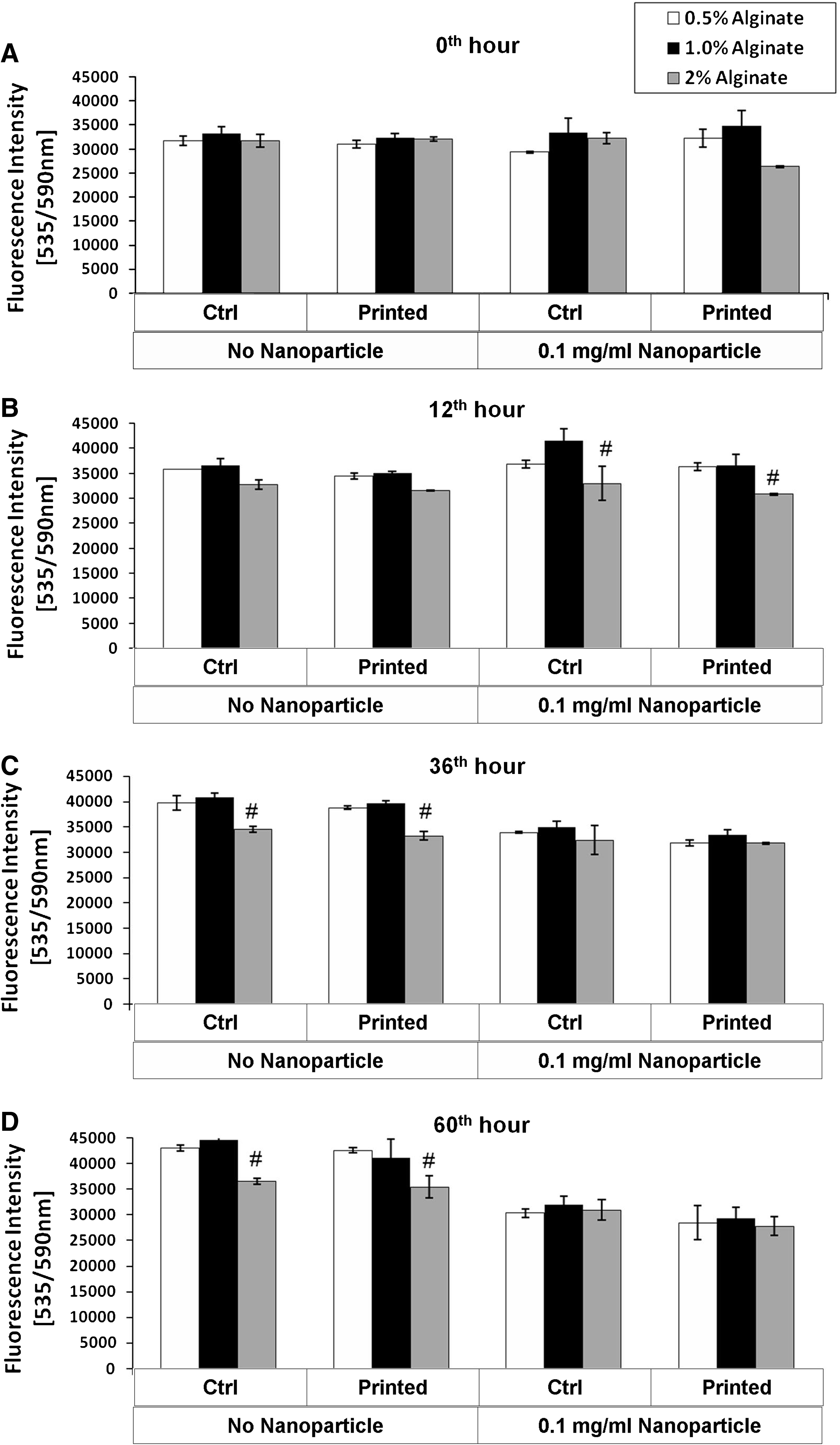
A higher viscosity alginate scaffold decreases cell viability; however, effect of timing depends on printing and nanoparticles. Cell viability for PAECs in 0.5%, 1%, and 2% (w/v) alginate and 0 or 0.1 mg/mL nanoparticles at 0 h (
Effect of cellular nanoparticle uptake on printed cell viability
Magnetically labeled cells, internally loaded with iron oxide nanoparticles, could be used to track and move cells printed within a tissue-engineered structure. The viability of nanoparticle-loaded cells was examined after printing in 1% alginate, with an initial dispensing pressure of 5 psi. Both control and printed samples without nanoparticles showed increased viability at time points up to 60 h. However, a steep decrease in cell viability was observed from 0 to 36 h for both control and printed cells loaded with either 0.1 or 1.0 mg/mL nanoparticles (Fig. 4A). Printed cells showed the most dramatic change, with a 40% decrease in the Alamar blue fluorescence when compared with printed cells without nanoparticles at 36 h. This viability change was in direct contrast to the lack of printing effect for samples with nanoparticles in the alginate. Although early cell viability was significantly decreased, there was no significant change at time points after 36 h, suggesting stabilization of the remaining cell population. When printing pressure was decreased to 2 psi, cell viability increased almost 20% (Fig. 4B).
Cell viability decreased for cells loaded with 0.1 and 1.0 mg/mL nanoparticles and the decrease was accentuated by printing. PAEC viability for cells printed in 1% alginate at a dispensing pressure of (
Nanoparticle and cell manipulation inside the alginate
The nanoparticles were magnetically manipulated within the alginate to determine if nanoparticles could be used to move bioactive factors after printing. The 1.0 mg/mL nanoparticles were homogenously distributed in 1%, 2%, and 3% (w/v) alginate, printed in bulk samples, and left as a viscous liquid or crosslinked with calcium chloride to form a gel (Fig. 5A, C, E, G, I, K). The nanoparticles printed in either 1% or 2% (w/v) alginate without calcium chloride moved toward the NdFeB magnet placed under the cell culture dish within a minute (Fig. 5B, D; arrows indicate nanoparticles at the magnet edge). However, no nanoparticle movement was observed in the 3% (w/v) alginate solution, likely because of the high alginate solution viscosity (Fig. 5F). When the samples were crosslinked with calcium chloride, nanoparticles similarly moved toward the magnet edge in the 1% and 2% (w/v) alginate, but not in the 3% alginate (Fig. 5H, J, L). However, the nanoparticles moved more slowly, and less spatial repositioning of nanoparticles was observed.
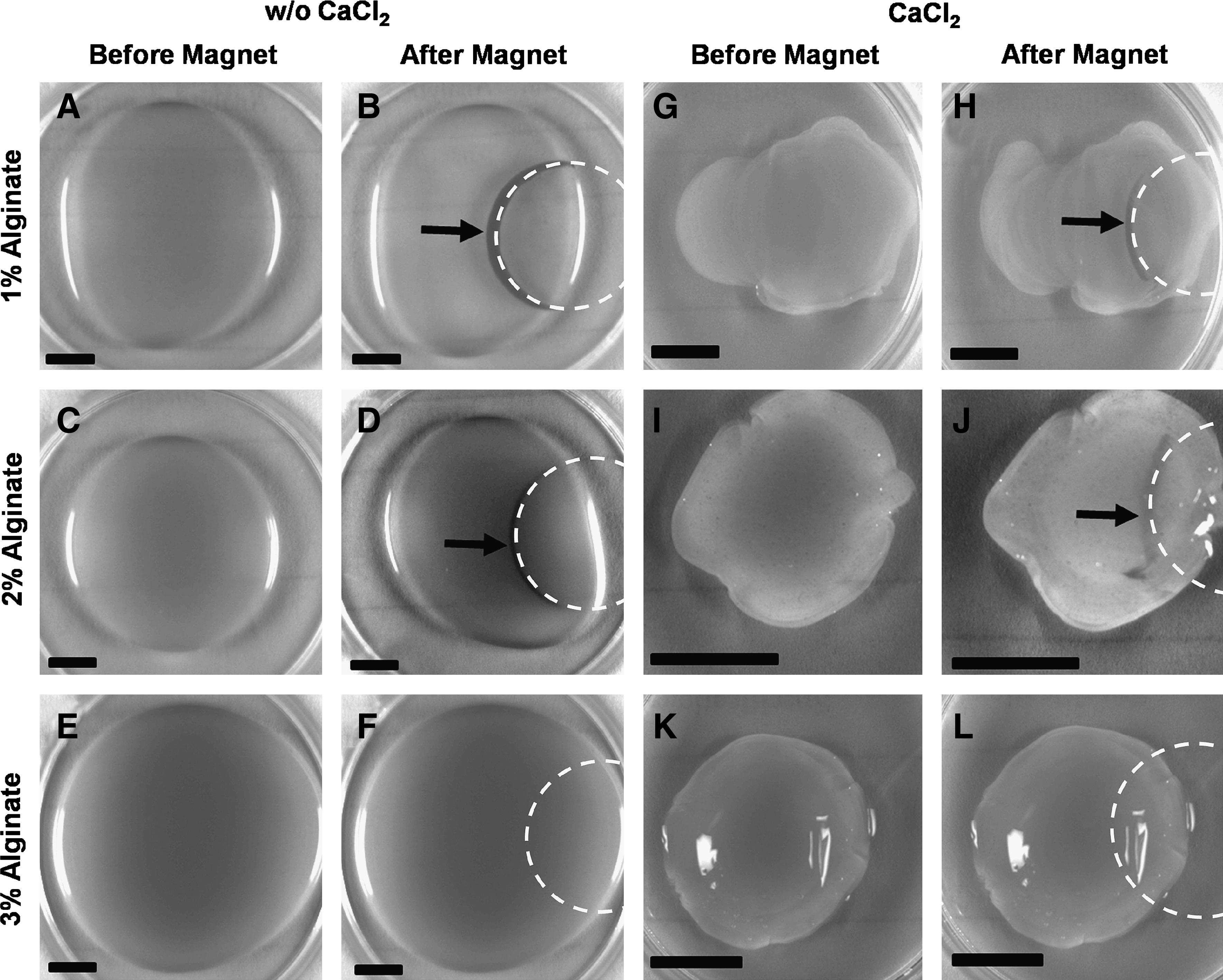
Bioprinted nanoparticles in the alginate scaffold move toward the magnet in a manner dependent on scaffold viscosity. The 1.0 mg/mL nanoparticles homogenously distributed in 1%, 2%, and 3% (w/v) alginate (
We next investigated if cells loaded with magnetic nanoparticles could be moved within the alginate biopolymer. PAECs magnetically labeled with nanoparticles were initially homogenously distributed in 0.5% and 1% (w/v) alginate (Fig. 6A, E, I; higher magnification in Fig. 6B, F, J). Magnetically labeled cells moved toward the NdFeB magnet placed under the cell culture dish (Fig. 6C, G, K). At higher magnification, individual cells were seen at the magnet edge (arrows in Fig. 6D, H, L). Isolated nanoparticles can also be seen in the alginate, which are likely artifacts of incomplete nanoparticle removal from the cell solution when it was mixed with alginate. Magnetically labeled cells continued to cluster at the magnet edge in the crosslinked alginate, but no movement was observed in alginate concentrations higher than 1%.

Magnetically labeled cells can be moved within the alginate scaffold using a magnet. PAECs loaded with 1 mg/mL nanoparticles homogenously distributed in 0.5% and 1% (w/v) alginate (
Specified patterns of nanoparticles and magnetically labeled cells were printed and moved using a magnetic field. The 1% alginate with iron oxide nanoparticles was printed in a pattern (Fig. 7A), and a magnetic field was used to move the nanoparticles to the printed pattern tips (Fig. 7B). The basic shapes (lines and rectangles) of either nanoparticles (Fig. 7C, D, G, H) or magnetically labeled cells (Fig. 7E, F) were moved to new locations while maintaining the original pattern.
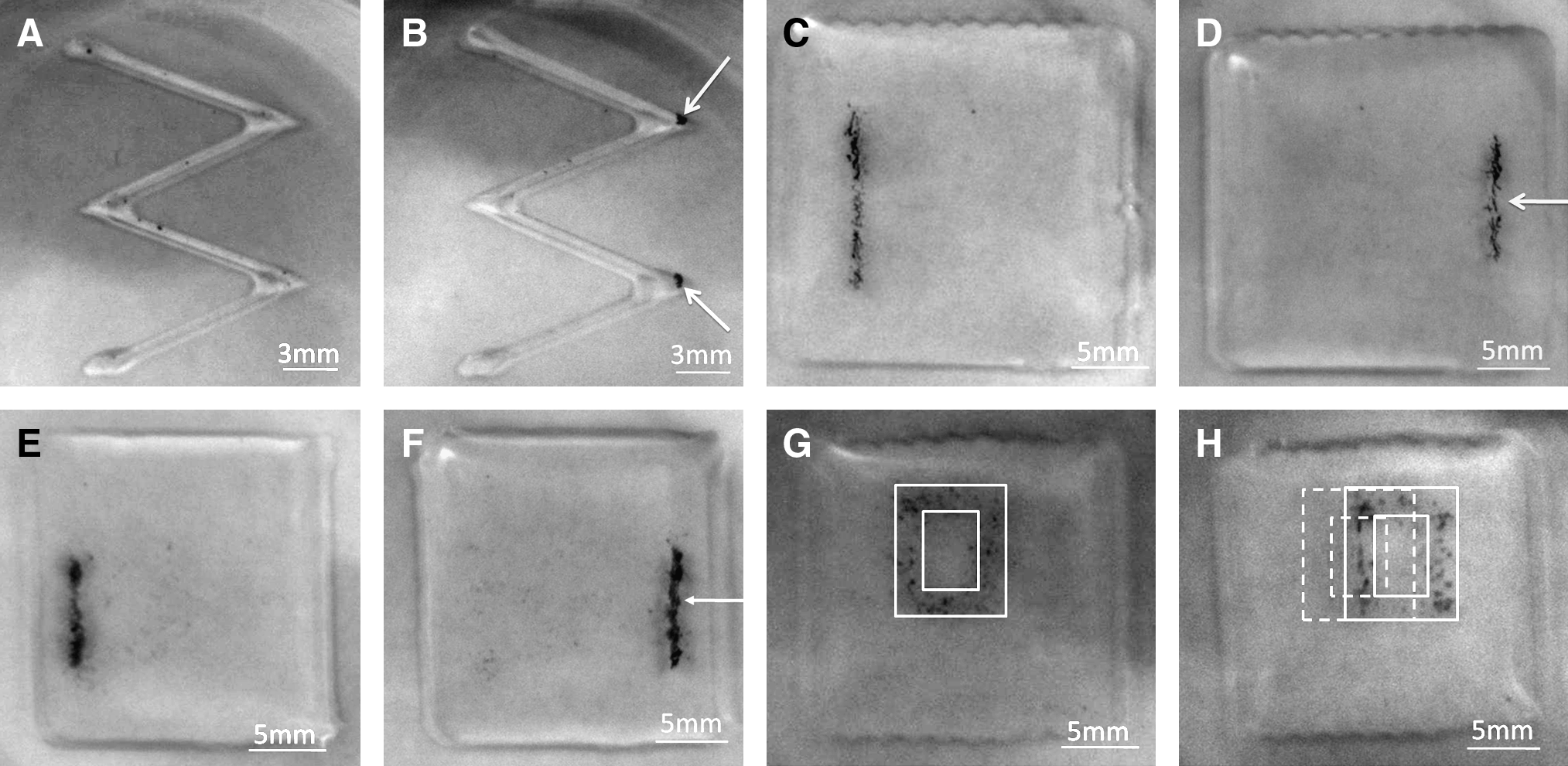
Printed shapes of nanoparticles or magnetically labeled cells in alginate were moved to new locations using a magnetic field. (
MicroCT scan of 3D deposited tissue scaffold
Magnetic nanoparticles printed within 3D alginate scaffolds were imaged by microCT to determine if nanoparticle printing would allow noninvasive tracking of bioactive factors and cell location in tissue-engineered structures. A nanoparticle–alginate prepolymer solution was encapsulated in alginate biopolymer solution using layer-by-layer deposition with the solid freeform fabrication-based direct cell writing system. Printed nanoparticle clusters were clearly visible by microCT scanning of the 3D tissue scaffold (Fig. 8, arrows).

The nanoparticles printed within a three-dimensional alginate biopolymer are visible by microcomputed tomography. Images represent sample cross sections in the (
Discussion
Enhanced nondestructive imaging of cellular and biochemical interactions within 3D tissues would advance the knowledge of tissue development, and the ability to precisely pattern cells and bioactive factors throughout the tissue growth process would improve fabrication of complex tissues. A combination of bioprinting, which allows initial patterning, and superparamagnetic nanoparticles, which allow tracking and repatterning, could help realize these tissue engineering goals. We now show that magnetic nanoparticles can be bioprinted for tissue engineering applications. Printing nanoparticles in the alginate biopolymer did not impact cell viability more than the presence of nanoparticles. Further, while printing cells loaded with nanoparticles did decrease cell viability, the viable cells were stabilized shortly after printing. Magnetically labeled biofactors and cells could be moved within the alginate structures in the presence of an externally applied magnetic field, or imaged nondestructively using microCT.
Sodium alginate was used in all experiments because it is nontoxic, remains as viscous liquid at room temperature, and crosslinks to form a gel under mild conditions. However, endothelial cells are unable to specifically interact with alginate, which prevents cell anchorage and attachment in the biopolymer. 25 The cells have a rounded morphology because they are encapsulated in, not adhered to, the polymer scaffold. As the printing system operates at room temperature, we are currently unable to print polymers such as collagen, to which the cells would attach, because collagen solidifies into a stiff gel at room temperature. In our experiments, alginate is advantageous because it maintains cell number without proliferation, which allows improved observation of cell death. In our future work, we will incorporate a cooling system into our bioprinting device to print collagen gels. We will then be able to study cell viability and proliferation after cells printed with nanoparticles have attached to the scaffold.
By bioprinting nanoparticles in the biopolymer, bioactive factors such as growth factors, antibodies, drugs, and genes conjugated to the magnetic nanoparticles can be precisely patterned within a 3D scaffold. Chemical coupling via amide or ester bonds has been used by others to conjugate bioactive factors to iron oxide nanoparticles. Linker molecules, including 1-ethyl-3-(3-dimethylaminopropyl) carbodi-imide hydrochloride (EDCI), N-succinimidyl 3-(2-pyridyldithio) propionate, or N-hydroxysuccinimide were used to attach targeting ligands and proteins to nanoparticle surfaces. 16 Insulin, lactoferrin, and ceruloplasmin were successfully conjugated to superparamagnetic iron oxide nanoparticles using EDCI. These nanoparticles were then targeted to cell surface receptors, thereby avoiding endocytosis, to achieve tissue and cell-specific drug targeting.26,27 Magnetic nanoparticles have also been dually conjugated, both with a ligand specific for a target cancer cell receptor and a cancer therapy drug. For instance, a radiolabeled anti-vascular endothelial growth factor monoclonal antibody was conjugated to magnetic nanoparticles to both target and deliver radiation therapy to liver cancer. 28 By chemically conjugating bioactive factors to the nanoparticles, it would be possible to initially pattern the bioactive factors in the scaffold and then move them during tissue growth. This technique could, for example, provide endothelial cells with a changing growth factor gradient to promote angiogenesis.
In this study, we observed decreased viability for cells exposed to both low and high nanoparticle concentrations in the alginate, independent of bioprinting. However, in our previous studies, endothelial cell viability was largely preserved in 2D culture up to an iron oxide nanoparticle concentration of 0.5 mg/mL. 29 This apparent contradiction might be related to differences in 2D and 3D endothelial cell culture. In 2D cultures, endothelial cells form a stable, confluent monolayer, whereas the same cells form tubes when grown in a 3D matrix. 30 The 3D angiogenic endothelial cell phenotype may be more susceptible to nanoparticle toxicity than the 2D monolayer. Cells may be able to uptake more nanoparticles in the 3D alginate structure because they are exposed to nanoparticles on all sides. In 2D culture, cells interact with nanoparticles only at their apical surface. The effect could also be specific to the alginate scaffold because cells do not attach to the alginate. These attachment-dependent cells may experience changes in critical cell functions when encapsulated, which coupled with functional changes caused by nanoparticle uptake may enhance nanoparticle cell toxicity. 31 Interestingly, long-term tests showed that cell viability is stable after 72 h, perhaps because all available nanoparticles in the vicinity of a cell have already been taken up.
The printing process itself did not impact endothelial cell viability when nanoparticles were mixed with the alginate. We hypothesize that cell viability is preserved because nanoparticles are free to move within the alginate without damaging cells during printing. In direct contrast, when nanoparticles were loaded inside cells, there were fewer viable printed cells than control cells but this decrease in viability was found to be related to the printing pressure. The system operates at printing pressures ranging from 1 to 40 psi. The majority of our experiments were conducted at a printing pressure of 5 psi, which was shown in previous studies to maintain cell viability. Only at pressures greater than 20 psi did cell viability decrease. 23 As dispensing pressure was lowered to 2 psi, the forces imposed on the cell as it moved through the nozzle decreased, which likely increased cell viability. However, the printing process at 2 psi dispensing pressure took more than twice as long, and so, for automated mass production, higher printing pressures such as 5 psi might still be preferred with the known loss in cell viability. At both pressures, cells without magnetic nanoparticles recovered in time from the mechanical perturbation and eventually reached a steady-state condition.
When cells uptake nanoparticles, they form vacuoles which disrupt the cell cytoskeleton. 26 The cells with a disrupted cytoskeleton may experience increased damage due to printing-induced forces. Alternatively, the nanoparticles inside cells may be more likely to damage the cell membrane through direct shear effects during the printing process. As the cells are pushed through the nozzle, the nanoparticles may break through the cell membrane or the nucleus, causing irreversible damage. It is also possible that the cells loaded with nanoparticles are already in a state of internal stress, perhaps because of reactive oxygen species. Intracellular reactive oxygen species generation is hypothesized to increase with nanoparticle uptake, leading to protein, DNA, and tissue injury and eventually cell death. 32 The addition of mechanical stress due to the bioprinting process may be more toxic when cells are already biochemically stressed by the internalized nanoparticles prior to the printing process. Both printing parameters and nanoparticle conditions will need to be optimized to minimize cell death.
Cell toxicity could alternatively be decreased while still allowing cell tracking, by attaching bioconjugated nanoparticles to the cell membrane. However, it might be difficult to manipulate cells with externally attached nanoparticles. The nanoparticles may detach during printing because of mechanical forces as the cell moves through the nozzle, or the nanoparticles may detach during cell movement through the scaffold because of viscous drag. Further, attaching nanoparticles to cells via a surface receptor could activate unwanted intracellular signaling, or prevent the cell from using that receptor to perform a particular function. Keeping these challenges in mind, in our future work, we will investigate whether printing and moving cells with membrane-attached nanoparticles is feasible and maintains cell viability.
Our studies revealed that cell viability decreased for samples printed in 2% (w/v) alginate when compared with 1.0% (w/v) alginate with and without any nanoparticles. The high solution viscosity may have exposed cells to higher printing forces during the cell-dispensing process. Cell membranes are highly fragile to mechanical loads, and excessive membrane perturbation can lead to cell death. 33 The initial cell viability change observed from 0 to 12 h in samples with nanoparticles in 2% alginate suggests that nanoparticles further increased the biopolymer viscosity. At later times (36 and 60 h after printing), the presence of nanoparticles inside the alginate overcame this initial cell viability decrease in the 2% alginate, and there was no difference among the different alginate samples. Even the cells without nanoparticles demonstrated decreased viability in the long term because of the 2% alginate, which was in contrast to the recovery process for cells in 1% alginate. This suggests that endothelial cell health is compromised in the stiffer gel; however, additional studies are needed for confirmation.
Although the cells in our experiments showed lower viability in stiffer gels, other studies have shown that cells prefer stiffer substrates. In 2D culture, cells form large, stable focal adhesions on stiff substrates, whereas cells form irregularly shaped, dynamic adhesions on softer substrates. 34 However, for 3D cell studies, cell migration speed and viability may depend not only on the substrate stiffness but also substrate adhesivity or cell–matrix adhesion availability. 35 Peyton and Putnam 36 found that when cell adhesiveness was reduced using an integrin-blocking antibody, the maximum cell migration speed shifted from stiffer to softer Matrigel substrates. Our printed cells are in 3D alginate gels to which they do not attach, and therefore, it is possible that unattached cells are more viable in softer alginate gels. There are rich opportunities for future studies to address mechanisms underlying these distinct differences in cell viability and migration in 2D and 3D environments and the relationship between substrate stiffness and cell–matrix adhesion.
The hybrid nanobioprinting system allows initial nanoparticle and cell patterning by computer-controlled printing, after which the nanoparticles and cells can be moved to a new location either in the initial pattern or in a new pattern defined by the magnetic field. We now also showed that nanoparticle movement depends on alginate viscosity. The nanoparticles moved toward the NdFeB magnet in 1% and 2%, but not 3% alginate. Movement of a single spherical magnetic nanoparticle at steady state in an external magnetic field is driven by the force due to the magnetic field gradient and opposed by the force due to viscous drag,37,38 which are given as follows:
where m, B, η, d, and v are the nanoparticle net magnetic moment, magnetic field, suspending medium viscosity, nanoparticle diameter, and instantaneous nanoparticle velocity, respectively. Considering a one-dimensional problem along the centerline of the magnet (x axis) at steady state, a force balance between equations (1) and (2) leads to a velocity given as
where Ms is the particle saturation magnetization and dB/dx is the magnetic field gradient along the central axis. As seen from equation (3), the nanoparticle velocity inside the alginate is inversely proportional to the medium viscosity and directly proportional to the magnetic field gradient. So in a higher viscosity biopolymer, a stronger magnetic field will be needed to move the same nanoparticle. Even though the nanoparticles did not noticeably move in 3% (w/v) alginate, and nanoparticle movement decreased with crosslinking, it may be possible to move these nanoparticles in the more viscous biopolymer with a stronger magnet. These nanoparticles could even be removed prior to tissue implantation, which would decrease any potential negative effects in vivo. When a magnet was placed next to a nanoparticle–alginate scaffold, the nanoparticles moved completely out of the alginate and attached to the magnet.
Magnetically labeling cells allows nondestructive imaging by microCT in the 3D tissue-engineered scaffold. The intracellular nanoparticle loading should be optimized to obtain maximum signal intensity while at the same time protecting cell viability. Our studies showed that it may be possible to qualitatively determine the nanoparticle or magnetically labeled cell density at a given location by microCT signal intensity. A sixfold increase in signal intensity was observed for 100,000 cells loaded with 1.0 mg/mL iron oxide nanoparticles when compared with the same cell number loaded with 0.1 mg/mL nanoparticles. However, excess nanoparticles that were not taken up by cells could also increase the microCT signal intensity. Nanoparticle loading parameters should be selected to minimize free nanoparticles, and any excess nanoparticles should be thoroughly washed away prior to cell printing. Magnetically labeled dead cells will light up on the microCT scans, which may decrease live cell tracking efficacy. An additional assay, such as Alamar blue or live/dead, may be needed to differentiate live from dead cells. Live magnetically labeled cells would also respond to biochemical signals by proliferating, moving, and forming 3D structures, which should help distinguish them from dead cells in microCT images.
Although cell viability was decreased with nanoparticle loading and printing, there is still potential by optimizing nanoparticle concentration and printing parameters to bioprint magnetically labeled cells. In tissue engineering, the development of complex 3D tissues requires various cell types, such as smooth muscle and endothelial cells for vascular systems or hepatocytes and sinusoidal endothelial cells for liver. However, cell–cell interactions are difficult to manipulate in coculture systems, even in 2D cultures. Ito et al. 39 used magnetic force to place magnetically labeled cells onto target cells and promote heterotypic cell–cell adhesion. The solid freeform cell writing system could enable assembly of 3D patterned tissue engineering constructs with various magnetically labeled cell types. Through microCT, both bioactive factors and cells could be noninvasively imaged within the tissue-engineered scaffold, which would allow longitudinal studies of tissue development. This hybrid nanobioprinting technique, which uses a combination of bioprinting and active magnetic patterning, could dramatically impact our ability to understand and recreate complex tissue development.
We have shown that a solid freeform fabrication system can be used to create magnetically functionalized 3D tissue scaffolds, which can be manipulated after printing using a magnetic field. This system could be used to add bioactive factors or specific cell types into a precisely patterned tissue-engineered scaffold, and later to move or remove these agents. Additionally, the bioactive agents or cells could be imaged within the tissue by MRI or CT. In our future work, we will improve printed magnetically labeled cell viability by exploring different nanoparticle sizes and shapes, as well as attaching nanoparticles to the cell membrane. The nanoparticles will be conjugated with bioactive factors, and cells will be printed in collagen gels with a cooled printing system. Finally, complex magnetic arrangements will be used to control cell movement and patterning after printing in the tissue-engineered construct.
Footnotes
Acknowledgments
This work was partially supported by the Mechanical Engineering and Mechanics Department of Drexel University. The authors thank Wonjin Jo for her assistance with the solid freeform fabrication cell writing system.
Disclosure Statement
No competing financial interests exist.