
Abstract
Bio-artificial liver support systems have been utilized as bridging devices to support acute and chronic liver injury. However, prolonged function of adult hepatocytes has not been achieved due to compromised proliferation and long-term survival of adult cells in vitro. As an alternative cell source, we investigated the potential of human fetal hepatocytes (hFH) in a four-compartment hollow fiber–based three-dimensional (3D) perfusion culture system. hFH were isolated from 17- to 19-gestational-week livers and cultured in the 3D perfusion bioreactors for 14 days. Metabolism activity, hepatocyte-specific gene expression, protein expression, and hepatic function were investigated. Increased glucose consumption and lactate production indicated cell proliferation in the bioreactor. The ratio of cytochrome P450 3A4 to 3A7 gene expression and the increase of the number of asialoglycoprotein receptor-positive cells indicated cell differentiation into mature hepatocytes. Histological and immunohistochemical analysis revealed reorganization of fetal liver cells. Hepatic function was further examined for ammonia metabolism and for albumin production using colorimetric assays and enzyme-linked immunosorbent assay, respectively. In contrast to conventional 2D culture, the 3D perfusion culture system induced functional maturation to hFH; these cells may be useful as an alternative cell source for extracorporeal liver support.
Introduction
One remaining key issue is identifying the right cell source to charge BALs with. There are several candidate cell types: adult human hepatocytes, xenogeneic hepatocytes (mainly porcine), and human hepatoblastoma cell lines, or immortalized human hepatocytes. Although porcine hepatocytes are more readily available,7,11,12 the concerns for potential zoonotic transmission, species differences, and immunological problems reduce enthusiasm for their clinical application.13,14 Human hepatoblastoma cell lines 15 and immortalized human hepatocytes 16 were suggested to overcome the shortage of adult human hepatocytes; however, their safety and biological efficiency are still major concerns. 17
Human fetal hepatocytes (hFH) are also considered as an alternative cells source, 18 as their intrinsic proliferative capacity and functional authenticity are attractive for BAL applications. There is no risk of zoonotic transmission and immunological problems such as those associated with xenogeneic hepatocytes. Since cells are not genetically modified, there is reduced risk of tumorigenicity by contrast to hepatoma cell lines or immortalized human cell lines. On the other hand, cell number availability and functional maturity represent issues of concern for fetal cells. Our goal was to investigate the culture behavior of hFH in a three-dimensional (3D) perfusion bioreactor developed for liver support. In this study, a downscaled (8-mL cell compartment size) 3D perfusion bioreactor was used to explore culturing 17–19-week-gestational-age hFH. As various authors have described, 3D culture and physical parameters such as flow improve the culture conditions for hepatocytes in vitro.19,20 Also, it has been previously described that cell density for hepatocyte culture in bioreactor culture plays a crucial role.21,22 Previous studies using the 4-compartment hollow fiber membrane bioreactor developed by our group demonstrated that the system is capable to maintain hepatic function of adult hepatocytes for a prolonged period of time. 19 In a recent study using mouse embryonic stem cells we demonstrated that these cells spontaneously differentiate and organize into in vivo–like structures under 3D perfusion culture conditions. 23 We hypothesized that a high-density 3D perfusion bioreactor system may support in vitro expansion of hFH while allowing maturation.
In this study, a total of six laboratory-scale high-density 3D perfusion bioreactor experiments were performed. An exhibiting 8-mL cell compartment was charged with 17–19-week-gestational-age hFH. To determine whether prolonged culture time is important to enhance the outcome, two different culture periods were chosen, 7 and 14 days, respectively, with three individual experiments for each culture period.
Materials and Methods
Bioreactor, tubing, and perfusion units
The bioreactor used was designed to simulate the hepatic vascular environment with hollow fiber technology.24,25 Bioreactors (0008/BR) and perfusion units (PS/2–8 mL) were prototyped by Stem Cell Systems (Berlin, Germany). Each bioreactor contains two bundles of hydrophilic hollow fiber microfiltration membranes (400 kDa cut-off) for transport of culture medium in a decentralized pattern, interwoven with one bundle of hydrophobic multi-laminate hollow fiber oxygenation membranes for transport of oxygen and carbon dioxide (CO2)26,27 (Fig. 1). Cells were cultured in the interstitial spaces between the fibers. The perfusion device contains two modular pump units, a heating unit, and a gas-mixing unit (Fig. 2). The perfusion tubing with bubble traps is made of standard medical-grade dialysis PVC (B. Braun, Melsungen, Germany). Sterilization is performed with ethylene oxide according to clinical standards.
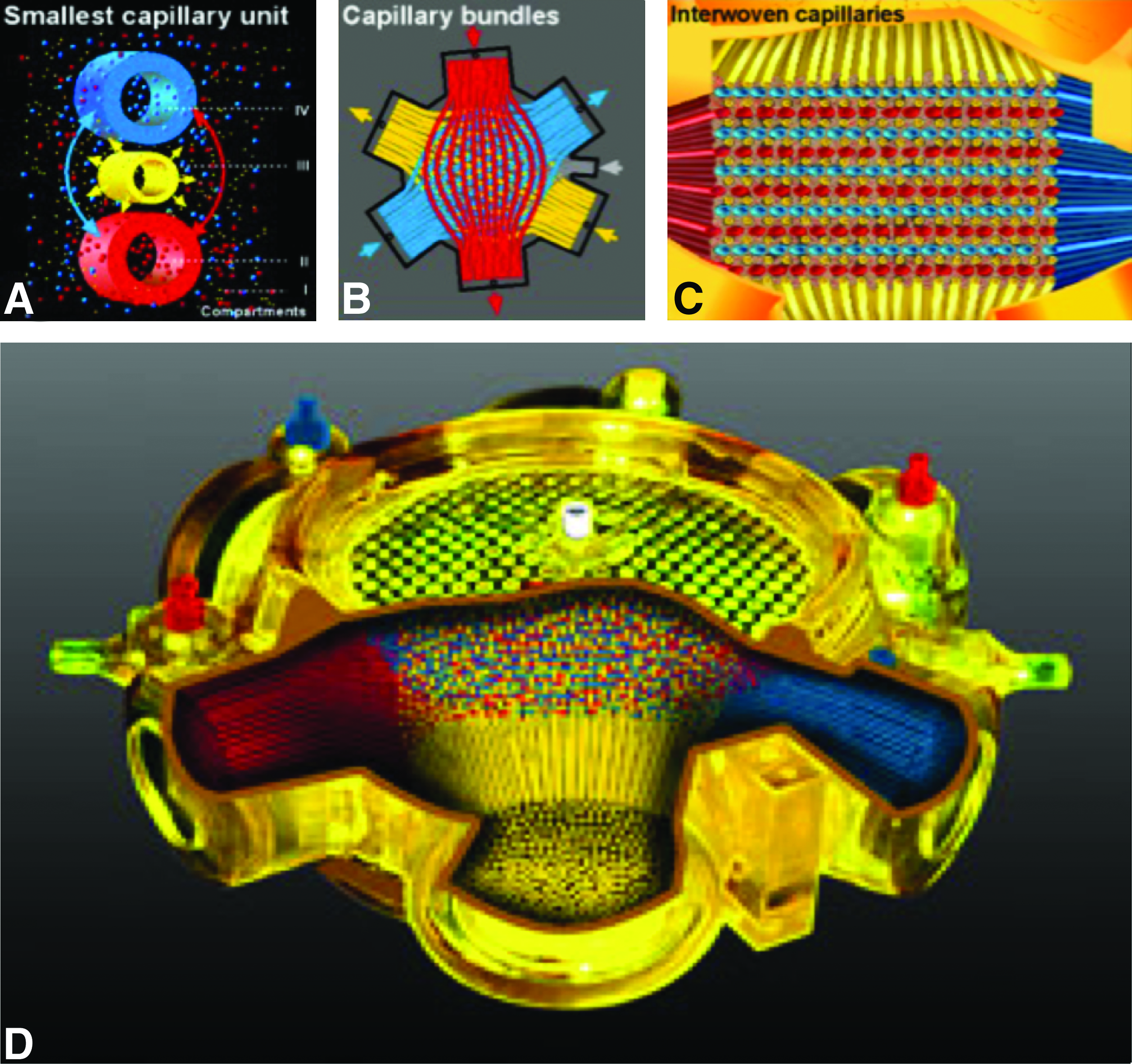
Bioreactor design technology. (
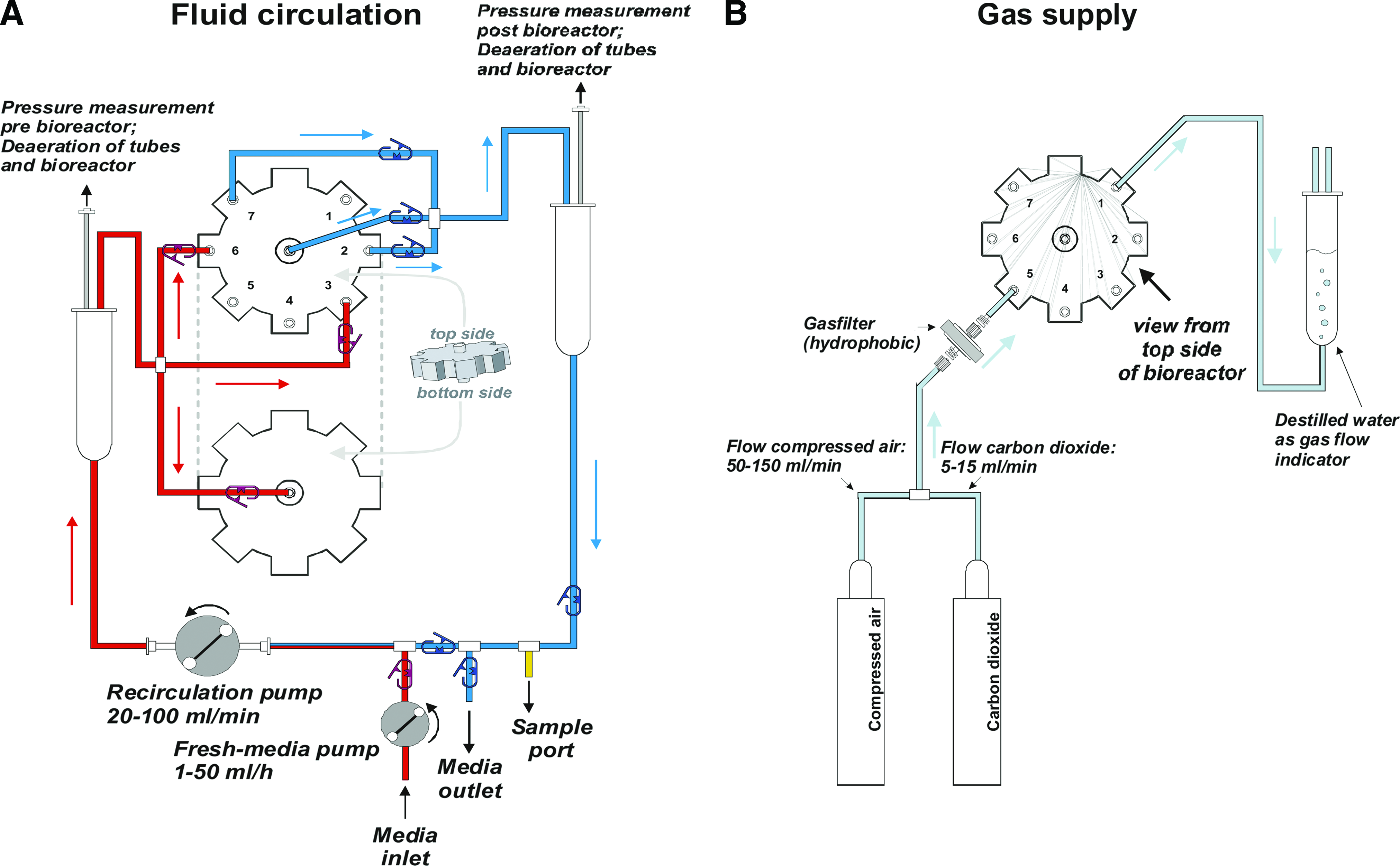
Fluid circulation and gas supply. (
Human fetal cell isolation and culture
Human fetal liver tissues of 17–19 week gestational age were obtained from the Allegheny Reproductive Health Center with internal review board approval of the University of Pittsburgh and prior written informed consent of the donor. To isolate hFH, fetal livers were enzymatically digested, followed by a density-based and cell-anchorage-dependent hepatocyte enrichment protocol. Briefly, the procured fetal liver tissues were placed into 0.5 M ethylene glycol tetra acetic acid solution and mechanically disrupted using a cell scraper. The tissue/cell suspension was then washed in phosphate-buffered saline with calcium (Ca2+) and magnesium (Mg2+) and incubated with 0.04% collagenase Type II plus DNase I (2000 U) (both from Sigma–Aldrich, St. Louis, MO) for 15–30 min at 37°C. All washing steps used centrifugation at 50 g.
After isolation, human fetal liver cells were seeded on EmbryoMax® ES Cell Qualified 0.1% Gelatin Solution (Millipore, Billerica, MA)–coated plates and incubated for 48 h at 37°C with 5% CO2, followed by phosphate-buffered saline washing to remove nonadherent cells and debris. Morphology confirmed that more than 90% of cells were hepatocytes, which were subsequently dissociated by incubation with trypsin–ethylenediaminetetraacetic acid for 3–5 min at 37°C. Cell count and viability was estimated using the trypan blue dye exclusion method.
Cell culture
2D culture
A total of 1 × 10⁶ cells were cultured on each conventional 100 mm plastic Petri dish, precoated with type I collagen (Biocoat; Becton Dickinson Labware, Bedford, MA). Cells were cultured in a humidified, 5% CO2, 95% air environment at 37°C. Culture medium was changed every day.
Bioreactor culture
Three independent bioreactor cultures were performed for each period (7 and 14 days). A total of 1 × 10⁸ cells were inoculated into each bioreactor. To provide extracellular matrix components' similar to collagen-coated dishes used for 2D culture, the collagen macroporous carrier Cultispher-S® (Sigma–Aldrich) was used. Due to a washout effect of the bioreactor, liquid collagen or gelatin coating solutions are not suitable. Preliminary experiments had been conducted to choose Cultispher-S, which supports the hFH settlement in the bioreactor. Since the diameter of Cultisphere-S particles exceeds the pore size of the hollow fiber membranes, these particles remain in the cell compartment. The Cultisphere-S beads were weighed and aliquoted. For each experiment, 0.2 g rehydrated beads were mixed and inoculated with 20 mL cell suspension.
To ensure even cell distribution, early prototypes with different cell inoculation tube configurations (distribution and numbers) were investigated by fat emulsion inoculation and subsequent observation of distribution within the cell compartment by magnetic resonance imaging. On the basis of these results, an optimal configuration was chosen: 16-cell inoculation tubes are incorporated throughout the cell compartment, providing even distribution of inoculated cell suspension.
Bioreactors were continuously perfused with culture medium at a recirculation rate of 30 mL/min. The volume of recirculation circuit is 50 mL. Fresh medium was continuously added from an external reservoir (Fig. 2A). Feed rate was set to a starting value of 2 mL/h, which equals one complete medium change in 24 h. To maintain a neutral pH and sufficient supply of cell nutrition, including glucose, the continuous fresh medium supply rate (pump speed) was adjusted (up to 8 mL/h) based on the daily measurements of pH and glucose concentration in the recirculation medium during bioreactor culture. The total flow of compressed air (medical grade, provided from the laboratory infrastructure) and CO2 (medical grade, conventional tank) is sterile filtered and mixed in the perfusion device, which is equipped with three rotameters to regulate gas flow (Fig. 2B). The gas flow rate was set to 35 mL/min, with a starting mixture of 95% air and 5% CO2. On the basis of the daily measurement of pH, the CO2 flow is adjusted to optimize the pH of the bicarbonate buffered culture medium. A handheld bedside analyzer iSTAT (Abbott Laboratories, Princeton, NJ) was used to measure glucose, lactate, pH, and CO2. At each end-point of the bioreactor culture, the upper bioreactor lid was opened and the cell mass, including the capillary layers, was cut from the surrounding potting and transferred into a sterile glass vessel for dividing into sections for further analysis.
Culture medium
Cells were cultured in Dulbecco's modified Eagle's medium/F-12 (Invitrogen, Grand Island, NY) supplemented with 10% fetal bovine serum (Invitrogen), 0.8 mg/L insulin, 5 mg/L transferrin, 0.003 mg/L glucagon (ITG Supplement; Biochrom AG, Berlin, Germany), 10−6 M dexamethasone (Sigma–Aldrich), 10 ng/mL human recombinant epidermal growth factor (BD Bioscience, Franklin Lakes, NJ), 2 mM L-glutamine (Invitrogen), 100 U/mL penicillin G, and 100 μg/mL streptomycin (Invitrogen).
Metabolic activity monitoring
The metabolic activity of the cells inside the bioreactors was evaluated every day by measuring glucose and lactate concentrations in the culture medium with iSTAT/Glu and iSTAT/CD4+ cartridges, respectively. The concentrations were adjusted by inoculated cell number and feeding rate to normalize individual bioreactor experiments. A total of six bioreactor cultures were performed, and the data were plotted using GraphPad Prism (version 5.0; GraphPad Software, La Jolla, CA).
Liver-specific gene expression
Cells were harvested before inoculation into the bioreactor (Pre-BR), 7 or 14 days after bioreactor (Post-BR), and after 2D static culture (2D culture). Total RNA was extracted from cells using the RNeasy® Mini Kit (Qiagen, Valencia, CA). Two micrograms of total RNA was used to synthesize complementary DNA with conventional reverse transcription (Promega, Madison, WI). Quantification of mRNA using real-time polymerase chain reaction analysis was performed using an ABI PRISM 7000 system (Applied Biosystems, Foster City, CA). The predesigned TaqMan probe and primer sets for albumin (Hs00609411_m1), alpha-1 antitrypsin (A1AT) (Hs00165475_m1), cytochrome P450 (CYP)3A4 (Hs00430021_m1), CYP3A7 (Hs00426361_m1), HNF4α (Hs00230853_m1), tyrosine aminotransferase (Hs00356930_m1), G6PC (Hs00609178_m1), and β-actin (Hs99999903_m1) were selected from TaqMan Gene Expression Assays (Applied Biosystems). Relative gene expression was analyzed using delta-delta-Ct (ddCt) methods. All values were then normalized to β-actin mRNA expression and are presented as relative increase of gene expression compared to freshly isolated hFH (day 0). Two-way analysis of variance test and Bonferroni posttest were performed to calculate p-value.
Flow cytometric analysis
To determine functional maturation of hFH, flow cytometric analysis was performed with an anti-asialoglycoprotein receptor (ASGPR) antibody. Before inoculation and after 14 days in culture, a total of 1 × 10⁶ cells were harvested from 2D static culture or 3D perfusion culture and incubated with monoclonal anti-ASGPR antibody (Cell Sciences, Canton, MA) for 1 h, followed by Alexa 488–conjugated goat anti-mouse IgG secondary antibody (Invitrogen, Carlsbad, CA) for 30 min. Mouse IgG1 isotype control antibody (Dako, Carpinteria, CA) was used as negative control.
Analyses were performed on a FACSCalibur system using Cell Quest software (Becton Dickinson, San Jose, CA). Forward and side scatter gates were set to include all viable cells. The percentage of positive cells was measured from a cut-off set using an isotype-matched control antibody using FlowJo software (Tree Star, Ashland, OR). Student's t-tests were performed using the Mann–Whitney U-test with a significance level of 95%.
Measurement of liver-specific functions
To determine the production of albumin, recirculation medium samples were taken every day and stored at −150°C until analysis. Human albumin concentration in the culture medium was determined by Human Albumin ELISA Quantitation kit (Bethyl Laboratory, Montgomery, TX) according to the manufacturer's instructions.
To evaluate the hepatic detoxification function, ammonia challenges were performed on day 4, 7 (n = 3), and 11 (n = 1) of the bioreactor culture cells. In brief, ammonium chloride (NH4Cl; Sigma–Aldrich) was injected into the medium circuit to a final concentration of 5 mM. After 30 min to allow for equimolarity, the cells were incubated for 4 h. During this period the system was operated without fresh medium supply to eliminate dilution. After incubation, samples were collected and urea concentrations were measured by using a colorimetric assay kit (Urea Colorimetric Assay Kit, QuantiChrom; BioAssay Systems, Hayward, CA).
Histological and immunohistochemical analysis
Bioreactor samples were harvested at each end-point of bioreactor culture (day 7 and 14) and immediately embedded into 2% low-melting agarose gel, after fixation in 10% formalin. The paraffin-embedded samples were cut into 4-μm-thick sections, and hematoxylin and eosin staining was performed to evaluate tissue architecture and organization. Immunohistochemistry (IHC) and immunofluorescence (IF) were performed on the paraffin-embedded samples. The antibodies used were polyclonal rabbit anti-human albumin (IHC; DakoCytomation, Carpinteria, CA), polyclonal rabbit anti-human α1 antitrypsin (IHC; Dako), monoclonal mouse anti-human cytokeratin (CK) 19 (IHC; CK19; Chemicon, Temecula, CA), and polyclonal goat anti-Ki67 antibody (IHC; Santa Cruz Biotechnology, Santa Cruz, CA), monoclonal mouse anti-human albumin (IF; Sigma–Aldrich), monoclonal rabbit anti-human CK8 (IF; Abcam, Cambridge, MA), monoclonal mouse anti-human CK19 (IF; Santa Cruz Biotechnology), and polyclonal rabbit anti-human CYP3A4 (IF; Cypex, Dundee, Scotland). Adjacent sections served as negative controls and were incubated with appropriate isotype control antibodies and secondary antibodies. For IHC, the 3,3′-diaminobenzidine Substrate Kit (Vector Laboratories, Burlingame, CA) was used to develop the slides and hematoxylin staining was performed as a nuclear counter staining. Secondary antibodies used for IF were polyclonal goat anti-mouse Alexa Flour 488–conjugated (Invitrogen) and polyclonal goat anti-rabbit Cy3–conjugated (Dianova, Hamburg, Germany). Nuclei were observed with 4′-6-diamidino-2-phenylindole staining.
The periodic acid-Schiff (PAS) staining system (Sigma–Aldrich) was used to observe glycogen in the tissue-like structure according to the manufacturer's instructions.
Statistical analysis
Data were obtained from two to six independent runs under each culture condition and are expressed as means and standard deviations. Statistical analysis was carried out using the statistic and graphic software Prism 5 for Mac OSX (GraphPad Software).
Results
Metabolic activity of fetal hepatocytes in a 3D perfusion bioreactor
Glucose consumption and lactate production of hFH increased continuously during the culture period, indicating cell proliferation and increasing total metabolic activity (Fig. 3A, B). Assuming that each individual cell consumes similar basal quantity of glucose, the overall increasing glucose consumption would reflect fetal hepatocyte proliferation. Overall, the metabolic activity in conjunction with the below described Ki-67 proliferation marker staining results indicates that the hFH were actively proliferating in the 3D bioreactor culture system.
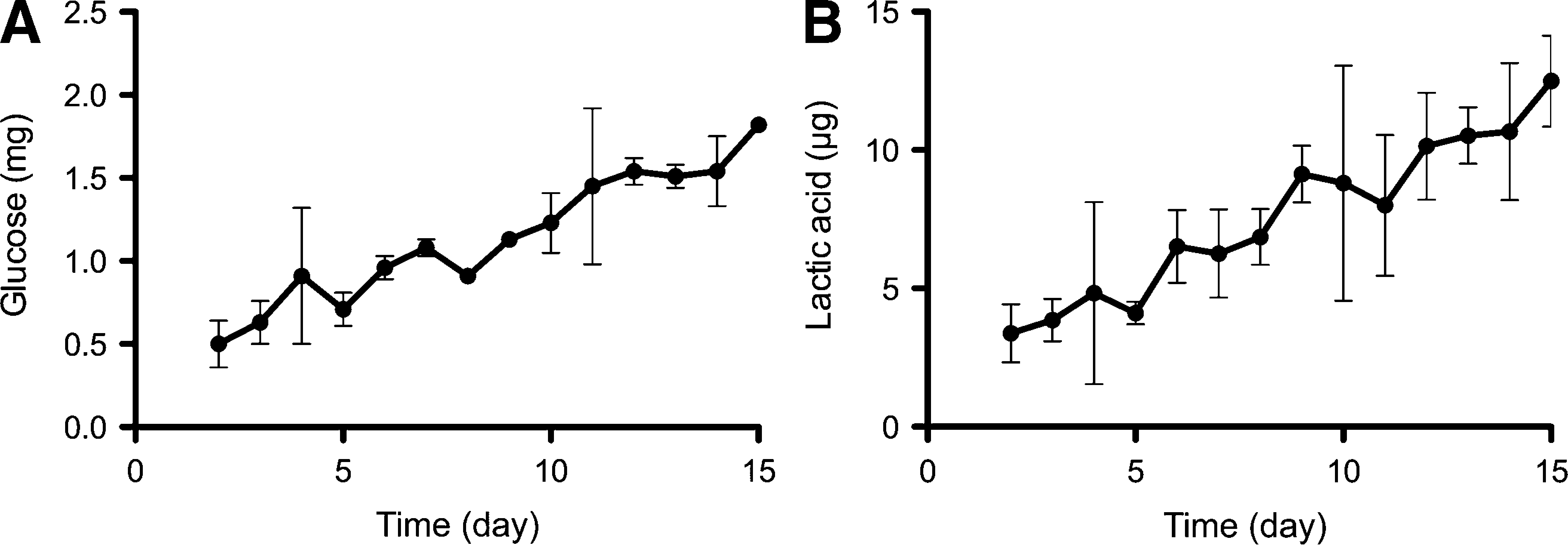
Time course of metabolic activity in a 3D perfusion bioreactor. Metabolic parameters glucose (
Hepatocyte-specific gene expression indicates spontaneous maturation
Quantitative real-time reverse transcriptase–polymerase chain reaction was performed to investigate the maturation of hFH during the 3D perfusion bioreactor culture. Expression of seven hepatic genes (HNF4α, albumin, A1AT, tyrosine aminotransferase, glucose-6-phosphatase, CYP3A4, and CYP3A7) was investigated. Overall, except for CYP3A4, expression of these genes under bioreactor culture conditions did not differ significantly between culture conditions. However, expression of all hepatic genes investigated tended to be higher if the cells were cultured in the 3D bioreactor than in 2D cultures. CYP3A4 gene expression was significantly increased in the 3D bioreactor cultured cells (Fig. 4A). The expression ratio of CYP3A4 and CYP3A7 was compared to evaluate hepatic maturation. The values of day 7 and 14 CYP3A4/3A7 ratio relative to the day 0 sample are depicted in Figure 4B. Although both 2D static and 3D perfusion culture conditions increased CYP3A4 expression, indicating progress of CYP3A7 to CYP3A4 conversion, the ratio was significantly higher in the 3D bioreactor cultured cells (p < 0.05, Fig. 4B). These data suggest that hFH spontaneously differentiated toward mature hepatocytes and that the 3D perfusion culture system induced more maturation than 2D static culture conditions.

Cytochrome P450 3A family genes expression ratio. (
Ratio of ASGPR-positive hepatocytes
The hFH show the presence of ASGPR as demonstrated by fluorescence-activated cell sorting. The anti-ASGPR antibody had been utilized to identify functional mature hepatocytes.28,29 Data from six 3D bioreactor culture experiments were studied and the ratio of functional hepatocytes was demonstrated (Fig. 5). Results were statistically analyzed with the Mann–Whitney test (n = 3). Before culture, freshly isolated hFH contained only a small number of ASGPR-positive cells (6.9 ± 3.5%). This percentage was dramatically increased after 14 days in 3D bioreactor culture (67.11 ± 9.88%), whereas 2D culture induced 25.0 ± 12.29% ASGPR-positive cells after 14 days (Fig. 5). The data confirm that the 3D perfusion culture system is superior to induce hepatic maturation than 2D static culture conditions.
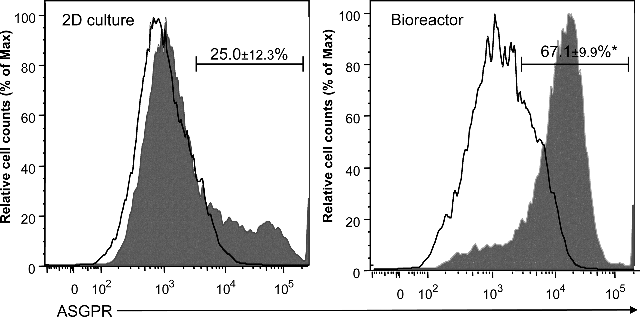
Flow cytometric demonstration of functional hepatocyte cell surface maker-positive cells in 3D perfusion bioreactor. For each analysis, hHF before and after 2D and bioreactor culture were aliquoted into individual tubes at a total of 1 × 10⁶ cells per tube. Analysis was performed using untreated cells, isotype control, and ASGPR-1 antibody. The representative histograms show fluorescent intensity versus percentage of maximum cell number of isotype control (line) and ASGPR-positive cells (shaded). ASGPR-positive cell ratios from three individual experiments are shown in the histogram (n = 3). Values are mean ± SD, *p = 0.0061.
3D perfusion bioreactor maintained hepatic function of hFH
Albumin production was measured as an indicator of hepatic synthetic function (Fig. 6). Albumin showed an initial increase, which could indicate that damaged cells release albumin in the culture adaptation phase, leading to false high levels of albumin. This was confirmed by high levels of lactate dehydrogenase (LDH) (as marker of cell death/disruption) (data not shown). After 5 days the albumin concentration was stable and did not drastically change for 14 days. Total production of albumin per 1 million inoculated cells was on average 3.4-fold higher under 3D conditions compared to 2D culture. Human albumin was not detected in the basal medium.

Change of parameters for hepatocyte-specific functions. (
Urea synthesis by hFH in the 3D bioreactor was investigated to monitor ammonia metabolism activity (Fig. 6B). The increase in activity was evaluated by comparing the levels of urea before and after a 4 h incubation with 5 mM NH4Cl (ammonia challenge test), which show an up to 4.3-fold increase of urea production. The results indicate that hFH in the 3D bioreactor maintained ammonia metabolism function even after 11 days in culture.
Histological and immunohistochemical analysis
Hematoxylin and eosin stain and PAS staining are shown in Figure 7. Immunohistochemical analysis showed that most cells stained positive for albumin and A1AT (Fig. 7B, D) as well as for PAS staining (Fig. 7F), which identifies glycogen storage function of the hepatocytes. Fetal hepatocytes restructured to a hepatic plate-like morphology in the bioreactor. There were only few areas that tested negative for albumin. Most likely, these cells represent nonparenchymal cells that can also be found in organs in vivo. To characterize these cells, the samples were stained with anti-CK19 antibody. CK19 is expressed exclusively in the biliary cells of the adult liver and in biliary lineage cells including bipotential hepatic progenitor cells of the fetal liver. We observed scattered CK19-positive cells and very few, but clearly CK19 positive, tubular structures in the cell mass (Fig. 8B, red arrow). These data indicate that some of the dissociated biliary lineage cells reoriented or proliferated to form bile-duct-like tubular structures within the human fetal liver cells. These tissue formations were not observed under conventional 2D culture.
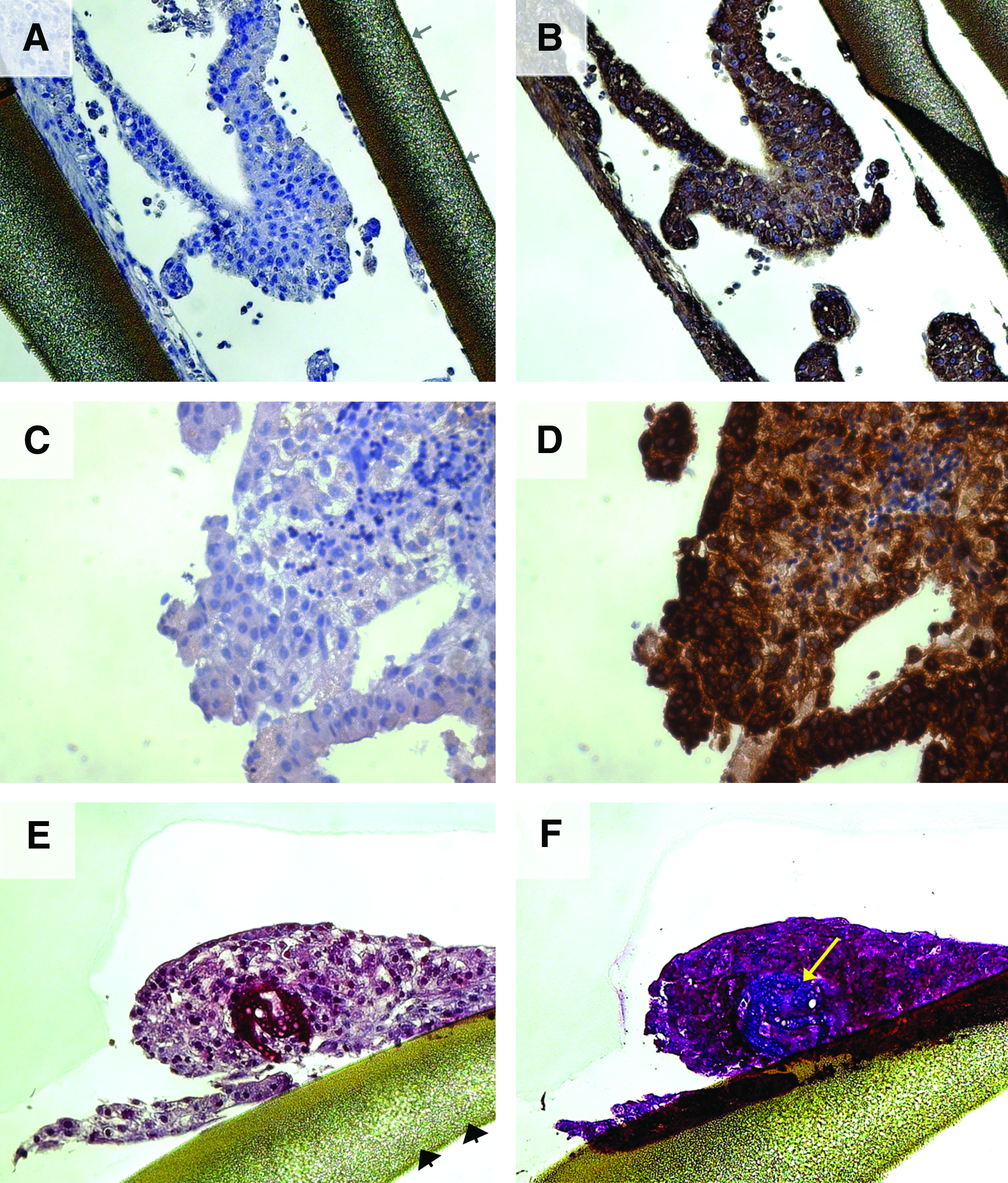
Expression of hepatic proteins after 3D perfusion bioreactor culture. Representative histological sections of 3D bioreactor cultured for 14 days. Paraffin-embedded samples were immunostained against human albumin (20 ×,

Reconstructed liver structure in a bioreactor. Immunohistological analysis with anti-cytokeratin (CK) 19 and Ki-67 (proliferation marker) antibodies. After 14 days in culture, hFH formed ductal structures in the 3D perfusion bioreactor (20 ×,
To observe the proliferating cells, Ki-67 staining was performed (Fig. 8D). Ki-67 is a cellular marker strictly associated with cell proliferation. 30 The protein is present during all active phases of the cell cycle (G1, S, G2, and mitosis), but is absent in resting cells (G0). Supporting the metabolic biochemical data, most of the hepatocyte-like cell nuclei were positive for the Ki-67 antibody (Fig. 8D).
IF analysis
IF double staining was performed with anti-human albumin/CYP3A4 (Fig. 9) and anti-human CK8/CK19 antibodies (Fig. 10) on day 14 samples. Most of the cells stained positive for albumin (Fig. 9A, D) and more than half of the albumin-positive cells were also positive for CYP3A4 (Fig. 9B, D). The observation indicates that hFH are functionally well maintained and induced further maturation in the 3D perfusion bioreactor. To further characterize the cells after cell culture, double staining for CK8 and CK19 was performed. Scattered CK19-positive cells were found (Fig. 10B, D) within the cell clusters that showed predominantly CK8-positive hepatic characteristics (Fig. 10A, D). Duct-like structures were not observed with the CK8/19 double-positive cells. Morphologically, the CK8/19 double-positive cells are small in size and have a low nucleus-to-cytoplasm ratio. Most likely, these cells represent bipotential liver progenitor cells that can give rise to hepatic epithelial and biliary cells.31–33 These results showed that some of the human bipotential liver progenitor cells were still maintained during the 14 days of 3D perfusion bioreactor culture.

Immunofluorescence analysis of the human fetal liver cells in the 3D perfusion bioreactor. Double immunostaining for human albumin (green, 20 ×,

Hepatic progenitor cells were preserved in the 3D perfusion bioreactor. Samples were taken from hFH after 14 days of culture, and double immunostaining for human CK8 (red, 40 ×,
Discussion
Bioengineering developments for extracorporeal temporary liver support offer technology advancements and provide improved culture conditions.34–37 One remaining biological limitation is the lack of a suitable cell source for BAL bioreactors meeting criteria such as safety, availability, and functionality. The goal of this work was to test whether the 3D perfusion culture system enables expansion and induces maturation of hFH, and if so, how it compares with the conventional 2D culture system. Due to the engineering structure of the bioreactor, which is designed in its clinical scale as a component of an extracorporeal liver support system to provide metabolic functions rather than producing removable cells, cell retrieval is very complicated. The dense 3D network within which hepatocytes are cultured and form tissue-like clusters makes it almost impossible to obtain exact cell numbers during and after the cell culture. Therefore, indirect methods such as monitoring of metabolic activity (glucose consumption and lactic acid production) and staining of cored biopsies (analog to clinical liver diagnostics) for the proliferation marker Ki67 were used to assess cell proliferation. Under the assumption that each cell's average glucose consumption and lactate acid production are stable, higher consumption of glucose and production of lactic acid was correlated to augmentation of cell mass in the bioreactor. Steady increase in metabolic activity was observed, suggesting that cells actively proliferated throughout the culture period. Staining for Ki-67, a proliferation marker, showed cell clusters that largely positive cells. Although we did not intend to induce terminal differentiation with exogenous growth factors, the hFH indeed spontaneously differentiated. The drug metabolism enzymes are considered as late-phase hepatocyte-specific genes 7 ; thus, the gene expression ratio of fetal and adult forms of CYP enzymes was used to evaluate hepatic maturation. 38 CYP3A7 is predominantly expressed in hFH and normally extinguished in adult hepatocytes, where CYP3A4, the adult counterpart, takes over.39,40 Although the maturation was not completed within 14 days, the tendency appears promising for further differentiation induction by additional culture condition optimization with exogenous growth factors. The spontaneous differentiation was also confirmed by expression of the ASGPR. The ASGPR is specific for desialylated glycoproteins and is expressed exclusively in hepatic parenchymal cells. 41 Specific expression has been used as a mature hepatocyte marker42,43 or utilized to distinguish healthy functional hepatocytes from hepatoma cells or nonfunctioning cells with nuclear medical approaches.44,45 Studied synthetic and excretory hepatic functions were also maintained. The inability of maintaining these functions for long-term in primary human adult hepatocyte culture is well documented.15,41 It has been shown that the 3D perfusion hollow fiber bioreactor technology enables maintenance of the hepatic function of primary adult hepatocytes over several weeks.25,46 Interestingly, hFH exhibit the prolongation of hepatic synthetic function in both 2D static and 3D perfusion bioreactor culture systems. There was no significant difference of the quantity of albumin mRNA expression between 2D and 3D culture, however, the actual quantity of secreted albumin was significantly higher in the 3D bioreactor culture. Since the albumin gene expresses relatively early phase of the liver development, the protein production might not be dramatically influenced by further maturation, resulting in the relatively stable production throughout culture. Nevertheless, continuous perfusion provides an additional advantage of the bioreactor system, which can easily be adjusted to the needs of cultured cells without compromising sterility by numerous medium changes. Future application of automated sensors, such as for pH, could completely automatize this process.
Histological examination of cultured cells revealed liver-tissue-like formation including CK19-positive duct-like structures, which were not observed under 2D conditions. The mixture of the nonparenchymal cells might positively influence the liver cells to reconstruct in vivo–like cell environments in the BAL system.47–49 The environment might play a key role to prolong fetal hepatocyte proliferation and differentiation. 50 According to our current results with PAS staining (glycogen storage) and anti-CYP3A4 immunofluorescent analysis, we did not observe zonal characteristics. Although the staining patterns were speckled, there were no clear centralized or radial distributions. In future experiments, more specific analysis will be conducted to characterize zonal functions in the 3D reconstructed liver-like structure. Although maturation of hFH cells was demonstrated, additional studies revealed a pool of bipotential progenitor cells that remained even after 14 days of culture in the bioreactor. Further studies, such as coculture with labeled cells, will be important to track their fate. However, a maintained pool of expandable progenitors could be an innate cell-replenishing source. In addition to the phenotypical maintenance, the 3D-bioreactor-cultured hFH exhibit functional parameter activities on detoxification function. One important aspect of BAL systems is the ability to eliminate ammonia. Challenging the cells with NH4Cl dramatically increased urea levels in the culture medium, indicating that hFH are capable of detoxification. 51 After the longer culture periods of 14 days, the capability to eliminate ammonia via the urea cycle increased significantly. Further studies, possibly be reseeding a defined number of cells after 3D culture and comparing detoxification to freshly isolated hFH, will be aimed to elucidate whether higher detoxification must be attributed rather to an increased cell number or functional maturation of the hFH in 3D culture.
In conclusion, the predominance of matured hepatic cells in combination with the maintenance of bipotential progenitor cells in the investigated culture periods indicates that dynamic 3D perfusion bioreactors are of interest to further study expansion and maturation of hFH for potential clinical applications. In a recently published study, Koyama et al. combined a static 3D culture with microporous carriers with soluble growth factors and showed a positive influence on the maturation of mouse fetal liver cells. 52 In future studies the combination of soluble factors with our 3D dynamic perfusion system will be of interest to investigate the possible synergistic effect of physical and biochemical factors on liver differentiation and maturation. Since the in vitro microenvironment enables tissue restructuring also with fetal liver cells, it appears feasible to provide bioreactors that yield a majority of functional cells desired for clinical applications and at the same time preserve a pool of stem cells as a source for self-renewal, which could significantly prolong usability of such a system.
Footnotes
Acknowledgments
Support for this study was granted by the University of Pittsburgh Medical Center. We thank Ms. Claire Keyes, and Ms. Sarah Dittoe from the Allegheny Reproductive Health Center, and the rest of the staff for their enthusiasm and diligence in supporting the project.
Disclosure Statement
No competing financial interests exist.