
Abstract
Microencapsulation-mediated cell therapy overcomes the immune incompatibility between donor and recipient in transplantation. The aim of this study was to investigate the effects of transplantation of microcapsules containing a mixture of rat hepatocytes and human fetal liver stromal cells (hFLSCs), engineered to produce basic fibroblast growth factor (bFGF), on acute liver failure (ALF) in mice. In vitro experiments showed that different combinations of microencapsulated rat's hepatocytes and stromal cells survive, grow, and function better in three-dimensional conditions. The metabolic activity of rat hepatocytes co-microencapsulated with hFLSCs, particularly when engineered to produce bFGF (FLSCs/bFGF), is significantly higher than that of microcapsules with rat hepatocytes alone. Intraperitoneal transplantation of the encapsulated hepatocytes with FLSCs/bFGF increased the survival rate and improved liver function of an ALF mouse model induced by a 70% partial hepatectomy in BALB/C mice. Moreover, dramatic liver regeneration was observed 2 days after transplantation in the group that received intraperitoneal transplantations of encapsulated hepatocytes with FLSCs/bFGF. Therefore, transplantation of encapsulated hepatocytes and hFLSCs/bFGF may be a promising strategy to treat ALF or related liver diseases.
Introduction
Both bioartificial livers used as liver assist devices and transplantation of hepatocyte suspensions are attractive strategies in the treatment of ALF.7–9 However, immune rejection is still a major problem for these approaches. To alleviate the problems of immunological rejection, microencapsulation-mediated liver cell transplantation was developed in recent years. Now intense attention is being given to this strategy for its immune isolation potency and its success in animal experiments.10,11 However, a major limitation for this method is the limited availability of high-quality hepatocytes and their lack of, or loss, of liver cell functions. Several reports have demonstrated that co-culture of hepatocytes with liver stromal cells can improve hepatocyte function and longevity.12,13 We also have found that rat hepatocytes cocultured with hepatic stellate cells transfected with hematopoietic growth factor exhibit a dramatic improvement in hepatocyte maintenance and development. 14 We developed a human fetal liver stromal cell (hFLSCs) line that stably expresses basic fibroblast growth factor (bFGF) (FLSCs/bFGF), 15 and we have found that FLSCs/bFGF increase survival, growth, and differentiation of hepatocytes. In this study, we used microencapsulation technology to co-culture rat hepatocytes with FLSCs/bFGF in vivo and then evaluated their therapeutic effects on ALF.
Materials and Methods
Animals
Wistar rats weighing 160 to 180 g and BALB/C mice weighing 22 to 25 g were maintained for more than 1 week before surgery. The animals were housed in the animal facilities at Beijing Institute of Pharmacology and Toxicology. Animals received care according to the Division of Laboratory Animal Medicine guidelines, which were approved by the Association for Assessment and Accreditation of Laboratory Animal Care. This study was approved by the Committee on Animal Ethics in the Care and Use of Laboratory Animals of the Beijing Institute of Transfusion Medicine. All procedures were performed under general (Ethel) anesthesia using sterile surgical techniques.
Instruments and chemicals
Testosterone, calcium chloride, sodium alginate, poly-L-lysine, N-(2-cyclohexylamino)ethanesulfonic acid (CHES), galactosamine, and phosphate-buffered saline were obtained from Sigma Chemical Co. (St. Louis, MO). Fetal bovine serum and HepatoZYME SFM were obtained from Gibco BRL, Gaithersburg, MD. A static electricity drip microcapsule generator was obtained from the Dalian Institute of Chemical Physics (Chinese Academy of Sciences, Dalian, China). Medium was analyzed using the Olympus AU 2600 automatic blood biochemical analyzer (Olympus Co., Tokyo, Japan).
Transfected stromal cells
A subline of human fetal-liver-derived cells (FLSCs) that had been previously derived in our laboratory 15 was generated by transduction of the hFLSCs with the recombined lentiviral vector pBPLV-bFGF, which we refer to as FLSCs/bFGF. Briefly, the pBPLV-bFGF plasmid was constructed by inserting the amplified human bFGF fragments into PstI/SalI sites of pBPLV (a gift from Luigi Naldini, Vita SaluteSan Raffaele University, Italy), with EGFP as a reporter gene. Then, the hFLSCs were isolated and identified by flow cytometry using antibodies against CD29, CD34, CD44, CD45, CD71, CD90, and CD105 conjugated to fluorescein isothiocyannate or phycoerthrin (Serotec, Oxford, United Kingdom). After the hFLSCs were infected with pBPLV-bFGF, the transgenic cell strains, hFLSCs/bFGF, were sorted and cultured. The control cell strain hFLSCspBPLV was prepared similarly, using hFLSCs infected with empty vector pBPLV, and the enzyme-linked immunosorbent assay to bFGF showed that hFLSCs/bFGF secreted 0.32 ng/mL bFGF in a 48-h period. 16 hFLSCs were from 14-week aborted fetus. The collection of fetal tissues was approved by the Committee on Human Ethics of the Beijing Institute of Transfusion Medicine and was according to National Regulations of China.
Isolation of rat hepatocytes
Rat liver cells were harvested using an in situ two-step ethylenediaminetetraacetic acid/collagenase digestion, as described previously. 17 After enrichment of cells through a Percoll density gradient, the cell viability was found to be greater than 90%, as judged by a trypan blue exclusion test. Finally, the cell density was adjusted to 5 × 10⁵/mL by GIBCO™ HepatoZYME-SFM (92008; Invitrogen, Carlsbad, CA).
Murine partial hepatectomy
A 70% partial hepatectomy (PH) was performed by removal of the median and left liver lobes, representing 67% to 70% of the original liver mass, as described previously. 18 Homeostasis at the surgical site was achieved with a 4-0 silk ligature. After the procedure, the dietary regimen remained unchanged, and animals were provided with 10% glucose water.
Encapsulation of cells in alginate-poly-L-lysine-alginate capsules
Cell encapsulation was performed using the static electricity drip technique, developed by investigators at the Dalian Institute of Chemical Physics of the Chinese Academy of Sciences. Briefly, 1 × 10⁸ freshly isolated hepatocytes were suspended in 10 ml of 0.9% sodium chloride containing 1.5% sodium alginate and antibiotics (1:100). Droplets of this suspension were gelled in a 1.1% CaCl2 solution. After washed in 0.1% CHES and 1.1% CaCl2, and then twice in 0.9% NaCl, the microspheres were coated with 0.05% (w/v) poly-L-lysine (Sigma Chemical Co.) for 10 min and washed with 0.1% CHES, 1.1% CaCl2, and 0.9% NaCl. They were then exposed for 4 min to 0.15% sodium alginate to form the outer layer of the membrane.19,20 The droplets were then washed twice in normal saline. The microcapsules were, on average, 500 μm in diameter.
Culture and measurement of the function of microencapsulated cells
Three kinds of microcapsules were used: group I, rat hepatocytes alone; group II, a mixture of hepatocytes and FLSCs; group III, ones containing a mixture of hepatocytes with FLSCs/bFGF. Microcapsules from each group were cultured in six-well plates, with four replicates per group. Each well contained 5 × 10⁵ hepatocytes that were co-cultured with the stromal cells in a 7:1 ratio (hepatocytes:stromal cells), mixed together, and then enclosed into microcapsules. There were 60 to 100 cells per capsule and ∼ 104 capsules per well. All of the microencapsulated cells were cultured for 5 weeks at 37°C in humidified air/5% CO2. The medium was replaced every other day. Cell morphological changes were observed under the microscope. Albumin (ALB) and alanine aminotransferase (ALT) in the medium were detected with an automatic blood biochemical analyzer. For functional assays, the medium was changed on the microcapsules, and the encapsulated cells were washed three times with phosphate-buffered saline. Testosterone (500 nm) was added and incubated with the microencapsulated cells for 2 h. Isovolumic acetonitrile (containing acetophenetidin as internal standard [IS]) was added into the cultures to terminate the reaction at 0 and 2 h of the incubation time, respectively; the microcapsules were then centrifuged at 14,000 rpm for 10 min, and the supernatant was collected to quantify residual testosterone using the LC-MS method, which evaluates the P450-CYP3A4 activities of hepatocytes. Each experiment was repeated three times using cells from three different preparations of hepatocytes.21,22
Transplantation studies
BALB/C mice were used as transplant donors and recipients. The mice were divided into five experimental groups with 20 mice in each group as follows. All were subjected to 70% PH, and all transplantations were done into the peritoneum. Group A mice were given a 70% hepatectomy only and used as a control; group B mice were transplanted with microcapsules alone; group C mice were transplanted with microcapsules containing only rat hepatocytes; group D mice were transplanted with microcapsules containing rat hepatocytes and FLSCs; group E mice were transplanted with microcapsules and containing rat hepatocytes with FLSCs/bFGF cells. Microcapsules containing approximately 1.25 × 10⁶ hepatocytes (with or without 0.18 × 10⁶ FLSCs or FLSCs/bFGF cells) were suspended in 1 ml of physiologic saline and injected into the abdomen using a 12G syringe needle. After the operation, all the mice were treated with penicillin, once each day for 3 days to prevent infection. After transplantation, the health and survival of the mice were monitored each day. At different times, blood samples were collected from each animal before sacrifice by collecting 100 to 300 μL of blood from the retro-orbital plexus (n = 3 per group for each time point). Microcapsules were then collected immediately by abdominal lavage. The formation of microcapsules and cells contained within the microcapsules were observed under the light microscope. Liver specimens were obtained at the specified time points and processed for histological examination. Paraffin-embedded sections (4 μm) were stained with hematoxylin and eosin, and immunocytochemistry was performed for proliferating cell and nuclear antigen (PCNA).
Statistical analysis
All values are expressed as the mean ± standard deviation. Statistically significant differences between two groups were determined using the t-test, and all of the statistical data were treated by analysis of variance test. The survival rates were analyzed using the Kaplan–Maier method and compared using a log-rank test. p-Values less than 0.05 were considered statistically significant.
Results
Morphology
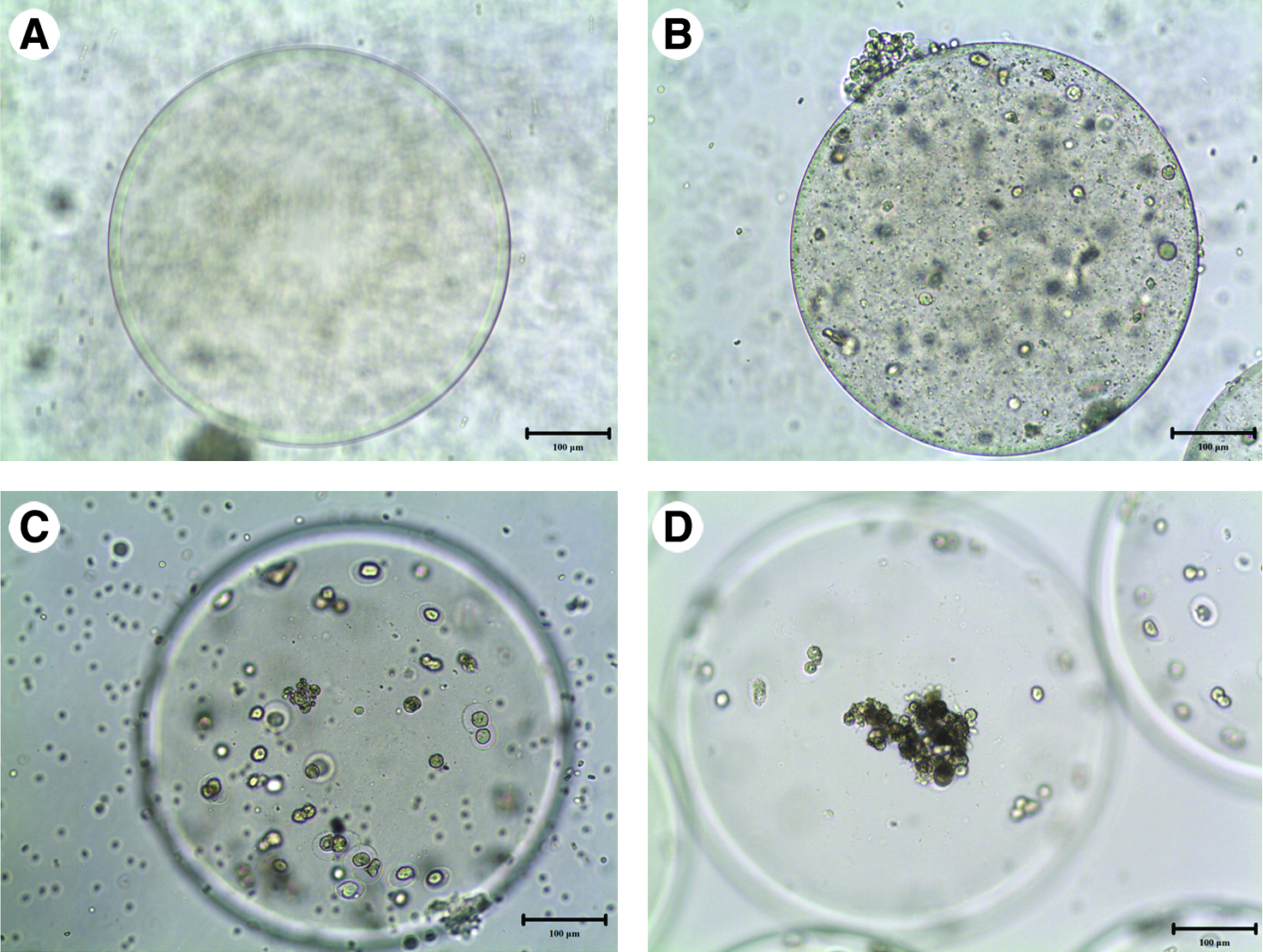
After culture of microencapsulated cells for 48 h, light microscopy revealed that microcapsules formed a pellet shape with a smooth surface. In group I, hepatocytes were circular and scattered in the microcapsules (Fig. 1A). In group II, both hepatocytes and FLSCs were spherical and contacted each other as small masses in shape. Contacts between small clumps were not detected (Fig. 1B). In group III, the FLSCs/bFGF cells were found as aggregates around the hepatocytes, and the proportion of cells within aggregates was higher. Small clumps of cells constantly assembled and formed larger clumps that appeared as liver tissue suspended in the microcapsule undergoing three-dimensional growth (Fig. 1C). In the larger clumps, expression of green fluorescent protein was detected in the FLSCs/bFGF cells under the fluorescence microscope (Fig. 1D).

Morphologic changes of different microencapsulated hepatocyte culture at 48 h time point. (
Hepatic functions in culture
The content of ALB and ALT in the supernatant of the culture medium was detected using an automatic blood biochemistry analyzer. During the first 3 days of culture, the microcapsules containing hepatocytes with FLSCs/bFGF (group III) produced distinctly more ALB than hepatocytes in the other two groups. After cultivation for 14 days, the ALB concentration produced in groups I and II declined and was lower than the ALB level on day 0. Although in group III the ALB level also slightly decreased, it remained at a higher level than that on day 0 and levels were sustained to the end of the study (Fig. 2A).
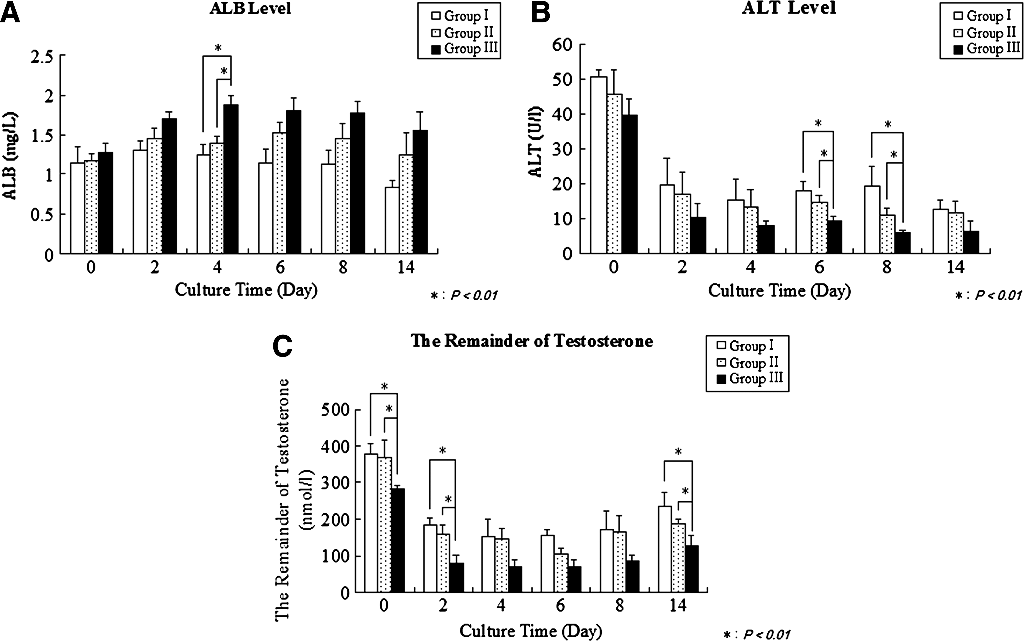
Evaluation of hepatic functions of different microencapsulated hepatocytes in culture. (
Hepatocytes in each of the groups released ALT. On day 2 of cultivation, the content of ALT activity in group III decreased significantly and was twofold lower than that observed immediately after encapsulation. There was no significant difference in ALT levels produced by the hepatocytes among the three groups after day 14 (Fig. 2B).
Hepatocytes had varying ability to testosterone depending on whether or not they were co-cultured with the FLSCs or the FLSCs/bFGF cells. By day 2, the residual testosterone in the culture supernatant of group III was significantly lower than the other two groups, whereas there was no statistically significant difference between groups I and II. By day 14, although the level of supernatant testosterone in groups I, II, and III significantly increased, its levels in group III were still lower than those of groups I and II, as well as the initial concentration (Fig. 2C). The statistical data show that the difference between the three groups has a statistical significance (in Fig. 2A, p < 0.001; in Fig. 2B, p = 0.001; in Fig. 2C, p < 0.001). To exclude the possible affect of the damaged/broken capsules and free cells in the culture medium, we seeded 5 × 10⁵ hepatocytes per each well, and the hepatocytes were co-cultured with the FLSCs or FLSCs/bFGF with the proportions of 7:1 for hepatocytes:stromal cells in monolayer. Then, there was equal number of cells per well by this way, and this part of results has been provided in Supplemental Figs. S1 and S2 (available online at
Transplantation study
In groups A, B, C, and D, the 72-h survival rates were 40%, 40%, 46.70%, and 60%, respectively. The mice demonstrated remarkable symptoms of liver failure revealed by activity obstacle, drowsiness, and coma. However, the mice in group E, those transplanted with microcapsules containing a mixture of hepatocytes and FLSCs/bFGF cells, did not show serious symptoms of liver failure compared with groups A, B, C, and D, and the survival rate was 86.70%. The 7-day survival rate in group E was twofold higher than that in the other groups. After 7 days, the survival rate was 80% in group E, 20% in group A as a control, 20% in group B, 26.70% in group C, and 40% in group D. The statistical data show that the difference between all groups is statistically significant (p = 0.0036). The mice in group E survived longer and showed fewer complications compared with the other groups (Fig. 3A).
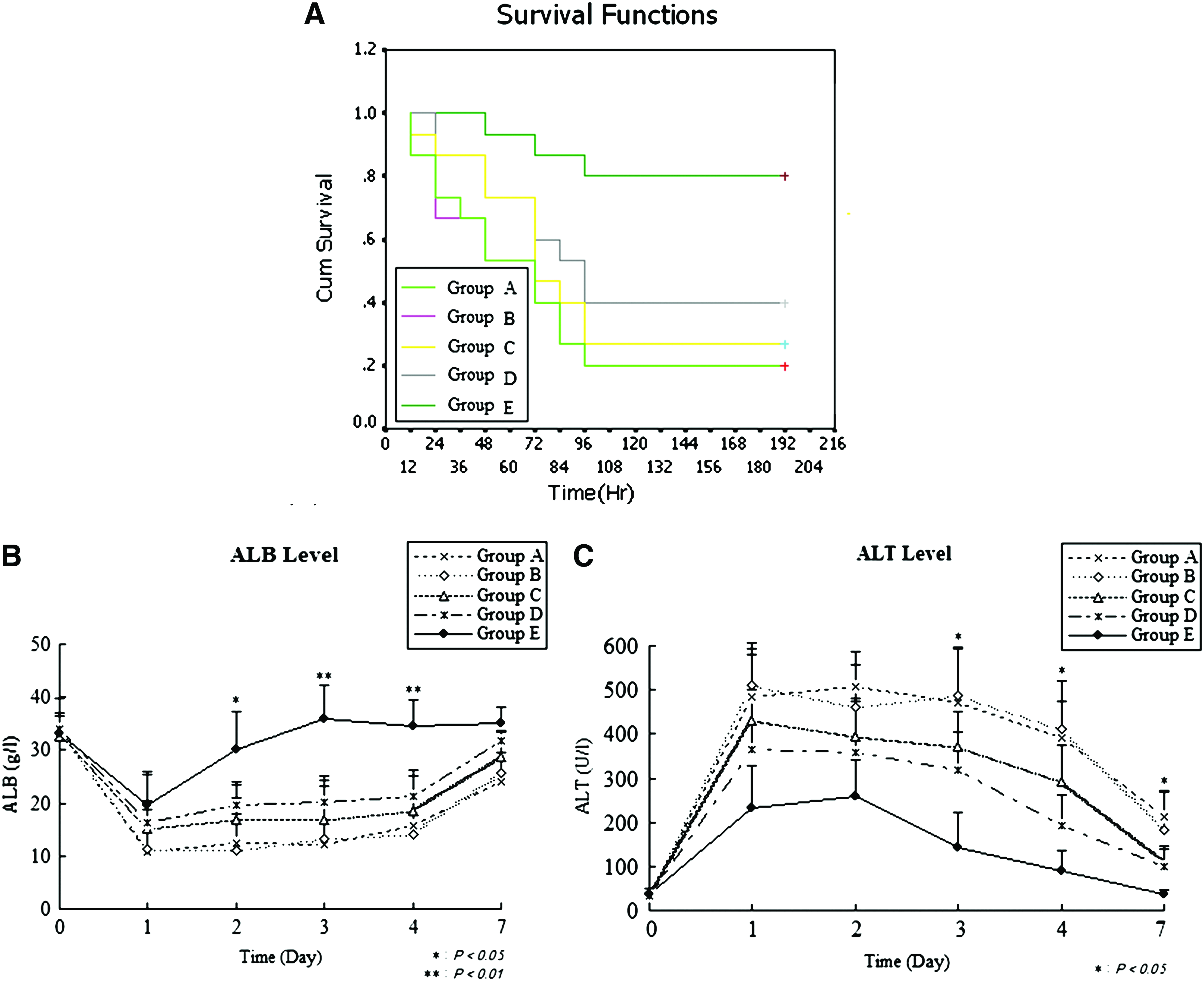
Evaluation of functionalities of different microencapsulated hepatocytes in transplantation recipient mice. (
Effect of intraperitoneal transplantation of microcapsules containing hepatocytes with and without stromal cells on liver function and regeneration
Two days after surgery, liver injury in mice reached a peak, and then gradually improved by the third day. Venous plasma ALT level increased to a peak of 259.5 ± 26.5 U/L at 2 day after the 70% PH in group E mice (70% hepatectomy with intraperitoneal transplantation of microcapsules with hepatocytes and FLSCs/bFGF cells), which was sixfold higher than the normal upper limit using plasma collected in the same fashion. This level gradually decreased to almost normal 7 days after transplantation, as shown in Figure 3C. However, compared with A, B, C, and D groups, the ALT levels were approximately twofold higher in group E, and they decreased more slowly thereafter (Fig. 3C). ALB levels decreased to 19.64 ± 6.41 g/L at 1 day after the 70% PH in group E, but returned to normal levels after 2 days. The ALB levels in group E at 2, 3, and 7 days after surgery were significantly higher than those in groups A, B, C, and D (Fig. 3B). In Figure 3B and C, the difference between all five groups is statistically significant (Fig. 3B, C, p < 0.001); specially, group E has a significant difference compared with the other groups (all p < 0.001).
No significant differences in the remnant lobe weight/body weight ratio were observed among the groups at 24 h after surgery. However, the mean ratio of the wet weight of the remnant liver to the body weight, which is a good indicator for liver regeneration, showed a significant increase in group E mice compared with that in the other groups at 72 h after surgery. Further, the restoration of the liver mass markedly increased with a statistical significance (p = 0.0072) in group E at 72 h (Fig. 4), which was not seen in other groups.
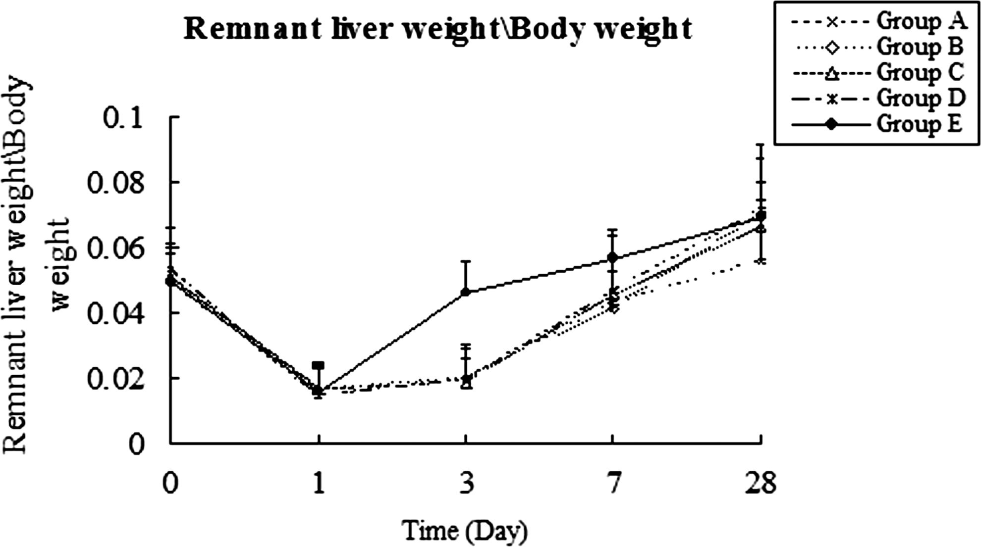
Effect of intraperitoneal transplantation of different microencapsulated hepatocytes on liver regeneration. Group A mice were subjected to 70% hepatectomy only and used as a control; group B mice were transplanted with microcapsules alone; group C mice were transplanted with microcapsules containing only rat hepatocytes; group D mice were transplanted with microcapsules containing rat hepatocytes and FLSCs; group E mice were transplanted with microcapsules and containing a mixture of rat hepatocytes with FLSCs/bFGF cells. All data are presented as mean ± SD of the mean.
Histopathologic and immunohistochemical observation of hepatic tissue
Seventy-two hours after 70% PH, the remnant livers appeared slightly yellow-white and tarnished, were clearly swollen, and displayed diminished flexibility, with a blunt and fragile surface in groups A, B, C, and D. Hematoxylin and eosin staining of hepatic tissue demonstrated massive necrosis by light microscopy; the normal structure of the hepatic lobule was absent. Hepatic parenchymal cells showed various types of degeneration, cytolysis, and fragmentation of cell nuclei, with an indistinct boundary between cells. An enlarged hepatic sinusoid and exfoliated vascular endothelial cells could be seen. There were large regions of light staining and obvious infiltration of inflammatory cells (Fig. 5A–D). The mice in group E had remnant livers with a slight luster, which were hypertrophic and displayed only mild swelling compared with the control group. The degree of liver tissue necrosis was significantly reduced; the structure of the hepatic lobule was slightly disordered, and the hepatic sinusoid enlarged irregularly (Fig. 5E). In the livers of group E mice, there were occasionally a small amount of degenerating or necrotic hepatocytes along with infiltration of a few inflammatory cells. Also observed were proliferating hepatocytes in the periportal areas. There were also a large number of binucleated cells.

Histopathologic examination (hematoxylin and eosin staining) of intraperitoneal transplantation of different microencapsulated hepatocytes 72 h after transplantation. (
Expression of PCNA in the tissue was detected using immunohistochemistry techniques to show the liver regeneration process after PH. Twenty-four hours after 70% PH, positive expression of PCNA had begun to increase rapidly in group E, which increased to a peak at 48 h and then gradually declined (Fig. 6E). The location of cells with nuclei positive for PCNA transitioned from the periportal zone, zone 1, at 24 h to zones 1 and 2 by 48 h, and to zone 3, the pericentral zone, by 72 h. The loss of cells with positive PCNA staining in the nucleus began also in zone 1 and transitioned to the cells in zone 3 by 96 h. In the livers of mice in the other groups, PCNA+ cells were not found before 72 h (Fig. 6A, B), or the expression levels were very low (Fig. 6C, D). Then, to quantify for comparative analysis, we also counted the positive cells of PCNA staining, the total cells, and the percentage of positive cells (Supplemental Fig. S3, available online at
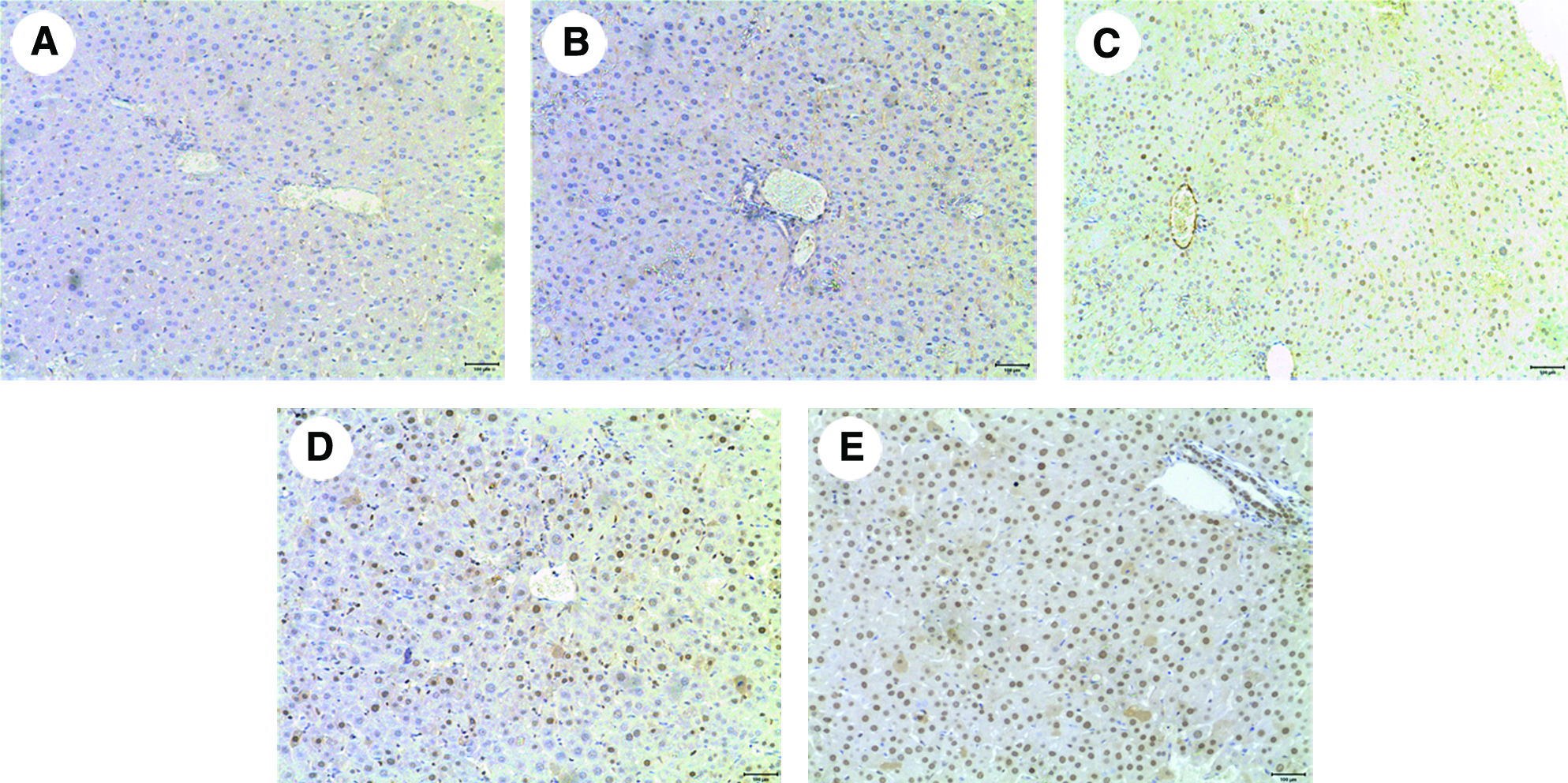
Immunohistochemical analysis (proliferating cell and nuclear antigen staining) of intraperitoneal-transplanted different microencapsulated hepatocytes 48 h after transplantation. (
Recovery of microcapsules
After 2 to 4 weeks of transplantation, the microcapsules were found in the peritoneum fluid or were found adhering to the liver capsule and collected by peritoneal lavage. The retrieval rate of cell capsules was about 89% ± 12% in group B. The retrieved microcapsules were found to cause little, if any, fibrosis. Although some of the microcapsules were damaged, others were still in good condition (Fig. 7A). In groups C and D, the retrieval rate was about 76% ± 35% and 78% ± 17%, respectively. Most of the microcapsules were found free within the peritoneal cavity, and a few of them were found adhering between the greater omentum and mesentery. The atrophy and/or rupture of cells and nuclear turbid cytoplasm were detected by light microscopy, which suggested that most cells had died (Fig. 7B, C). However, in group E, many capsules were found together at the edge of the injured liver and/or adhering to the liver surface. The retrieval rate was about 77% ± 24%. In the microcapsules, the hepatocytes and FLSCs/bFGF were aggregated with each other (Fig. 7D). The cell membranes were intact, and there were rich granules in the cytoplasm, indicating that the cells were still alive.
Morphologic examination of microcapsules recovered at 2 weeks after transplantation. (
Discussion
Recently, hepatocyte cell transplantation (HCT) has become very promising as a new and effective strategy for treating liver failure. The therapeutic effect of HCT has been dramatic in experimental animal model of rats with acute hepatic failure.23–25 As for acute liver injury, HCT can provide metabolic compensation, promoting the regeneration of damaged liver, supporting adequate liver function, even helping patients to overcome the difficulties of ALF, and enabling them to survive to OLT. 26 In some cases, liver transplantation was no longer required; HCT was able to fully restore the liver functions. However, acute immune reaction is inevitable in the process of allogenic liver cell transplantation or xenotransplantation and affects the success rate of HCT. In attempts to address this issue, some researchers have used the microencapsulation technology of alginate–poly-L-lysine–alginate microcapsules, which can effectively prevent the occurrence of immune rejection.27,28–32
In the present study, we mixed hepatocytes with FLSCs in microcapsules. The FLSCs play an important role in maintaining the survival and proliferation of hepatocytes via paracrine signaling. 33 As a result, the functions of hepatocytes improved significantly. Microcapsules containing both hepatocytes and FLSCs/bFGF cells resulted in increased hepatic function compared to those with hepatocytes alone. The alginate-poly-L-lysine-alginate capsules serve as immune barriers, with ideal permeability, and also create favorable three-dimensional structures in which cell-to-cell interactions are enhanced. 34 FGF2 (also known as basic FGF or bFGF) and other growth factors secreted by transgenic FLSCs/bFGF cells may play an important supporting role in both cells. The molecular weight of bFGF and other liver regeneration factors from FLSCs is lower than 100,000, making them able to easily permeate the membrane of the microcapsules. FGF2 has a pleiotropic activity in the regulation of growth and proliferation of a wide variety of mesenchymal, endocrine, and neural cells. It is also a potent angiogenic factor acting as a mitogen by autocrine modulation of cell growth and transformation. Increasing evidence supports a role for FGFs in liver growth and regeneration.33,35,36 Our results also showed that FLSCs/bFGF stimulates DNA synthesis in injured livers, suggesting that bFGF stimulates liver regeneration in vivo. 35 The mitogenic effect of bFGF on fetal and adult hepatocytes in vitro has also been reported. The presence of heparin binding growth factors in the liver during embryogenesis and liver regeneration suggests a role for these proteins in hepatocyte growth. 33 FGF was also found to be expressed in the early stages of carbon tetrachloride liver intoxication in endothelial cells and major blood vessels. 36 Transplantation of collagen-coated fibers coated with this growth factor into the peritoneum of rats stimulated the formation of organoid structures containing vascular lamina. Therefore, in addition to stimulation of hepatocyte proliferation, bFGF may also exert indirect effects by improving the blood supply.37,38 However, the therapeutic use of transgenic cells would expose patients to the risk of insertional mutagenesis. Research efforts have thus focused on potential strategies for reducing or eliminating the risk of insertional mutagenesis. Liver regeneration after liver failure in mice was significantly increased in mice transplanted with the microcapsules containing transgenic FLSCs/bFGF and hepatocytes. These microcapsules proved to be beneficial to the long-term survival of the mouse. In addition, the role of immune isolation of the microcapsule has been further confirmed in the present study with the application of microencapsulation technology for allogenic transplants. In vivo experiments, final location of capsules of group E to the injured liver is very interesting. The FLSCs homing to the disease niche is one potential mechanism for this observation.
The findings from our study suggest that microcapsules containing both hepatocytes and FLSCs/bFGF cells could yield high-quality cell products for large-scale culture of hepatocytes and application that could be used in a bioreactor for a bioartificial liver. Because the function of liver cells can be significantly enhanced by the transgenic FLSCs/bFGF, the number of hepatocytes required for a bioartificial liver could be substantially reduced. It is possible that co-culture of hepatocytes with hepatic nonparenchymal cells could form a similar structure and function of hepatic lobules. Therefore, the therapeutic effect of bioartificial livers can be markedly enhanced in the treatment of patients with ALF. It is not yet known whether these factors in the application of a bioartificial liver will result in adverse reactions in humans. Although this has not been observed in animal experiments, it warrants an in-depth study in the future. On the basis of our result, we believe that not only the number of cells but also the level of hepatic function is important for the support of severe liver damage. Nevertheless, the issue of the optimal cell number required to improve a patient's fulminant hepatic failure status needs to be further studied for future clinical applications.
In summary, intraperitoneal transplantation of microencapsulated hepatocytes co-cultured with FLSCs/bFGF cells can improve the survival rate of animals with ALF, block immune rejection, and better protect the hepatocytes. These studies contribute toward our understanding of conditions for the maintenance of hepatocytes and their functions, and provide a new direction for the clinical treatment of ALF, and new strategies that may replace OLT in the future.
Footnotes
Acknowledgments
The authors are grateful to Professor Kimberly Mace at Healing Foundation Centre for Tissue Regeneration at University of Manchester, Professor Guangping Gao at University of Massachusetts Medical School, and Professor Lola M. Reid at the University of North Carolina School of Medicine at Chapel Hill for helpful comments and critical revision of this article; to Professor Hirotoshi Miyoshi at University of Tsukuba at Japan for his comments; and to Professor Dan Feng at Department of Medical Statistics at Chinese PLA General Hospital for her help in statistical analysis. This work was supported by National High Technology Research and Development Program of China (No:2006AA02A107) and the Major State Basic Research Program of China (No:2005CB522702).
Disclosure Statement
No competing financial interests exist.
References
Supplementary Material
Please find the following supplemental material available below.
For Open Access articles published under a Creative Commons License, all supplemental material carries the same license as the article it is associated with.
For non-Open Access articles published, all supplemental material carries a non-exclusive license, and permission requests for re-use of supplemental material or any part of supplemental material shall be sent directly to the copyright owner as specified in the copyright notice associated with the article.