
Abstract
Introduction:
Tissue engineering of human nasal septal chondrocytes offers the potential to create large quantities of autologous material for use in reconstructive surgery of the head and neck. Culture with recombinant human growth factors may improve the biochemical and biomechanical properties of engineered tissue. The objectives of this study were to (1) perform a high-throughput screen to assess multiple combinations of growth factors and (2) perform more detailed testing of candidates identified in part I.
Methods:
In part I, human nasal septal chondrocytes from three donors were expanded in monolayer with pooled human serum (HS). Cells were then embedded in alginate beads for 2 weeks of culture in medium supplemented with 2% or 10% HS and 1 of 90 different growth factor combinations. Combinations of insulin-like growth factor-I (IGF-1), bone morphogenetic protein (BMP)-2, BMP-7, BMP-13, growth differentiation factor-5 (GDF-5), transforming growth factor β (TGFβ)-2, insulin, and dexamethasone were evaluated. Glycosaminoglycan (GAG) accumulation was measured. A combination of IGF-1 and GDF-5 was selected for further testing based on the results of part I. Chondrocytes from four donors underwent expansion followed by three-dimensional alginate culture for 2 weeks in medium supplemented with 2% or 10% HS with or without IGF-1 and GDF-5. Chondrocytes and their associated matrix were then recovered and cultured for 4 weeks in 12 mm transwells in medium supplemented with 2% or 10% HS with or without IGF-1 and GDF-5 (the same medium used for alginate culture). Biochemical and biomechanical properties of the neocartilage were measured.
Results:
In part I, GAG accumulation was highest for growth factor combinations including both IGF-1 and GDF-5. In part II, the addition of IGF-1 and GDF-5 to 2% HS resulted in a 12-fold increase in construct thickness compared with 2% HS alone (p < 0.0001). GAG and type II collagen accumulation was significantly higher with IGF-1 and GDF-5. Confined compression modulus was greatest with 2% HS, IGF-1, and GDF-5.
Conclusion:
Supplementation of medium with IGF-1 and GDF-5 during creation of neocartilage constructs results in increased accumulation of GAG and type II collagen and improved biomechanical properties compared with constructs created without the growth factors.
Introduction
In general, tissue engineering of cartilage involves forming tissue from a limited number of cells. Autologous tissue engineering involves harvesting tissue from a donor, digesting the extracellular matrix (ECM), and isolating chondrocytes. Chondrocytes are usually then expanded in monolayer culture to greatly increase the cell number. During monolayer expansion the cells take on fibroblastic characteristics, a process called dedifferentiation.1,2 Expanded cells are then cultured in a three-dimensional (3D) scaffold such as alginate, agarose, or polyglycolic acid, inducing redifferentiation and production of ECM.3–5 The capacity of cells to redifferentiate and form cartilaginous tissue becomes impaired with increasing levels of expansion.2–5 Variables such as medium composition, growth factors, cell seeding density, 3D scaffold properties, and physical stimulation influence the ability of expanded cells to redifferentiate and produce functional cartilaginous ECM.
A number of growth factors have been evaluated individually and in limited combinations to assess their ability to induce redifferentiation and production of ECM by human nasal septal chondrocytes. Insulin-like growth factor-I (IGF-I), transforming growth factor β (TGFβ)-1, TGFβ-2, bone morphogenetic protein (BMP)-2, and BMP-7 have all been shown to improve chondrogenesis during 3D culture either in serum-free medium or in medium supplemented with fetal bovine serum (FBS).6–8 We recently reported that culture of human nasal septal chondrocytes with pooled human serum (HS) results in increased proliferation and chondrogenesis compared with FBS. 9 The goal of this study was to identify growth factor combinations that improve chondrogenesis during 3D culture of human nasal septal chondrocytes in the medium supplemented with HS.
Materials and Methods
Study design
The study design had two parts. In part I a high-throughput screen evaluating the chondrogenic effects of many combinations of growth factors was performed. In part II, the top performers from part I were analyzed in more detail by creation of neocartilage constructs using the alginate-recovered chondrocyte (ARC) method.5,10
Part I
Eight growth factors were selected for testing in part I based on previous reports and pilot data in our own lab. Ninety different combinations of IGF-1, TGFβ-2, BMP-2, BMP-7, BMP-13, growth differentiation factor-5 (GDF-5, also known as BMP-14 or cartilage-derived morphogenetic protein-1), insulin, and dexamethasone were tested with both 2% HS and 10% HS. The concentrations tested are shown in Table 1, along with selected references regarding the use of these growth factors for tissue engineering. Concentrations tested were based on previous reports in the literature and pilot data in our lab (not shown). All possible combinations of IGF-1, BMP-2, BMP-7, BMP-13, GDF-5, and insulin were tested (63 conditions). The ability of TGFβ-2 and/or dexamethasone to augment a subset of combinations was tested in the remaining 27 conditions.
BMP, bone morphogenetic protein; DEX, dexamethasone; GDF-5, growth differentiation factor-5; IGF-1, insulin-like growth factor-I; INS, insulin; TGFβ, transforming growth factor β.
Cell-associated glycosaminoglycan (GAG) accumulation during 3D culture over 2 weeks was chosen as a marker of chondrogenesis during part I. GAG was chosen over collagen and other matrix components because of ease of measurement for the high-throughput assay and because GAG accumulates in greater quantity than collagen during early culture in alginate.5,10,11 The cell-associated portion of GAG was assessed because of its usefulness for creation of scaffold-free ARC constructs.
Cell isolation
Nasal septal cartilage specimens were collected from three subjects undergoing routine septoplasty or septorhinoplasty at the University of California–San Diego Medical Center or VA San Diego Medical Center. The subjects in part I were a 40-year-old man, a 53-year-old woman, and a 62-year-old man. The study was approved by the Institutional Review Board at each institution. Specimens were stored in sterile saline for transportation to the lab. Cartilage specimens were dissected free of perichondrium and diced into approximately 1 mm3 pieces. They were then digested by incubation at 37°C with 0.2% Pronase type XIV (Sigma, St. Louis, MO) in medium (composed of Dulbecco's modified Eagle's medium (DMEM)/F-12, 0.4 mM L-proline, 2 mM L-glutamine, 0.1 mM nonessential amino acids, 10 mM HEPES, 100 U/mL penicillin G, 100 μg/mL streptomycin sulfate, and 0.25 μg/mL amphotericin B) for 60 min followed by incubation with 0.025% collagenase P (Roche Diagnostics, Indianapolis, IN) in the medium for 16 h. Suspensions of digested cartilage were then passed through a 70 μm filter, washed, and centrifuged to isolate the chondrocytes. Cells were resuspended in the cell culture medium (composed of low-glucose DMEM, 0.4 mM L-proline, 2 mM L-glutamine, 0.1 mM nonessential amino acids, 10 mM HEPES, 100 U/mL penicillin G, 100 μg/mL streptomycin sulfate, and 0.25 μg/mL amphotericin B). The number of cells isolated was quantified by staining an aliquot of the cell suspension with trypan blue and counting with a hemocytometer.
Monolayer culture
Isolated chondrocytes were seeded at a density of 5000 cells/cm2 into T-25 culture flasks (Corning, Inc., Corning, NY). The low seeding density was chosen to allow for a large monolayer amplification of cell numbers before transferring to alginate beads. 12 The monolayer cell culture medium was supplemented with 2% HS (pooled human AB serum; Gemini Bioproducts, Woodland, CA), TGFβ-1 (1 ng/mL), fibroblast growth factor (FGF)-2 (5 ng/mL), and platelet-derived growth factor-BB (10 ng/mL). A minimum of 1 mL medium per 106 cells per day was added and changed every 2–3 days. After 15 days, at which point confluence was achieved, cells were released from monolayer using 0.05% trypsin (Life Technologies, Grand Island, NY). Chondrocytes were counted with a hemocytometer after centrifugation and washing.
Culture in alginate
Cells were then suspended at a density of 4 × 10⁶ cells/mL in a solution of 1.2% low-viscosity alginate (Kelco LV, San Diego, CA). Droplets of the alginate–chondrocyte suspension were polymerized in 102 mM CaCl2 for 5 min to form beads. 3 The volume of each bead was approximately 10 mm3 (40,000 cells). After washing with 0.9% saline, the beads were transferred to culture medium (DMEM/F-12, 25 μg/mL ascorbate, 0.4 mM L-proline, 2 mM L-glutamine, 0.1 mM nonessential amino acids, 10 mM HEPES, 100 U/mL penicillin G, 100 μg/mL streptomycin sulfate, and 0.25 μg/mL amphotericin B). Two beads were transferred to each well of a 96-well culture plate (Corning, Inc.). The alginate–chondrocyte beads were then cultured for 2 weeks in culture medium supplemented with either 2% or 10% HS alone or in addition to one of 90 combinations of the eight growth factors tested (a total of 182 conditions). For each condition, two replicates per subject were maintained. The culture medium was changed every 2–3 days.
Termination of alginate culture
After washing the beads, the alginate was depolymerized with 55 mM sodium citrate for 15 min. After centrifugation, the supernatant containing the far-removed matrix was removed. The pellet containing the cells and their cell-associated matrix (CAM) was digested with 0.5 mg/mL proteinase K (PK; Roche Diagnostics). 13
Quantitative assay for GAG
The GAG content in the PK digests was determined using the dimethyl-methylene blue reaction. 14 Aliquots of each sample were mixed with dimethyl-methylene blue dye. Absorbance at 525 nm was measured on a spectrophotometer and compared with a plot of standards made from shark chondroitin sulfate type C (Sigma).
Part II
Nasal septal cartilage from four subjects was obtained. The median age for donors in part II was 43 years (range, 23–51). There were three male and one female donors. Chondrocytes were harvested, isolated, expanded in monolayer, and suspended in alginate beads as described above. Alginate beads from each donor were then divided and cultured in culture medium for 10 days in one of five conditions: (1) 10% FBS, (2) 2% HS, (3) 10% HS, (4) 2% HS + IGF-1 + GDF-5, and (5) 10% HS + IGF-1 + GDF-5. Growth factor concentrations were the same as in part I. Previous studies have found that excessive CAM can interfere with successful seeding of ARC constructs. The duration of bead culture in part II was shortened to 10 days based on amount of CAM observed in part I. 10 The culture medium was changed every 2–3 days.
Formation of ARC constructs
Chondrocytes and their CAM were then recovered from the alginate beads as described above. The recovered cells were resuspended in culture medium (supplemented according to the experimental condition) at a cell density of 4 × 10⁶ cells/mL. This suspension was used to seed multiple 12-mm-diameter transwell clear polyester membrane cell inserts (Corning, Inc.) at a density of 5 × 10⁶ cells/cm2. Four transwells were seeded per subject per condition. Cultures were maintained for 4 more weeks with culture medium changes every 2–3 days. For each condition, the seeded inserts continued to receive the same culture medium and supplementation as was used during alginate culture (10% FBS, 2% HS, 10% HS, 2% HS + IGF-1 + GDF-5, or 10% HS + IGF-1 + GDF-5). At the end of the culture period constructs were removed from the culture inserts, weighed, and measured.
Quantitative assay for GAG and DNA
Two constructs from each condition for each subject were used for biochemical analysis. These constructs were divided in half, and each half weighed. A half of each construct was digested with PK in phosphate-buffered disodium ethylenediamine tetraacetate for 16 h. 13 GAG was measured in the PK digests as described above. DNA was quantified using the PicoGreen double-stranded DNA reagent (Molecular Probes, Eugene, OR) as described previously. 13 Fluorescence of mixtures of 100-μL digested samples with 100 μL of PicoGreen reagent was measured. Fluorescence values were converted to DNA quantity using standards of DNA from calf thymus in the appropriate buffer solution. Cellularity of the digests was estimated using the value of 7.7 pg DNA per chondrocyte. 15
Quantitative assay for collagen
Type I and II collagen were quantified in a half of the constructs by enzyme-linked immunosorbant assay using the human type I and native type II collagen kits (Chondrex, Inc., Redmond, WA). 16 Briefly, collagen was digested with 10 mg/mL pepsin dissolved in 0.05 M acetic acid at 4°C overnight followed by incubation with 1 mg/mL pancreatic elastase at 4°C for 16 h. Separate type I and type II collagen enzyme-linked immunosorbant assays were performed following the manufacturer's instructions. Samples and the provided standards were diluted with buffer from the kit. Capture antibodies were added to the plates and incubated overnight at 4°C. Samples were then added and incubated at room temperature for 2 h. The appropriate detection antibody was then added and allowed to incubate for 2 h. After washing, samples were incubated with streptavidin peroxidase for 1 h. After rinsing, the color reaction was initiated by adding o-phenylenediamine and urea–H2O2. The reaction was terminated by adding 2 N sulfuric acid. The optical density was read at 490 nm with an Emax precision microplate reader (Molecular Devices, Sunnyvale, CA). Collagen content was normalized to DNA content to estimate collagen accumulation per cell. Uniform DNA per wet weight was assumed. DNA and collagen assays were performed on separate halves from the same construct.
Histochemistry and immunohistochemistry
Constructs were analyzed by histochemistry and immunohistochemistry for localization of GAG and collagen types. Samples to be used for histochemistry were placed in OCT compound and frozen. They were then sectioned in a cryostat at 5 μm thickness. For histochemical localization of GAG, slides from each sample group were stained with 0.1% alcian blue in buffer (0.4 M MgCl2, 0.025 M sodium acetate, and 2.5% glutaraldehyde, pH 5.6) overnight, and destained with 3% acetic acid until clear. Immunohistochemistry was performed with the Vectastain Elite ABC kit (Vector Laboratories, Burlingame, CA), a peroxidase-based detection system. Mouse monoclonal antitype collagen I and collagen II antibodies (Chondrex, Inc.) were used. Sections were counterstained with methyl green nuclear stain (Vector Laboratories). Specimens were examined using bright-field microscopy using a Nikon Eclipse TE 300 microscope (AG Heinze, Irvine, CA) equipped with SPOT RT camera (Diagnostic Instruments, Burlingame, CA).
Biomechanical testing
Compression testing was performed on two constructs from each condition for each subject, using methods previously described.17–19 A metal punch was used to isolate a disk measuring 9.6 mm in diameter from the center of each construct. The disks were placed between two porous platens in a radially confining chamber and subjected to equilibrium compression tests to determine the compressive modulus.
Statistical analysis
Data are represented as mean ± standard error of the mean. Statistical analysis was performed using SAS Analyst 9.1 (SAS Institute, Cary, NC). Differences in proliferation, DNA, GAG, collagen, and confined compression modulus were assessed using mixed models with fixed effects for serum condition and random effects for subject. If a mixed model identified an overall significant effect, Tukey's HSD method was used to compare individual groups. An α level of <0.05 was used to determine significance.
Results
Part I
During the single monolayer cell passage, the cell number increased a mean 50-fold (range, 48–55; final cell density, 2.52 × 10⁵ ± 0.12 × 10⁵ cells/cm2). After 2 weeks of alginate bead culture, GAG accumulation was greater for each of the 10% HS conditions compared with 2% HS. For the serum-only controls there was 25.8 μg GAG per bead with 10% HS and 7.0 μg GAG per bead with 2% HS (p < 0.0001). As shown in Table 2, IGF-1 and GDF-5 were present in each of the top 10 conditions containing 10% HS, with additional growth factors having negligible impact. On the basis of this observation, IGF-1 and GDF-5 were selected for further testing in part II. In part I, none of the top 30 conditions contained dexamethasone.
The top 30 performing 10% HS conditions are shown, in addition to results for select individual growth factors. Conditions are sorted from the highest accumulation of GAG to the lowest in the presence of 10% HS.
GAG, glycosaminoglycan; HS, human serum.
Part II
As shown in Figure 1, ARC construct weight and thickness varied significantly with the medium condition, with the addition of IGF-1 and GDF-5 resulting in significant increases compared with serum alone for both 2% HS and 10% HS conditions (see Table 3 for p-values). Biochemical properties of the constructs are shown in Figure 2. The amount of cellular DNA in the constructs varied significantly by the medium condition, although there were no significant differences between the 10% HS alone, 2% HS + IGF-1 + GDF-5, and 10% HS + IGF-1 + GDF-5 conditions (Fig. 2A). The amount of GAG and type II collagen per mg of tissue was significantly greater for the 2% HS + IGF-1 + GDF-5 condition, as compared with each other condition (Fig. 2C, D and Table 3). GAG accumulation per cell was also significantly greater in the 2% HS + IGF-1 + GDF-5 condition, as compared with each other condition (p < 0.02). GAG accumulation per cell was 0.318 ± 0.042 ng per cell in the 2% HS + IGF-1 + GDF-5 condition, compared with 0.233 ± 0.048 ng per cell in the 10% HS + IGF-1 + GDF-5 condition. Type I collagen accumulation was below the limit of detection for the assay (0.08 μg/mL) for all samples. Immunohistochemistry revealed dense staining for type II collagen in the growth factor conditions, with little staining for type I collagen (Fig. 3).
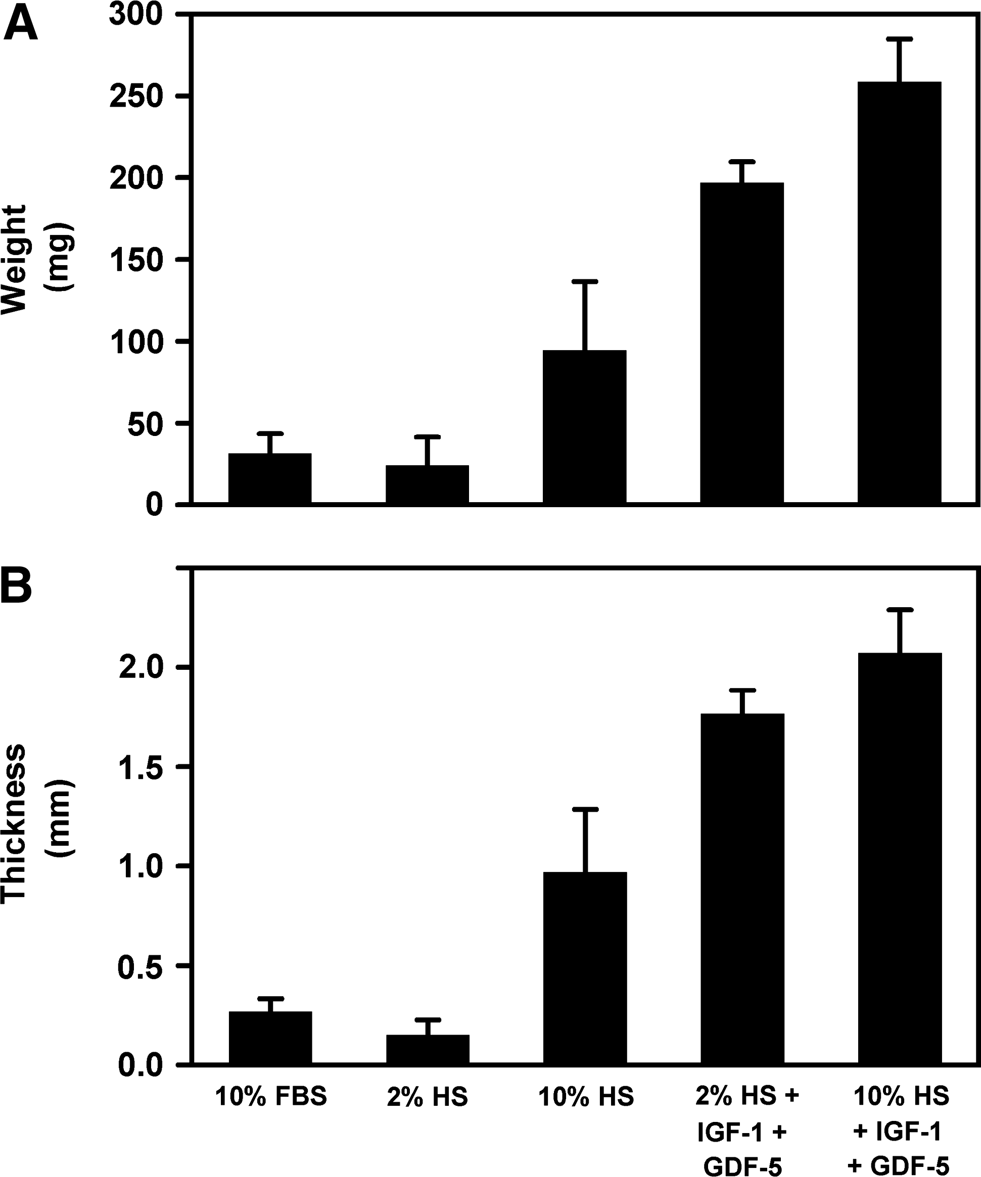
Weight (
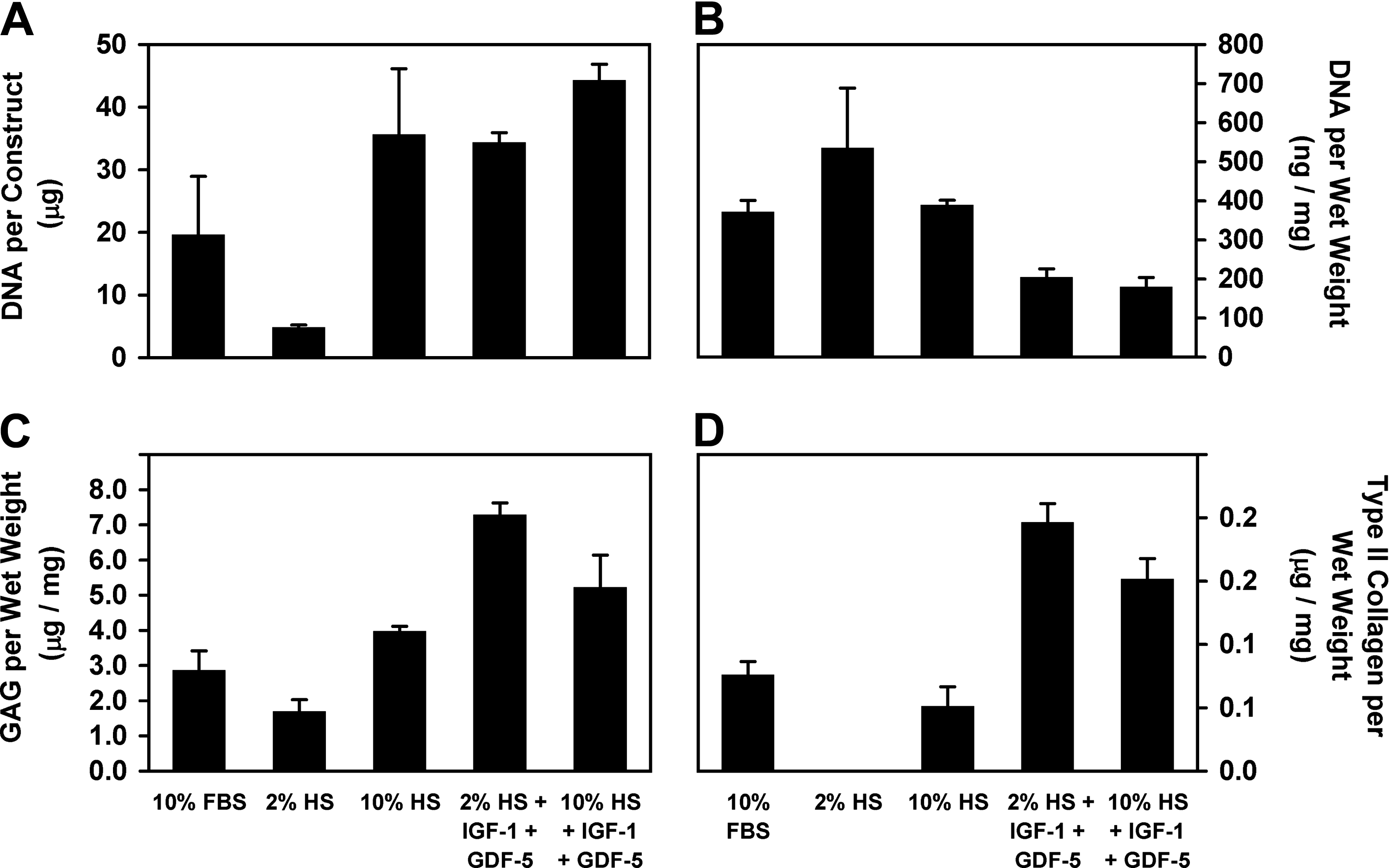
Biochemical properties of the alginate-recovered chondrocyte constructs for each of the five conditions in part II. DNA per construct (
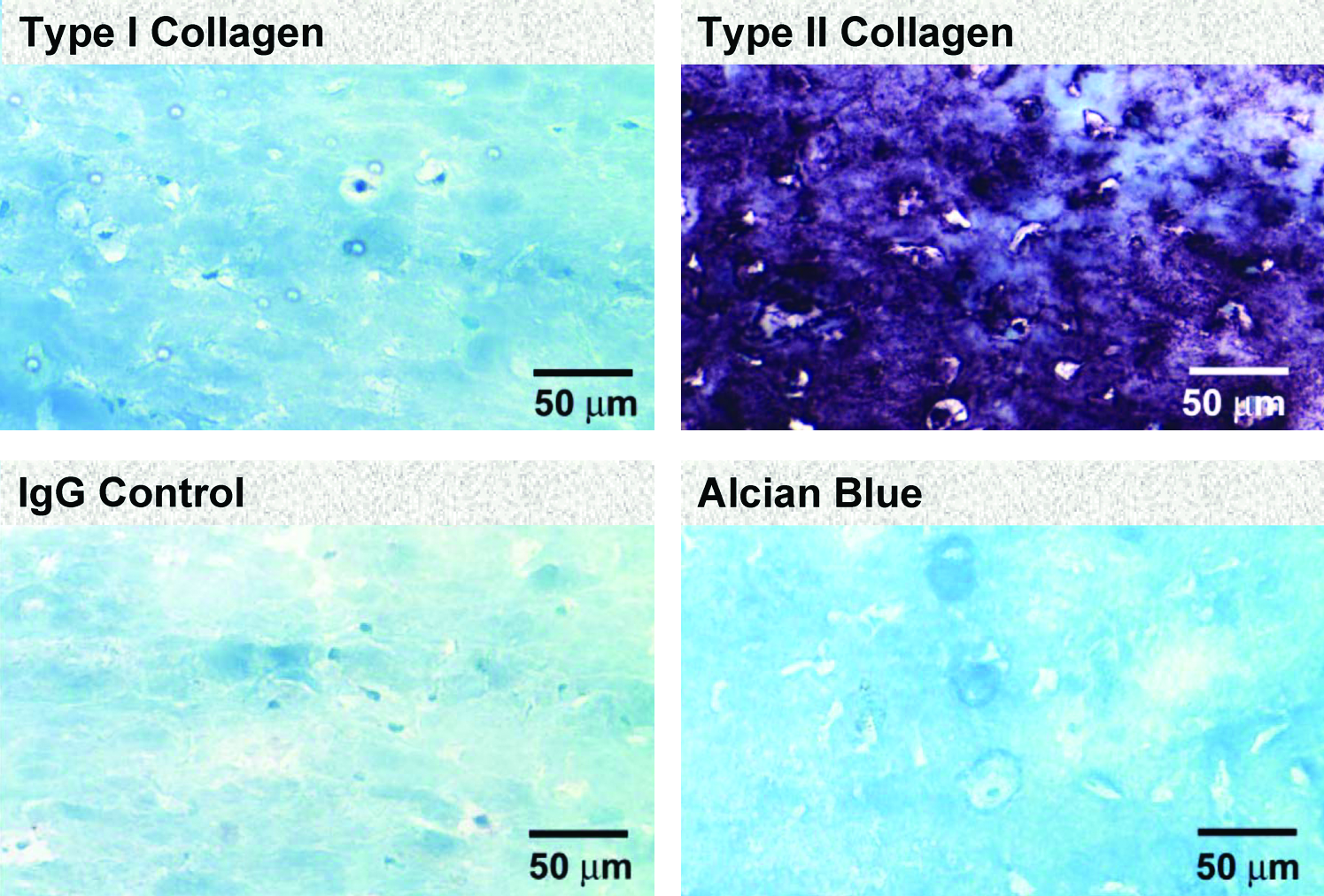
Immunohistochemistry staining of neocartilage constructs created with 2% HS + IGF-1 + GDF-5. There was abundant staining for type II collage with minimal staining for type I collagen and nonspecific IgG. Alcian blue staining reveals abundant GAG. Color images available online at
Significant values are in bold. For each variable, the p-value for the overall model was <0.0001. Individual conditions were compared using Tukey's method. Insufficient sample was available for measuring type II collagen in the 2% HS condition.
WW, wet weight; FBS, fetal bovine serum; I.S., insufficient sample.
The biochemical findings were in agreement with the qualitative handling properties of the tissue (Fig. 4). Only constructs in the growth-factor-containing conditions had sufficient strength to complete accurate compression testing. As shown in Figure 5, the confined compression modulus, HA, was significantly greater in the 2% HS + IGF-1 + GDF-5 condition (p < 0.0001), compared with the 10% HS + IGF-1 + GDF-5 condition.
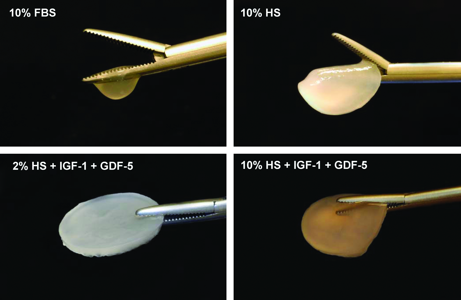
Photographs of representative constructs. The 2% HS alone constructs had insufficient rigidity to handle. Color images available online at
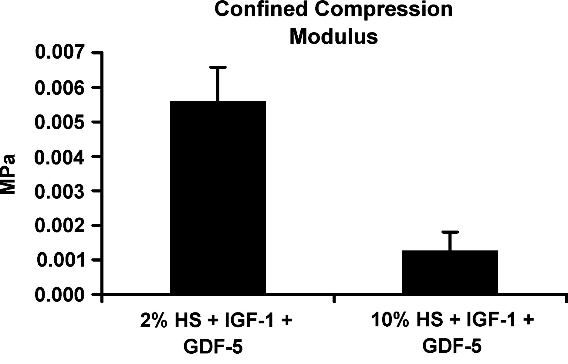
Confined compression modulus for neocartilage constructs created with IGF-1 and GDF-5. The confined compression modulus was significantly greater in the 2% HS + IGF-1 + GDF-5 condition compared with the 10% HS + IGF-1 + GDF-5 condition (p < 0.0001).
Discussion
The addition of IGF-1 and GDF-5 to medium supplemented with HS promotes chondrogenesis in human nasal septal chondrocytes after expansion of cells in monolayer. Neocartilage constructs created using the ARC method with IGF-1 and GDF-5 had increased amounts of GAG and type II collagen relative to controls without the growth factors. The mechanical properties of the constructs created with the growth factors were also more favorable.
The chondrogenic effect of GDF-5 on cultured human nasal septal chondrocytes has not been reported previously. GDF-5 is known to be an important regulatory factor during the embryologic development of the appendicular skeleton and plays a role in the healing of long bone fractures.20,21 Variations in the GDF-5 gene in humans have been associated with the development of osteoarthritis. 22 Bai et al. reported that GDF-5 induced the differentiation of mesenchymal stem cells into chondrocytes and promoted increased accumulation of GAG and type II collagen during pellet culture. 23 Chubinskaya et al. reported that addition of GDF-5 resulted in a 1.5-fold increase in proteoglycan accumulation in cadaveric adult human articular chondrocytes cultured in alginate beads for 9 days, compared with controls without growth factors. 24 Their study also evaluated BMP-2, -4, -6, -7, and cartilage-derived morphogenetic protein-2. They found that BMP-7 resulted in the greatest chondrogenesis during alginate bead culture, whereas BMP-7 performed relatively poorly during part I of our study. Hicks et al. also found a strong chondrogenic effect for BMP-2 and BMP-7 during bead culture of human nasal septal chondrocytes. 8 The use of HS instead of FBS is one potential reason for the differences between this study and prior studies of the chondrogenic effects of growth factors during 3D culture. In addition, nasal septal cartilage may respond differently to specific growth factors than articular cartilage.
Several studies have demonstrated the ability of IGF-1 to improve proliferation of human nasal septal chondrocytes during monolayer and 3D culture.25–28 Richmon et al. found increased GAG accumulation during monolayer culture of human nasal septal chondrocytes with IGF-1. 29 van Osch et al. reported increased GAG accumulation and type II collagen during alginate bead culture with a combination of IGF-1 and TGFβ-2. 7 This combination also increases GAG accumulation during culture of nasal chondrocytes on biodegradable scaffolds. 30 The addition of TGFβ-2 did not show a consistent favorable impact during part I of this study.
The components in HS that result in improved chondrogenesis relative to FBS remain to be characterized. Epidermal-derived growth factor, platelet-derived growth factor-AB, and IGF-1 have been proposed as important components of HS that may affect chondrocyte proliferation and chondrogenesis.31,32 Prior studies found 10% HS to be superior to 2% HS during alginate bead culture of human nasal septal chondrocytes. 9 However, in this study 2% HS actually outperformed 10% HS in the presence of IGF-1 and GDF-5 during the formation of ARC constructs.
Although the results in this study are encouraging, additional work is needed to create clinically useful neocartilage constructs. The amount of GAG per mg of wet weight tissue in the ARC constructs cultured for 1 month with 2% HS + IGF-1 + GDF-5 was approximately 5.5-fold less than that previously reported for native human nasal septal cartilage (5.22 ± 0.92 μg/mg for the ARC constructs vs. 28.7 ± 9.2 μg/mg for native tissue). 33 Total collagen content measured by hydroxyproline assay in human nasal septal cartilage has been reported as 87.1 ± 20.1 μg/mg. 33 Given that immunohistochemical studies have confirmed that type II collagen is the predominant collagen found in native nasal septal cartilage, 34 0.197 ± 0.015 μg of type II collagen per mg of tissue seen in the 2% HS + IGF-1 + GDF-5 ARC constructs is likely considerably less than that found in native tissue. The compressive modulus HA in native human septal cartilage compressed in the medial axis has been reported as 0.44 ± 0.04 MPa, 19 compared with 0.0056 ± 0.0012 MPa measured in the 2% HS + IGF-1 + GDF-5 constructs.
Several limitations to this study exist. Assessment of possible temporal or sequential effects of growth factor addition during the engineering of neocartilage constructs was beyond the scope of this study. A distinct temporal pattern of expression of BMP receptors during chondrocyte culture has been demonstrated. 35 It is possible that introducing the right growth factors at the right time may significantly improve chondrogenesis. Another limitation, which could affect the ability to generalize these results, is that a single lot of pooled HS was used. Pooling of serum from many donors should minimize lot-to-lot variation. A study of the ability of IGF-1 and GDF-5 to enhance chondrogenesis during culture of human nasal septal chondrocytes with autologous HS is underway. Finally, dose effects of IGF-1 and GDF-5 combinations on the creation of ARC constructs were not assessed in this study. Further evaluation of dose–response profiles for combinations of these growth factors is planned for the future.
The use of xenogenic or allogenic materials during engineering of autologous constructs intended for reimplantation in humans introduces the risks of immunogenic response and transmission of infection. In this study, the addition IGF-1 and GDF-5 to medium supplemented with pooled HS was shown to improve chondrogenesis during the creation of neocartilage constructs using a scaffold-free system, relative to medium supplemented with pooled HS alone or FBS. Culture of chondrocytes in autologous HS augmented with chondrogenic recombinant human growth factors may allow the creation of clinically useful constructs while minimizing the risks of immunogenic response and transmission of infection.
Footnotes
Acknowledgments
This project was supported by a grant from the Research Foundation of the American Academy of Facial Plastic and Reconstructive Surgery (T.H.A.), a Veterans Administration Merit Grant (D.W.), and NIH R01 AR044058 (R.L.S.). This project was presented at the annual meeting of the American Academy of Facial Plastic and Reconstructive Surgery, Washington, DC, 2007.
Disclosure Statement
No competing financial interests exist.