
Abstract
Recently, hydrogels (alginate, agarose, polyethylene glycol, etc.) have been investigated as promising cartilage-healing materials. To further improve cell–material interactions or mechanical properties of such hydrogel scaffolds, many materials (such as ceramics or carbon nanotubes) have been added to produce composites with tailored properties. In this study, rosette nanotubes (RNTs, self-assembled nanotubes built from DNA base pairs), hydrogels, and cells (specifically, fibroblast-like type-B synoviocytes [SFB cells] and chondrocytes) were combined via a novel electrospinning technique to generate three-dimensional implantable scaffolds for cartilage repair. Importantly, results of this study showed that electrospun RNT/hydrogel composites improved both SFB cell and chondrocyte functions. RNT/hydrogel composites promoted SFB cell chondrogenic differentiation in 2 week culture experiments. Further, studies demonstrated that RNTs enhanced hydrogel adhesive strength to severed collagen. Results of this study thus provided a nanostructured scaffold that enhanced SFB cell adhesion, viability, and chondrogenic differentiation compared to nanosmooth hydrogels without RNTs. This study provided an alternative cartilage regenerative material derived from RNTs that could be directly electrospun into cartilage defects (with SFB cells and/or chondrocytes) to bond to severed collagen and promote cell adhesion, viability, and subsequent functions.
Introduction
To treat cartilage defects, a key step is the repair of its damaged extracellular matrix. The cartilage extracellular matrix is a three-dimensional (3D) scaffold composed of several different macromolecular constituents (such as collagen and glycosaminoglycans [GAGs]) that mediate chondrocyte (cartilage-forming cell) adhesion, proliferation, and subsequent functions.5,6 Surgeons have developed several methods to achieve such a goal. For example, autologous chondrocyte implantation/transplantation (ACI/ACT) is a cell-based therapy already in clinical use. 7 For ACI/ACT, chondrocytes are seeded onto synthetic8–10 or naturally derived10,11 scaffold matrices that are sutured into the joint. However, current histological and clinical data of ACI/ACT are in conflict and are highly controversial. One of the top concerns associated with these methods is the lack of cellular and consequently tissue bonding between engineered cartilage and natural tissue.6,27
Novel scaffolds, rosette nanotube (RNT)/hydrogel composites, were studied here. As the bioactive component of the composites, RNTs are a new class of biomimetic self-assembled supramolecular structures. The RNT basic building block is a G∧C heteroaromatic bicyclic base featuring the Watson–Crick donor–donor–acceptor hydrogen bonding array of guanine and the acceptor–acceptor–donor array of cytosine.12–14 G∧C undergoes a hierarchical self-assembly process under physiological conditions to form a six-membered supermacrocycle held by 18 hydrogen bonds. Because of base stacking interactions and hydrophobic effects, the rosettes form a stable stack with a diameter of ∼3.5 nm and an inner channel 11 Å in diameter (Fig. 1). Because nanophase materials (i.e., materials with at least one dimension <100 nm) can mimic the dimensions of constituent components of natural connective tissues (e.g., collagen, GAGs, and hydroxyapatite), they have great potential for promoting cell adhesion. For example, nanostructured surfaces have been shown to enhance select protein adsorption from serum, resulting in better cell functions and tissue regeneration.15,28,29 Some earlier studies also showed that RNTs mimic the natural nanostructure roughness of bone/cartilage tissue and create a surface environment that improves protein (e.g., collagen and fibronectin) adsorption and thus enhances cell adhesion and subsequent cell functions.15–17

RNTs (with a lysine side chain) undergo spontaneous self-assembly under physiological conditions. RNTs, rosette nanotubes. Color images available online at
To produce such scaffolds, electrospinning was chosen for this study. Electrospinning has been explored as a simple and effective tissue engineering fabrication method for producing nano- to microscale fibers.18–20 Because of the variety of organic and inorganic fibrous materials that could be fabricated using electrospinning, many have recognized its potential in tissue engineering.21–23 However, RNTs themselves are not suitable for electrospinning and are mechanically too weak to encapsulate cells compared with a natural cartilage extracellular matrix; thus, here, hydrogels were chosen to combine with RNTs. For example, water-soluble polysaccharide hydrogels (such as alginate and agarose) are well known for their natural chemical similarity to GAGs (one of the major components in cartilage) and biocompatibility properties for cell encapsulation.24,25 Although alone they usually cannot be electrospun, previous studies have demonstrated that polyethylene glycol (PEG) or polyvinyl alcohol (PVA) can be used to blend with such hydrogels (e.g., alginate) to perform electrospinning.19,26
Moreover, the current state-of-the-art in tissue engineering utilizes prefabricated electrospun scaffolds, which are then seeded with cells. For example, for ACI/ACT, the shape and size of the prefabricated scaffolds are designed well before surgery without prior knowledge of the size or geometry of the cartilage defect. In this study, a new method to electrospin cells with RNT/hydrogel composites was explored, aimed at fabricating scaffolds and seeding cells simultaneously at the clinical bedside.
This study investigated a novel RNT/hydrogel composite, formed by an in situ electrospinning method, and demonstrated that electrospun RNTs/hydrogel composites with cells are promising for treating cartilage damage and deserve further attention. Results showed that the nanostructured roughness created by RNTs on submicron hydrogel fibers enhanced synovial fibroblast (SFB) cell and chondrocyte viability and adhesion. Especially, RNTs with hydrogel composites induced SFB chondrogenic differentiation as indicated by histology stains and GAG content. Moreover, mechanical adhesive tests showed that RNTs increased hydrogel adhesive strength to model sutured collagen sheets, thus improving the properties of PEG (an electrospinning additive 19 ) as a tissue sealant. 30
Materials and Methods
Mechanical adhesive testing and contact angle measurements
Collagen casings #320 purchased from Nippi were cut into 10.0 ± 0.5 cm × 2.0 ± 0.05 cm × 0.050 ± 0.002 mm pieces and cleaned with ethanol (70%). PEG (MW ∼20,000; Sigma) was dissolved in double-distilled water or Dulbecco's modified Eagle's medium (DMEM; Invitrogen) with/without 10% serum to a concentration of 0.2 mg/mL. RNTs (aqueous solution at a final concentration of 0.05 and 0.1 mg/mL) were added to the PEG solution. The RNT-PEG solution (50 μL) was added to collagen casing pieces on a 2 × 2 cm2 adhesive area. Two pieces of collagen casings were then combined at the adhesive area and heated to 60°C for 10 min. After 24 h, the maximum tensile load was recorded. An Instron 5882 mechanical testing system was used to separate the two pieces of collagen casings at a speed of 0.5 cm/min. The tensile strength was recorded by a 500N-load sensor connected to a computer.
To understand why RNTs enhanced PEG adhesive property, PEG either with or without RNTs (at a final concentration of 0.05, 0.1, and 0.5 mg/mL) was placed on the collagen casings mentioned above and contact angles were measured using a static contact angle meter (KRÜSS, FM40). An auto pipette was employed with a meter to ensure that the volume of the sample (20 μL) remained constant across the collagen casings. The contact angle was measured approximately 30 s after the solution droplet was placed on the surface.
Material fabrication
For electrospinning experiments, four different hydrogel solutions were prepared separately in serum-free DMEM: (1) PVA (10% w/w; Sigma); (2) PVA (10% w/w) and PEG (2% w/w); (3) PVA (5% w/w), alginate (3% w/w), and PEG (1% w/w); and (4) PVA (5% w/w), alginate (3% w/w), PEG (1% w/w), and an RNT solution (0.01 mg/mL). 2-Hydroxy-1- [4- (hydroxyethoxy) phenyl]-2-methyl-1-propanone (photocrosslinker; Sigma) was dissolved in each solution (0.05% w/w). Each solution was placed in a 10 mL syringe and sterilized. Glass slides were sonicated gradually in acetone, ethanol, and deionized water, and then they were arranged on a grounded rectangular aluminum foil (static collector). The positive lead from a high-voltage supply was clipped onto the external surface of the needle. The tip-to-collector distance was 12 cm. A long-wavelength UV lamp (∼365 nm and ∼8 mW/cm2) was directed toward the area between the tip and the collector (Fig. 2).
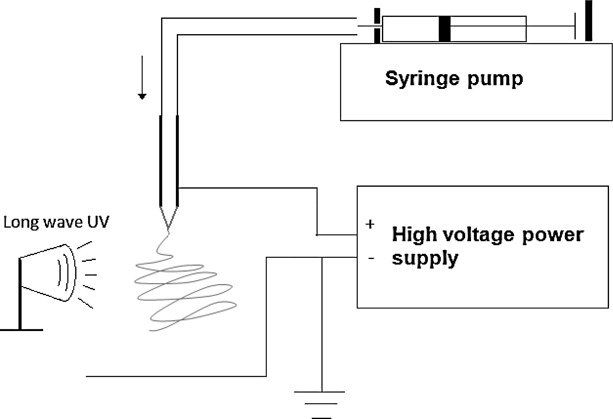
Electrospinning device. High voltage power was applied between a syringe and the aluminum foil collector. Then, a hydrogel solution was injected for electrospinning, while long-wave UV light was applied to crosslink the hydrogel fibers.
The electrospinning system operating at 20 kV (GAMMA) was placed in a laminar flow hood and sterilized under short-wave UV light for 24 h. SFB cells and chondrocytes (primary cells obtained from the knees of pigs as described below) were mixed with each solution mentioned above to a density of 0.5 million cells/mL. They were then electrospun with the hydrogel into fibers at a flow rate of 2 mL/h using an automatic syringe pump (Harvard Apparatus). The fibers were electrospun for 45 s while being exposed to long-wave UV light (to initiate the hydrogel crosslinking process) and collected on glass slides. The glass slides were then placed into Petri dishes containing DMEM cell culture medium with 10% fetal bovine serum (Invitrogen).
Material characterization
Transmission electron microscopy (TEM) was utilized to examine the morphology of RNTs before and after the mechanical adhesive tests. A carbon-coated 400-mesh copper grid (EM Sciences) was floated on the viscous PEG solution supplemented with 0.1 mg/mL RNTs. Another copper grid was rinsed with water and allowed to adhere onto the collagen casing surfaces after the tensile test. The grids were then placed on a droplet of 2% aqueous uranyl acetate (EM Sciences) for 20 s for negative staining. The grid was then blotted and dried with filter paper. TEM images were recorded at a magnification of 10,000× to 235,000× on a Philips EM410 operating under an acceleration voltage of 100 kV.
To further determine that the structures observed by TEM were indeed those of RNTs, the hydrogels on collagen casings or copper grid surfaces were also analyzed by infrared spectroscopy (Mattson). The spectra were recorded in the 400–4000 cm−1 range using KBr pellets.
In addition, to verify the presence of RNTs and the accessibility of their amino acid side chains, an ATTO-TAG CBQCA (3-(4-carboxybenzoyl) quinoline-2-carboxaldehyde) assay (Molecular Probes) was utilized to confirm the presence of lysine side chains. Briefly, a dimethyl sulfoxide solution of (3-(4-carboxybenzoyl) quinoline-2-carboxaldehyde) (10 μL, 5 mM), a solution of KCN (5 μL, 20 mM), and sodium borate buffer (135 μL, 0.1 M, pH 9.3) were mixed, and then cast onto the electrospun hydrogel fibers. The samples were kept at room temperature for 1 h before observation using fluorescence microscopy (Leica).
Cell harvest, culture, and assays
SFB cells and chondrocytes were isolated from synovial membranes or articular cartilage from knee joints of 4-month-old female pigs, respectively, as previously described, 31 and digested with 0.1% trypsin and 0.4% collagenase II. SFB were isolated by magnetic bead separation using Dynabeads CD14 (Invitrogen). High-glucose DMEM (Invitrogen; 4.5 g/L D-glucose, L-Glutamine, and 110 mg/L sodium pyruvate) supplemented with fetal bovine serum (10%), ITS (1%; BD Biosciences) + Premix, penicillin (100 U/mL; BD Biosciences), streptomycin (100 μg/mL; BD Biosciences), L-glutamine (2 mM; BD Biosciences), and amphotericin (2.5 μg/mL; BD Biosciences) was used to culture the cells under standard cell culture conditions (a sterile, 37°C, humidified, 5% CO2/air environment).
After hydrogels were electrospun with cells on glass coverslips (as mentioned in the Material Fabrication section), cells were cultured for 1 day, and then the glass slides were removed from the medium and washed thrice with phosphate-buffered saline. To determine the adhesion density and viability of the SFB cells and chondrocytes after electrospinning, the LIVE/DEAD Viability/Cytotoxicity Kit (Molecular Probes) was used. Briefly, a phosphate-buffered saline (10 mL) was mixed with a calcein AM (5 μL, 4 mM) and EthD-1 solution (20 μL, at 2 mM) to make the working solution. This solution (0.5 mL) was added directly onto the substrate surfaces so that all cells were covered in the solution. After incubating for 30 min, living cells were stained fluorescent green, and dead cells were stained fluorescent red. A fluorescence microscope was used to observe the live and dead cells.
For SFB cell differentiation studies, hydrogels (2% low-gelling temperature agarose) were mixed with SFB cells at a seeding density of 11 million cells/mL with/without 0.001 mg/mL RNTs. Hydrogel/RNT/cell composites were cultured under standard cell culture conditions as mentioned above with the chondrogenic medium (high-glucose DMEM, supplemented with 1% ITS + Premix, 100 U/mL penicillin, 100 μg/mL streptomycin, 2 mM L-glutamine, 2.5 μg/mL amphotericin B and 50 μg/mL ascorbic acid, 0.1 mM nonessential amino acids, 0.4 mM proline, and 10 ng/mL rhTGF-β1; R&D Systems). About 100 nM of dexamethasone (Sigma) was added for the first week of culture. Dynamic culture was established at 65 rpm on an orbital shaker. The hydrogel composites were harvested after 14 days of culture. Pellets were lyophilized overnight and digested (over night, 60°C) in a 0.3 mL papain solution (125 μg/mL papain, 10 mM D-cysteine, 10 mM EDTA, and 100 mM phosphate). DNA content was measured fluorometrically using a Picogreen dsDNA assay kit (Invitrogen). 32 GAG concentration was measured spectrophotometrically by a 1-9-dimethylmethylene blue dye assay, using bovine chondroitin sulfate as a standard. 33 For histology stains, hydrogel composites with SFB cells were fixed with a 1:1 methanol:acetone solution at 4°C, embedded in paraffin, sectioned at 5 μm, and stained with alcian blue/nuclear fast red.
Statistical analyses
Data were expressed as mean ± standard deviation. Statistical analyses were performed using a Student's one-tailed t-test, with p < 0.05 considered statistically significant.
Results
Mechanical adhesive testing and contact angle studies
Results from the mechanical adhesive testing showed that as the RNT concentration in the PEG solution increased, the adhesive strength of PEG on the collagen casings increased (Fig. 3). Specifically, PEG mixed with RNTs of the highest concentration (0.1 mg/mL) gave the largest adhesive strength on the severed collagen casings. Importantly, PEG and RNT solutions dissolved in the serum-supplemented medium gave higher adhesive strengths than PEG dissolved in the serum-supplemented medium only or PEG and RNTs dissolved in the serum-free medium. To understand why RNTs with PEG improved the adhesive strength of severed collagen casings compared to PEG alone, contact angles of the PEG solution with and without RNTs were studied (Fig. 4). With increasing RNT concentrations, the contact angles of the hydrogel (PEG) solution on collagen casings decreased. Specifically, the PEG solution with 0.1 mg/mL RNTs had the smallest contact angles (thus greatest interaction) of all the samples. Such results suggest that RNTs helped increase hydrogel surface wetting and infiltration into the collagen casings, thus causing an increase in adhesive strength.
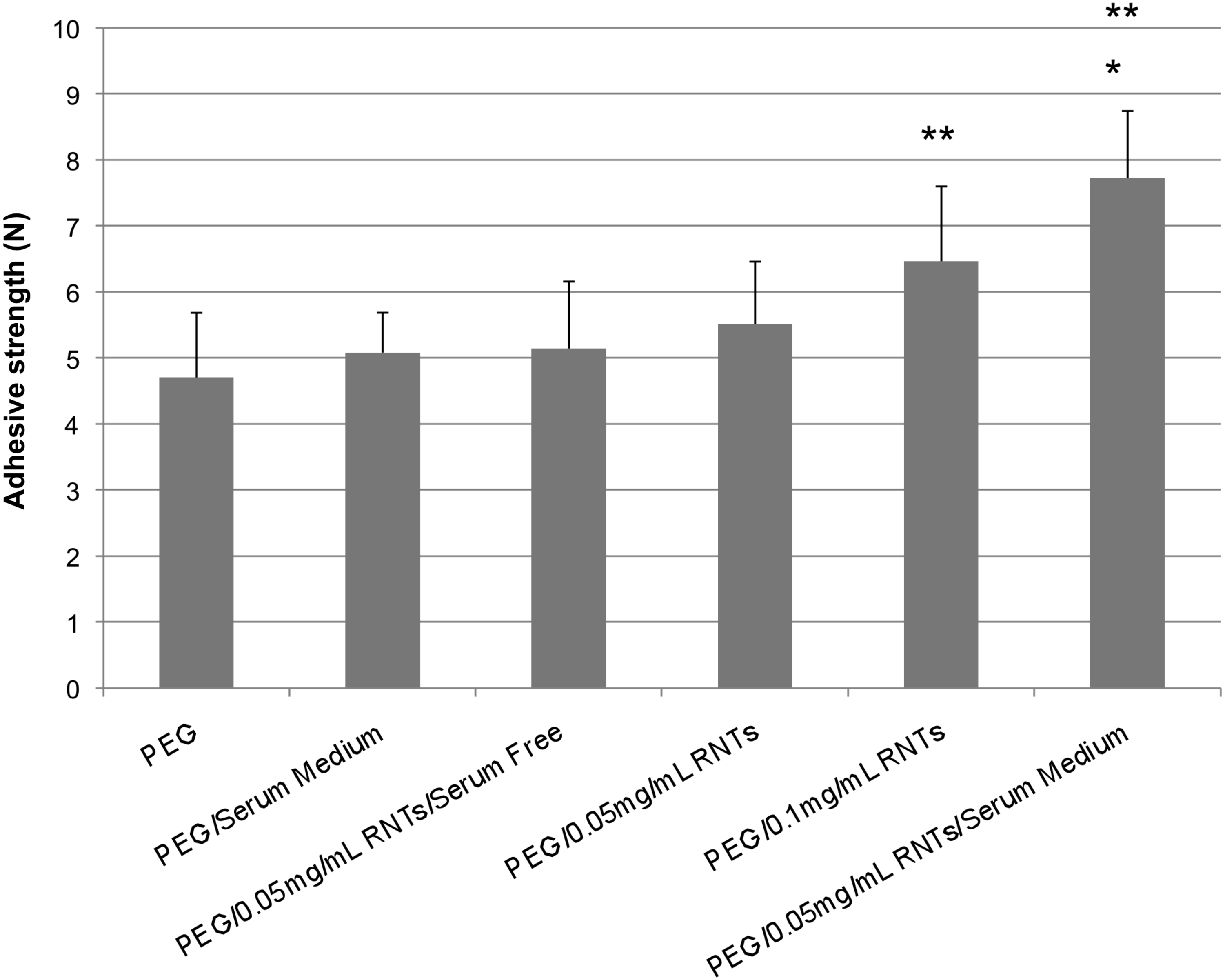
Increased adhesive strength of PEG in the presence of RNTs to hold severed collagen casings together. Data are mean ± SEM (n = 9). **p < 0.05 compared to PEG only. *p < 0.05 compared to PEG in the serum-supplemented medium and PEG with 0.05 mg/mL RNTs in the serum-free medium. PEG, polyethylene glycol; SEM, standard error of the mean.

Decreased contact angles of the PEG solution in the presence of RNTs on collagen casings. Data are mean ± SEM (n = 9). **p < 0.05 compared to controls (PEG only).
Material characterization
The morphology of RNTs on collagen casings was investigated by TEM, which showed that before the tensile tests, even without heat, 0.01 mg/mL of RNTs were able to self-assemble into nanotubes in the PEG-containing hydrogel mixture (Fig. 5). Moreover, RNTs aggregated into larger ribbons (Fig. 5A) with diameters of 50–200 nm (a single RNT has a diameter of ∼3.5 nm). 14 These larger nanotube ribbons may have increased the mechanical adhesive strength of RNTs to hold the severed collagen casings together under a larger tensile force. After heating the RNT-PEG solution at 60°C for 10 min, RNTs assembled into longer nanotubes and ribbons and some fracture points were found during the tensile tests (Fig. 5B). These results support the notion that RNT-containing hydrogels adhered strongly to the collagen casings and thus increased the adhesive strength of PEG.
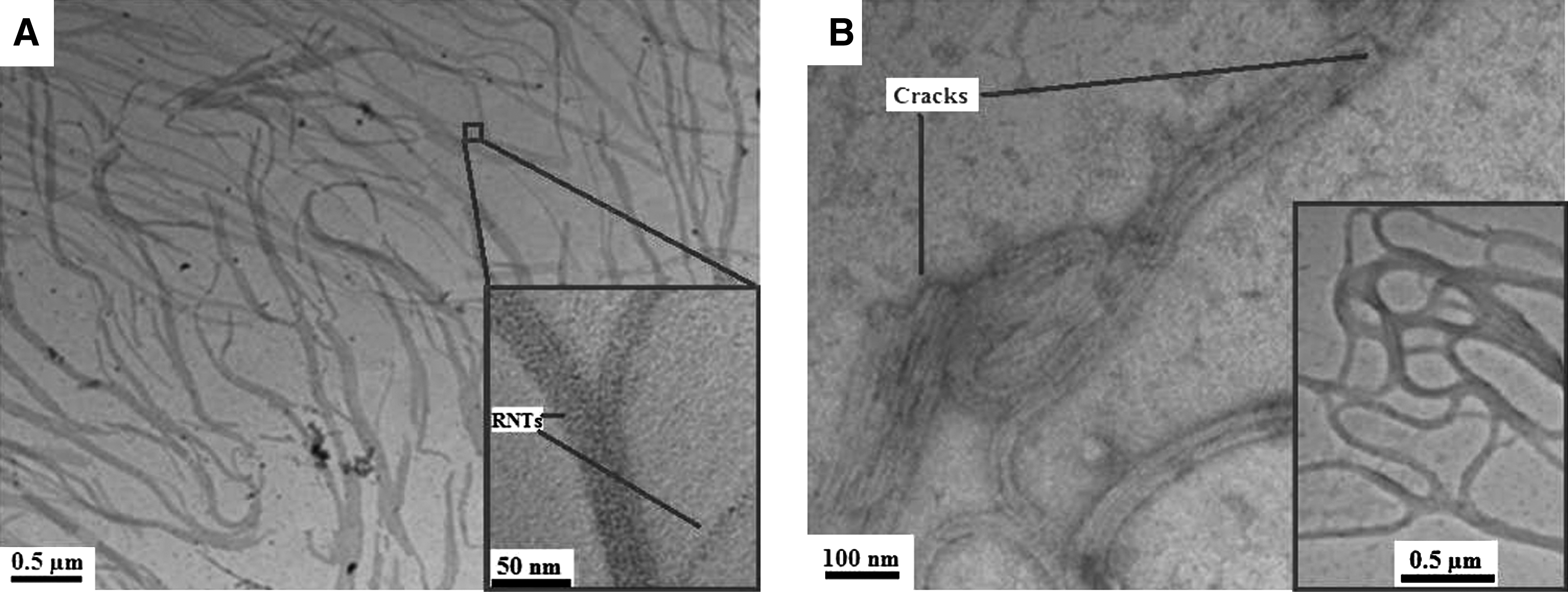
Transmission electron microscopy pictures of the unheated RNT/PEG solution on a transmission electron microscopy grid (
To further confirm that the structures observed using TEM were RNTs, IR spectroscopy was applied to examine the surface of the collagen casings after tensile tests (Fig. 6). IR vibrations around 3200 and 1700 cm−1 (Fig. 6B) corresponded to the N–H and the C = N/C = O groups found in RNTs. Thus, RNTs did indeed adhere to the collagen casing surface, in agreement with the TEM data.
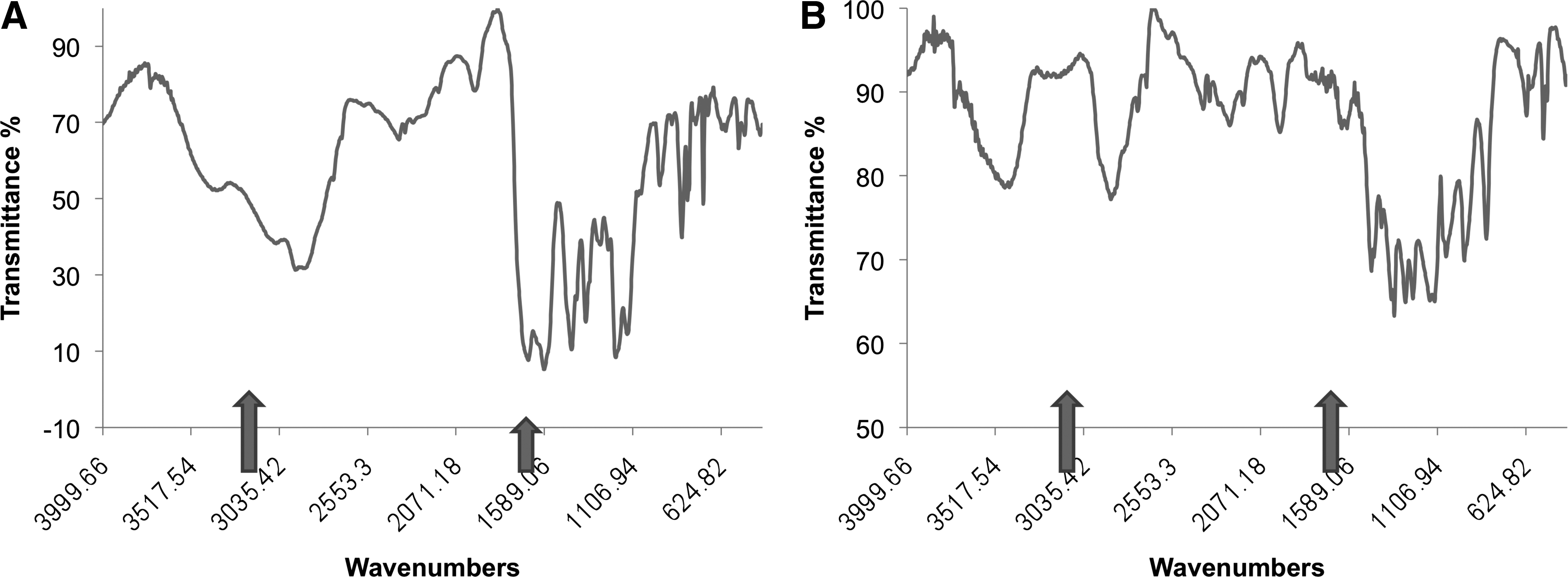
IR spectra of collagen casing surface residues after a tensile test (
After electrospinning, the lysine side chains of RNTs were labeled with a fluorescent probe to allow for the observation of the RNT distribution in the hydrogel composite. Fluorescence microscopy imaging (Fig. 7A) showed that RNTs were present on the surface of the electrospun fibers but not deep inside the fibers. In contrast, electrospinning fibers without RNTs (Fig. 7B) did not show any significant fluorescence. Moreover, these results confirmed the accessibility of the amino groups from RNTs on the surface of the hydrogel fibers.
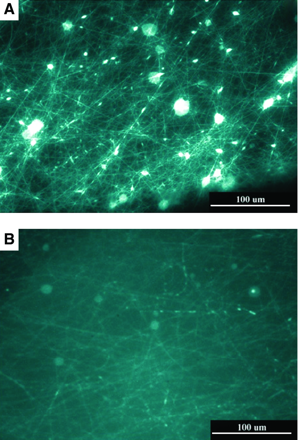
Electrospun hydrogel fibers viewed under a fluorescence microscope (
To investigate the surface roughness of the hydrogel composite, the PVA/alginate/RNT hydrogel was electrospun into submicron fibers with diameters in the 200–700 nm range. TEM imaging of this material clearly showed striations corresponding to RNTs on the electrospun fibers (arrows, Fig. 8A). In contrast, hydrogel composites without RNTs (Fig. 8B) were featureless. These observations suggested that RNTs may behave as a novel nanoscaffolding on the surface of the hydrogel fibers with the ability of exposing bioactive functional groups (e.g., amino groups) to cells at the biologically relevant nanoscale dimension.

Electrospun hydrogel fibers (
Cell viability, adhesion, and differentiation assays
It was estimated that fibers spun within a 1–1.2-cm-diameter circle had a cell seeding density of 3000∼4000 cells/cm2. In addition, because the diameter of the electrospun fibers was below 1 μm, which is well below the average size of chondrocytes and SFB cells, the cells could adhere only on the surface of the fibers rather than fully penetrate inside the fibers. Chondrocyte and SFB cell viability (live cells divided by total cell number) on the PVA/alginate/PEG or PVA/alginate/PEG/RNT samples were higher after 1 day of cell culture compared to PVA or PVA/PEG-only samples (Fig. 9). These results suggested that alginate and RNTs enhanced SFB cell and chondrocyte viability after 1 day of culture. Chondrocyte and SFB cell adhesion density on the PVA/alginate/PEG/RNT samples after 1 day of cell culture was the highest compared to all other scaffolds (specifically, the PVA, the PVA/PEG, and the PVA/PEG/alginate composites without RNTs) investigated (Fig. 10). These results suggested that RNTs in the electrospun hydrogels enhanced SFB cell and chondrocyte adhesion, thus improving hydrogel cytocompatibility properties.
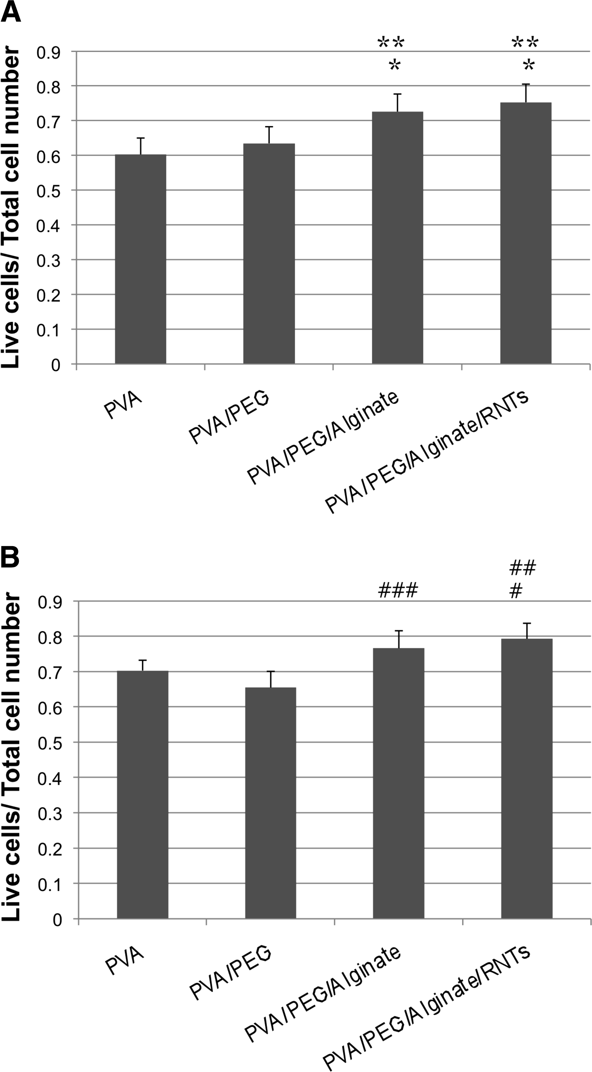
Chondrocyte (
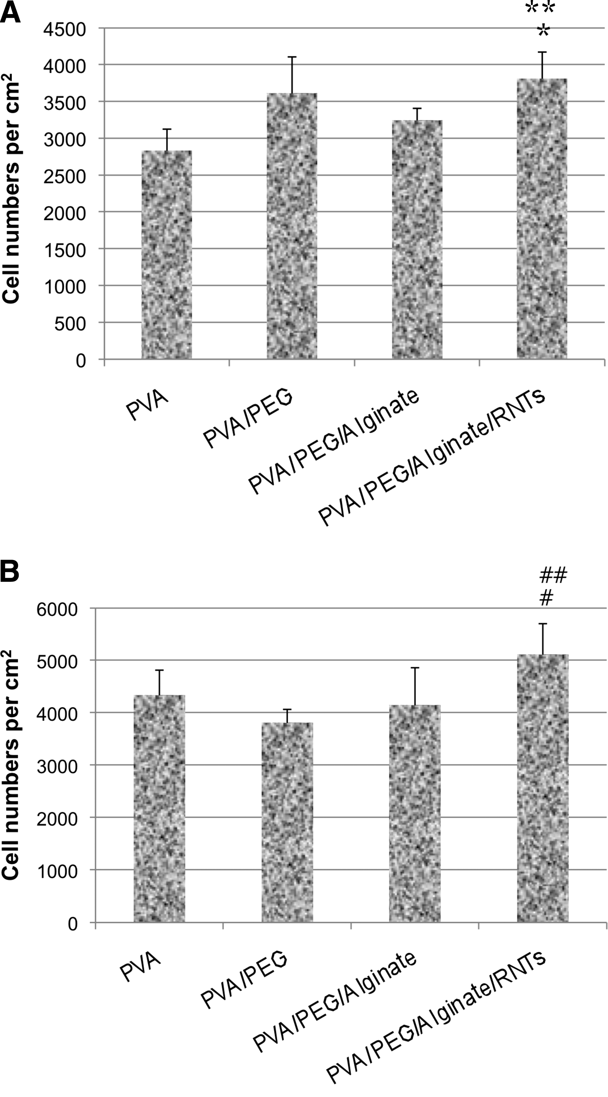
Chondrocyte (
For chondrogenic differentiation studies, as shown in Figure 11A, from histology stains, SFB cell nuclei were stained with red-purple and hydrogel composites (especially, the area around SFB cells in the hydrogel composites with RNTs) were stained with blue-green, demonstrating a positive result for GAG content, which qualitatively indicated chondrogenic differentiation from SFB cells. As seen in Figure 11B, quantitative results showed that the hydrogel/RNT composite had a higher GAG content per unit DNA than without RNTs, suggesting that RNTs improved SFB cell chondrogenic differentiation. Interestingly, hydrogel composites with lower RNT concentration (0.001 mg/mL) had a higher GAG content per unit DNA compared to 0.01 mg/mL RNTs (corresponding to references,34–36 details will be discussed in the following section). Such results correlated with the aforementioned experiments demonstrating that RNTs increased SFB cell adhesion.
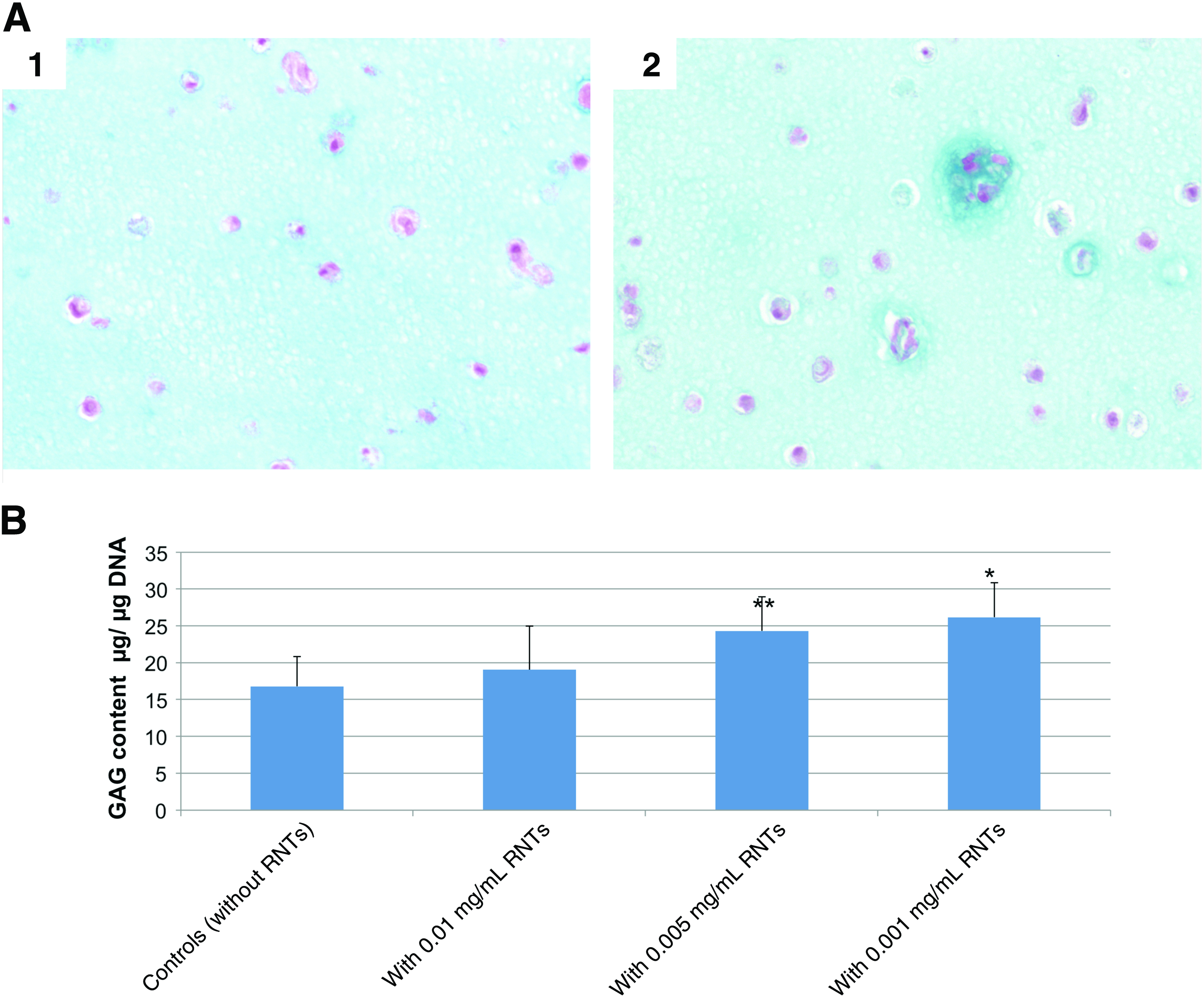
(
Discussion
One critical property for engineering cartilage tissue scaffolds is the ability to bond to the host tissue, potentially serving as an adhesive between severed tissue to initiate a healing process. Although RNTs are not mechanically strong, it was reasoned that in combination with other materials (e.g., hydrogels), their stickiness may be improved. In this manner, this study provided the first evidence that PEG/RNT composites dissolved in the serum-supplemented medium resulted in a higher adhesive strength to collagen casings compared to PEG alone placed in the serum-supplemented medium or RNTs alone placed in the serum-free medium. Such results suggested that RNTs may bond strongly to some proteins in serum to aid in bridging severed tissue, which is in agreement with an earlier report showing that RNTs enhanced osteoblast (bone-forming cell) adhesion and long-term functions 15 by improving initial fibronectin adsorption from serum.
To further understand the properties of RNTs that may have improved bonding strength to collagen casings, contact angle studies were conducted. As suggested by the decreased contact angles of the PEG/RNT composite on collagen casing surfaces, this improvement in adhesive strength may be due to an improved infiltration into the collagen casings of the hydrogel in the presence of RNTs. Coupled with data from previous studies, 15 RNTs may have promoted collagen (a major constituent in cartilage) adsorption.
As stressed in the beginning of this section, other than bonding with host tissue, it is also critical to promote proper functions of cells (such as the growth of chondrocytes or chondrogenic differentiation from SFB cells) in hydrogel scaffolds after implantation.37–50 Since natural articular cartilage usually has a low cell density with little self-regeneration properties,2,4,5 it is critical to implant enough cells to maintain relatively high cell viability for cartilage defect repair. For this reason, cell viability in electrospun hydrogel composites was studied here. Results showed that both chondrocytes and SFB cells were viable after electrospinning, which suggested the promise of using such techniques to fabricate hydrogels with living cells. Importantly, hydrogel composites with RNTs increased chondrocyte and SFB cell viability compared to PVA alone, which confirmed earlier results highlighting the ability of RNTs and biocompatible hydrogels (like alginate) to improve cytocompatible properties of traditional materials.12,13,43–45 Moreover, for the first time, SFB cells were electrospun into RNT-supplemented scaffolds to serve as a pluripotent cell source to populate cartilage defects.
Many have noticed that mesenchymal stem cells (e.g., SFB cells), like many anchorage-dependent cells (e.g., chondrocytes), must first adhere to a surface for further cell migration, proliferation, differentiation, and long-term functions.34–36 Thus, this study focused not only on cell viability but also on cell adhesion and differentiation. Previous reports demonstrated enhanced cell adhesion on nanofibers as a result of their high surface-to-volume ratio and high degree of biologically inspired surface roughness.12–16 In this study, the advantages of combining alginate with RNTs using electrospinning was demonstrated via increased numbers of chondrocytes and SFB cells compared to non-RNT-supplemented materials. As shown by TEM and cell adhesion results, RNTs were well aligned on hydrogel fibers, thus imparting nanoroughness to the submicrometer fiber meshes to enhance the interaction between such materials and chondrocytes and SFB cells. In cell differentiation studies, hydrogel composites with RNTs had higher GAG content per unit DNA compared to controls, which was consistent with SFB cell adhesion experiments. Such results provided evidence that RNTs promoted cell–material interactions to enhance SFB cell adhesion and subsequent functions, such as GAG synthesis. One reason, as emphasized before, is the potential increased select protein adsorption on the nanorough RNT surfaces. Another reason could be the positively charged lysine side chains on RNTs, since previous studies have suggested that low concentrations (0.001 mg/mL) of polylysine with positive charges could enhance the interaction between mesenchymal stem cells and materials to induce chondrogenic differentiation. 36 However, higher concentrations of polylysine (0.01 mg/mL) showed minor toxicity to mesenchymal stem cells. 36 This could also explain the results that lower concentrations (0.001 mg/mL) of RNTs promoted SFB cell chondrogenic differentiation better than higher concentrations of RNTs (0.01 mg/mL).
For cartilage tissue engineering applications, other than biological effects, two important considerations from the material science point of view are how to routinely fabricate consistent biocompatible scaffolds and how to properly seed cells (such as chondrocytes and SFB cells). In this context, electrospinning is a versatile technique that can be used to fabricate scaffolds with a wide range of properties. Because electrospun scaffolds usually resemble the nanostructured environment of the natural extracellular matrix, electrospinning scaffolds provides a better environment for cartilage cell attachment and proliferation compared to conventional scaffolds (such as those made via traditional cast-mold processes). 37
The synthesis of nanometer fibrous polymeric biomaterials, on the other hand, has also emerged as an important technology given its relevance to mimicking protein orientation in cartilage, skin, and the vasculature. 38 Electrospinning is also a promising technique considering that cells can be simultaneously spun with polymers, thus potentially allowing for extreme control over spatial cell placement to further mimic natural tissue architecture. Although voltages applied during electrospinning are in the kV range, proteins and cells can remain viable under these conditions, 39 thus allowing for electrospinning hydrogel fibers with live cells directly into the desired damaged tissue site. Therefore, the present research made a novel improvement to conventional electrospinning processes by creating new materials and combining scaffolds with cells to achieve a uniform material that could be easily inserted into cartilage defects.
Because electrospinning is governed by solution properties (i.e., viscosity and conductivity) and external parameters (i.e., applied voltages and flow rates of the solution), aqueous solutions cannot be electrospun unless their viscosity and conductivity are adjusted. Alginate is usually not electrospun because of its relatively high viscosity and low conductivity. At slightly higher polymer concentrations (enough to generate fibrous structures), the solution becomes so viscous that it cannot be ejected by electrostatic forces even at high voltages.49,50 Thus, to ensure that the final solution has the appropriate viscosity and conductivity to produce micron- to nanosized fibrous scaffolds, low-viscosity/high-conductivity cell-containing aqueous solutions were added in this study to high-viscosity/low-conductivity solutions of an alginate gel, PVA, and PEG. The resulting solution with the appropriate viscosity and conductivity was then amenable to electrospinning.
Of course, for tissue engineering applications, the biocompatibility and biodegradability properties of electrospun scaffolds are critically important. Not only should the engineered biomaterial be cytocompatible, but it should also be biodegradable over a desired period of time. Because alginate hydrogels are known to be biocompatible and biodegradable (as well as readily available), they were selected in this study. Further, PVA and PEG are nontoxic water-soluble polymers.40,41
In the present electrospinning process, although PVA could be crosslinked under long-wave UV (365 nm) light exposure, 40 the electrospun crosslinked fibers began to dissolve after 3 days in the cell culture medium. To control the time course of the dissolution process, cross-linking times may be optimized, various concentrations of the crosslinker may be used, and functionalized PVA/PEG may be employed.40,42 However, these optimization experiments were not the aim of this report but rather will be the focus of future studies.
Conclusions
To the best of our knowledge, this study is the first to electrospin cells directly in a biodegradable hydrogel/RNT composite. Here, it was shown that not only were live cells successfully electrospun into a hydrogel, but also 3D scaffolds for tissue engineering applications were formed with improved adhesive and cytocompatibility properties. These results demonstrated that RNTs increased the adhesion strength to model severed collagen and can be directly electrospun with synovial cells and chondrocytes into a 3D fiber mesh at the site of cartilage damage. RNTs may accomplish this by increasing the surface energy of hydrogel composites to better infiltrate tissue and by providing nanostructured surface features. Especially important to this study, such nanostructured RNTs promoted SFB chondrogenic differentiation. Thus, RNTs provided a supportive matrix for cartilage precursor cells that could in the future be used to promote chondrogenic differentiation for cartilage tissue engineering applications.
Footnotes
Acknowledgments
This study was supported by Hermann Foundation, the Office of Research and Development, RR&D Service, Department of Veterans Affairs, the National Research Council of Canada, and the Natural Science and Engineering Research Council of Canada.
Disclosure Statement
No competing financial interests exist.