
Abstract
Context:
Tissue engineering of mandibular articular condyles encounters many challenges, especially restoring adequate mechanical strength that is correlated to matrix production by the tissue-engineered mandibular condyles (TEMCs). Low-intensity pulsed ultrasound (LIPUS) has been shown to enhance cell expansion, differentiation, and matrix production by different cells.
Objective:
This study evaluated effect of daily LIPUS treatment (in vitro and in a pilot in vivo study) for 4 weeks on matrix production and functional integration of the TEMCs in rabbits.
Methods:
Bone marrow stromal cells were isolated from the femoral bones of skeletally mature New Zealand rabbits, expanded, and differentiated into chondrogenic and osteogenic lineages. Animals employed in the in vivo study were divided into four groups: (1) TEMCs and LIPUS treatment; (2) TEMCs without LIPUS treatment; (3) empty scaffold and LIPUS treatment, and (4) empty scaffolds without LIPUS treatment.
Results:
In vitro results showed that LIPUS enhanced chondrogenic and osteogenic differentiation of bone marrow stromal cells. The in vivo study showed that LIPUS led to better structural formation (namely, new osteogenic and chondrogenic tissue formation) and integration of the newly formed tissues and original condylar bone than those without LIPUS treatment. LIPUS resulted in a small amount of tissue regeneration in the empty scaffolds, whereas empty scaffolds without LIPUS treatment showed no signs of repair.
Conclusions:
The preliminary results of this pilot study suggest that LIPUS can enhance TEMCs both in vitro and in vivo.
Introduction
The temporomandibular joint (TMJ), like other synovial joints, is susceptible to different degenerative diseases collectively affecting over 10 million individuals in North America.3–9 In these disorders, structural destruction of the TMJ necessitates surgical replacement. The current TMJ replacement techniques utilize bone/cartilage grafts and artificial materials.10–12 Despite certain levels of reported clinical success, autografts are associated with donor-site morbidity, unpredictable clinical outcomes, and a relatively high incidence of re-operation.13–15 Alloplastic and xenoplastic grafts are associated with potential transmission of pathogens, immunorejection, and unpredictable clinical outcomes.16–18
Tissue engineering efforts for reconstruction of the mandibular/articular condyles are increasingly employed to restore the lost function. These efforts utilize osteoblasts and chondroblasts/chondrogenic cells from different tissue/cell sources.19–24 However, these efforts have been faced with problems, including insufficient numbers of stem cells to be differentiated into two types of cells (chondrogenic and osteogenic), different bone ingrowth patterns, 22 different rates of gel degradation and matrix production, and inferior mechanical properties for clinical use. 21 Also, a relatively longer time is required for integration of tissue-engineered constructs for osteochondral repair; for example, 3–6 months in rabbit femoral heads, 23 6–12 months in horses, 24 and up to 9 months in sheep 25 are typical. It has been suggested that mechanical stimulation may provide better functional integration of the engineered articular condyle. 25 In addition, the long time needed for in vitro construct preparation may be complicated by other tissue culture problems, such as infection and inconsistency due to routine laboratory and human errors. Thus, tissue-engineered mandibular condyle (TEMC) research is in need of a new method to overcome these deficiencies. Of the aforementioned problems, the lack of mechanical strength is considered one of the major road-blocks to cartilage tissue engineering.26,27
Mechanical strength of the TEMC is thought to be related to matrix production, and a number of techniques have been used to enhance the matrix production. Pulsed electromagnetic fields have been shown to increase chondrocyte and osteoblast-like cell proliferation.28,29 Bioreactors have been used to enhance the material properties of tissue-engineered cartilage constructs,30–36 and cyclic compressive loading has been demonstrated to induce phenotypic changes between cartilaginous and osseous tissues.37–39 Further, mechanical stimulation enhances vascular endothelial growth factor that is important for angiogenesis and bone formation in the mandibular condyles. 40 However, the pulsed electromagnetic fields, bioreactors, and mechanical application machines are not clinically applicable.
Low-intensity pulsed ultrasound (LIPUS) enhances periosteal cell expansion 41 and stimulates bone marrow stromal cell (BMSC) expansion and differentiation into chondrocytes.42,43 LIPUS was shown to enhance early and late osteoblastic differentiation gene expressions such as alkaline phosphatase. This enhanced osteogenic differentiation by LIPUS is suggested to be due to activation of early differentiation genes (c-jun, c-myc, COX-2, Egr-1, and TSC-22) as well as the osteogenic marker genes (osteonectin and osteopontin, phosphorylation, 44 and activation of ERK1/2 and p38 MAPK pathways). 45 Further, LIPUS enhanced bone morphogenetic proteins expression in bone cells, 46 expansion of BMSC and human umbilical cord perivascular stem cell,47,48 and growth of mandibular condyle in animals and human.49–51 Finally, LIPUS has been shown to enhance angiogenesis 52 and minimize apoptosis of human stem cells in vitro. 53 The optimum LIPUS application for bone healing and growth stimulation has been reported to be 20 min/day for 3–4 weeks. 54 Different scaffolds for tissue engineering articular condyles have been proposed in the literature and biological scaffolds composed of extracellular matrix (ECM) have been successfully employed in in vivo tissue engineering. 55 Despite all of this research, little is known about the potential effect of LIPUS on TEMC in vitro or in vivo. Consequently, the aim of this study was to evaluate the effect of LIPUS on tissue engineering mandibular condyle both in vitro and in vivo. Our working hypothesis was that LIPUS will enhance BMSC differentiation into chondrogenic and osteogenic cells in vitro as well as enhance maturation and functional integration of the TEMC in vivo in rabbits.
Materials and Methods
All the procedures in this protocol were approved by the Animal Care Committee at the University of Alberta, Canada. Twelve skeletally mature white New Zealand rabbits were chosen and were divided into four groups of three animals each. Group 1 received TEMC in addition to daily 20 min of LIPUS treatment for 4 weeks; group 2 received only TEMC; group 3 received empty scaffold in addition to daily 20 min of LIPUS treatment; group 4 received only empty scaffolds. Empty scaffolds with or without LIPUS applications were used as negative controls for the differentiated cells and/or LIPUS application. All animals underwent an initial short surgery to aspirate bone marrow from both femoral bones according to a previously described method. 56 For each rabbit, bone marrow from both femur bones was pooled and the BMSCs were isolated by their adherence to the base of the tissue culture flasks. BMSCs were allowed to multiply for 2 weeks until they reached confluence; then, the cells for each rabbit were expanded until passage two (P2) was reached. P2 cells were induced to differentiate into either chondrogenic or osteogenic cells using special conditioning media. Chondrogenic medium contained Dulbecco's Modified Eagle Medium (DMEM), 10% fetal bovine serum, 1% penicillin/streptomycin, and Fungizone (as a base medium) in addition to 0.1 mM nonessential amino acid, 50 μg/mL ascorbic acid-2-phosphate, 10 nM dexamethasone, 5 μg/mL insulin, and 5 ng/mL transforming growth factor-β1. Osteogenic medium contained base medium in addition to 50 μg/mL ascorbic acid-2-phosphate, 10 nM dexamethazone + 7 mM glycerol phosphate + 1 μg/mL bone morphogenetic protein-2. LIPUS was applied using Exogen ultrasonic appliance (Exogen Inc.). The devices were donated by Exogen Inc. LIPUS was applied for 10 min/day for 4 weeks to both chondrogenic and osteogenic cells to enhance their differentiation.
In vitro study
Subgroups of chondrogenic and osteogenic cells from each rabbit were studied for the effect of LIPUS application on their differentiation in vitro. For this experiment, chondrogenic- and osteogenic-driven BMSCs were divided into LIPUS treatment and control (no LIPUS treatment) groups. Both groups were further subdivided into cell culture subgroup and collagen-seeded subgroup. In the collagen-seeded subgroups, cells were seeded (at a density of 200,000 cells/5 mm3) into collagen sponges (Helistat™; Dental Implant Technology, Scottsdale, AZ). The LIPUS treatment group was treated using ultrasound devices that produce 1.5 M Hz pulses that are repeated at 1 kHz and produce power of 30 mW/cm2 (SmileSonica, Edmonton, AB, Canada). LIPUS treatment was performed for 10 min/day for 4 weeks according to the previously recommended protocols.47,48
After 4 weeks, the cell culture subgroups were harvested and processed for quantitative real time polymerase chain reaction (qPCR) analysis for specific gene expression, namely, collagen II and osteopontin. Gene expression was performed using SYBR Green PCR master mix. The amplification mixture contains 1 μL RNA, 2 μL of each primer, and 12.5 μL of the components of the SYBR Green PCR core reagents kit (Applied Biosystems, Foster City, CA) in a final volume of 25 L. Real-time polymerase chain reaction was performed using the ABI Prism 7700 sequence detection system (Applied Biosystems) with the following cycle conditions: 2 min at 50°C, 15 min at 95°C, and 40 cycles of 15 s at 94°C, 30 s at 60°C, and 30 s at 72°C. Glyceraldehyde 3-phosphate dehydrogenase was used as an internal control in each run. Normalized fluorescence was plotted against cycle number (amplification plot), and the threshold, as suggested by software, was used to calculate Ct (cycle at threshold). Results of the real-time polymerase chain reaction are expressed as Ct and the expression levels of specific genes. At the end of 4 weeks, the collagen-seeded subgroup was fixed in 10% paraformaldehyde and evaluated by histological, histochemical, and immunostaining examinations. Histological examination was performed using hematoxylin and eosin–stained sections, while histochemical staining included Safranin-O and von Kossa staining for both chondrogenic and osteogenic cell staining. Immunostaining was performed by aggrecan staining for chondrogenic and calcein staining for osteogenic-driven BMSCs.
In vivo experiment
The chondrogenic and osteogenic differentiated cells were seeded into Helistat sponges that were inserted into biodegradable ECM scaffolds to form TEMC (Fig. 1). The collagen sponges were used to support chondrogenic and osteogenic cell differentiation and matrix production in vitro to facilitate the in vivo implantation of the differentiated cells into the condylectomy sites. The biodegradable ECM scaffold was prepared as previously described. 55 Briefly, the urinary bladders of market-weight (6 months of age) pigs were harvested immediately after euthanasia. The bladders were cleaned of excess connective tissues and immersed in 1.0 N saline to remove the urothelial cells on the luminal surface. The tunica serosa, muscularis externa, and tunica submucosa were removed by mechanical delamination. The remaining tunica propria and basement membrane were then decellularized by serial washes in hypotonic saline, 0.1% peracetic acid/4% ETOH, and numerous rinses in saline. The resulting material was referred to as urinary bladder matrix (UBM). The UBM ECM was then lyophilized. Six sheets of the UBM ECM material were laminated by vacuum pressing with placement of a central accumulation of comminuted (powdered) ECM. This local accumulation of powdered ECM was placed between the third and fourth layers, effectively creating a pillow. The construct was then terminally sterilized with ethylene oxide. The central pillow region of the construct served as the meniscal substrate site, while the adjacent multilaminate sheets served as suture anchoring points.
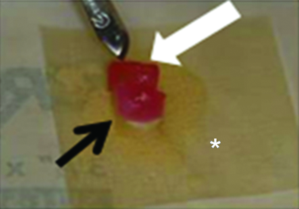
Chondrogenic-loaded collagen sponge (white arrow) and osteogenic-cell-loaded collagen sponge (black arrow) were added together and folded with the extracellular matrix pillow (*) before implantation into the excised condyles in rabbits. Color images available online at
The left TMJ condyle in each rabbit was excised under general anesthesia (ketamine [70 mg/mL] and xylazine [10 mg/mL] at a rate of 1 mL/kg intramuscularly) according to the previously published technique. 57 The right condyle in each rabbit was left untouched to serve as a self-control for each rabbit and to allow the rabbits to eat after surgery. Postsurgical care included antibiotics (Baytril 5 mg/kg) twice a day subcutaneous for 3–4 days and analgesics (Metacam 0.2 mg/kg) once a day for 2 days subcutaneous and then 0.1 mg/kg once a day as needed. Rabbits were fed a soft diet for a few days and then they resumed a normal diet. The tissue-engineered condyles were implanted into the amputated TMJ articular condylar defect and fixed appropriately with absorbable sutures to the surrounding muscles and with calcium sulfate and calcium phosphate (ProDense; Wright Medical Technology Canada Ltd., Mississauga, ON, Canada) (Fig. 2). ProDense is an injectable calcium sulfate and calcium phosphate mixture that has a unique triphasic resorption profile that provides an environment for the direct deposition of bone by binding growth factors and by providing a slow-resorbing matrix that propagates healing across the defect. 58 In groups 3 and 4, the amputated TMJ articular condyles were replaced with empty scaffolds and were fixed by the bone graft substitute as in groups 1 and 2. Groups 1 and 3 were treated daily for 20 min with LIPUS for 4 weeks, whereas groups 2 and 4 did not receive LIPUS. After 4 weeks, all rabbits were euthanized by intravenous injection of sodium pentobarbital 0.5 mL/kg and the mandibles were dissected. All rabbit mandibles were first scanned by microCT. After scanning, both the right and left condylar areas were carefully cut and processed for histological examination. Histomorphometric analysis was performed on three sections of each condyle or TEMCs of each rabbit in all groups. The analysis of new tissues (bone, cartilage/chondrogenic tissue, and fibrocartilage/fibrous tissue) was performed in regions with the highest number of bone trabeculae determined subjectively. Four adjacent high-power (X40) microscopic fields (100 μm2 each) were analyzed. Bone trabeculae were automatically counted in the selected microscopic fields with the use of image analysis software (Metamorph Version 6.1r1). Images were automatically corrected for brightness and contrast, and converted into 8-bit grayscale, and an automated counting of bone trabeculae was performed. Statistical analysis was performed with nonparametric analysis using (Mann–Whitney U-test) to analyze differences between the groups using SPSS statistical package (version 17; Chicago, IL). The level of significance was set to 95%, and α was set at 0.025 (0.05/2 groups in each comparison).

Photograph showing the ProDense added after implantation of the TEMCs. Black arrow refers to condylectomy surgical site, white arrow refers to the TEMC in place of the condylectomy site. Color images available online at
Results
Figure 3 shows the chondrogenic and osteogenic differentiated cells after 4 weeks with or without LIPUS treatment as evaluated histochemically by Safranin-O (for chondrogenic differentiation) and by von Kossa (for osteogenic differentiation). LIPUS enhanced chondrogenic and osteogenic differentiation as noted by the increased Safranin-O and von Kossa staining in the LIPUS-treated samples. Figure 4 shows immunostaining of the chondrogenic (for reaction to aggrecan) and osteogenic (for reaction to osteocalcin) BMSCs. LIPUS enhanced chondrogenic and osteogenic matrix production by the chondrogenic and osteogenic BMSCs in vitro. Figure 5 and Table 1 show gene expression by osteogenic cells with or without LIPUS treatment. LIPUS enhances collagen II and osteopontin expression in vitro.
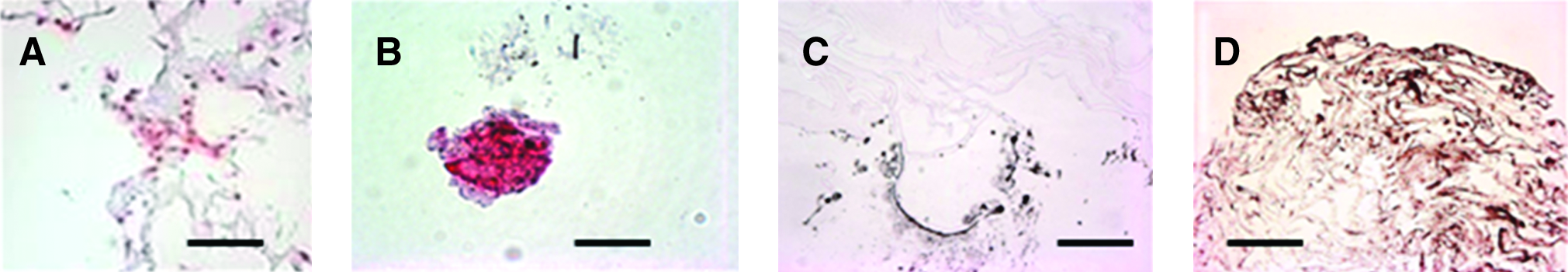
In vitro chondrogenesis and osteogenesis of BMSCs in samples of collagen scaffolds. (

Immunostaining of chondrogenic (
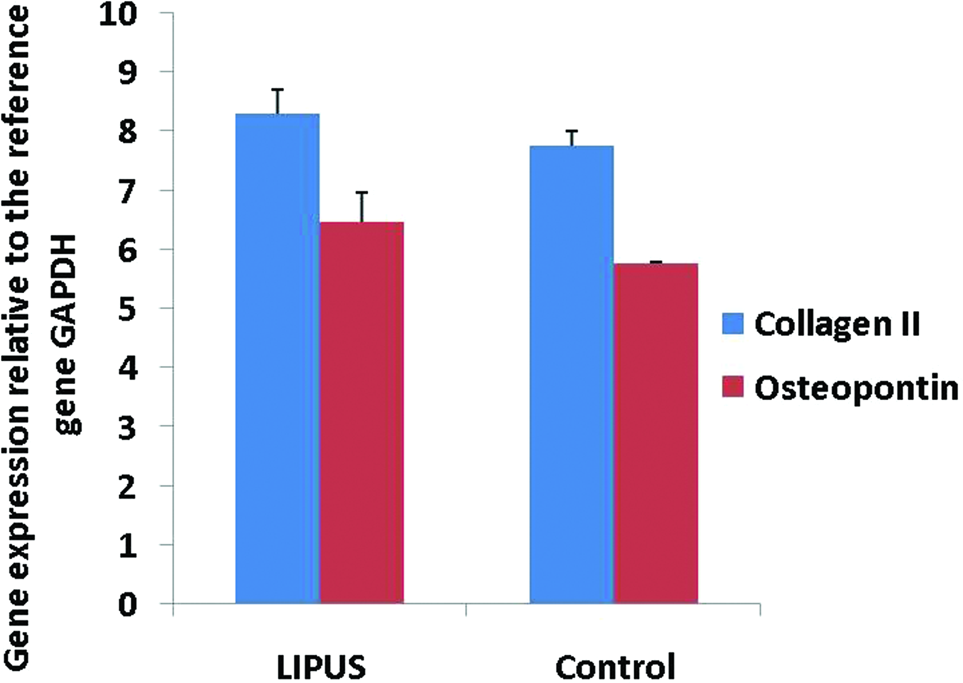
Quantitative real time polymerase chain reaction (qPCR) results of LIPUS-treated (20 min/day) osteogenic differentiated BMSCs for 4 weeks and controls. LIPUS-treated osteogenic cells expressed more osteopontin and collagen type II genes (equalized to GAPDH), which is indicative of enhancing osteogenic differentiation of BMSCs effected by LIPUS. Both graphs represent results of performing qPCR on nine samples (three trials in triplicate). There was a significant increase in collagen II gene expression by LIPUS than in the control group (p = 0.009), and also there was a significant increase in the osteopontin gene expression by LIPUS compared to that in the control group (p = 0.004). GAPDH, glyceraldehyde 3-phosphate dehydrogenase. Color images available online at
Nonparametric analysis (Mann–Whitney U-test). Significance level α = 0.05/2 groups = 0.025. Gene expression is presented as percentage to the reference gene glyceraldehyde 3-phosphate dehydrogenase. There is a statistically significant increase in collagen II and osteopontin gene expression by LIPUS compared to non-LIPUS-treated samples. LIPUS, low-intensity pulsed ultrasound.
From the in vivo study, Figure 6 shows histological evaluation of the left condylar area in each group compared to the normal condyle. LIPUS enhanced chondrogenic and osteogenic matrix production in TEMC but not in empty scaffolds. Figure 7 shows microCT evaluation of the left condylar sites compared to the normal right condyles in each group. Figures 8 and 9 show chondrogenic (by Safranin-O) and osteogenic (by von Kossa) staining of the TEMC with or without LIPUS treatment in addition to the empty ECM scaffolds. LIPUS enhanced matrix production by the chondrogenic and osteogenic differentiated BMSCs. Also, the empty ECM scaffold (collagen sponges not loaded with chondrogenic and osteogenic differentiated BMSCs) with or without LIPUS did not show any replacement of the excised mandibular condyles. Figure 10 and Table 2 show histomorphometric analysis of bone trabeculae, cartilage/chondrogenic tissue, and fibrocartilage/fibrous tissue in all groups. The LIPUS-treated TEMCs showed bone morphometric measurement similar to normal condyles as there was no statistical difference in the amount of bone formation between normal condyle and the TEMC + LIPUS group. TEMC without LIPUS showed less bone formation (bone volume [BV]/TV = 1.1 ± 0.07) when compared to LIPUS-treated TEMCs (BV/TV = 1.23 ± 0.46) or the normal condyle (BV/TV = 1.23 ± 0.41); however, these differences were statistically insignificant. Also, EMC with LIPUS showed more chondrogenic tissue formation than TEMS without LIPUS or normal condyle; however, this increased cartilage formation was not statistically significant. ECM scaffolds with or without LIPUS did not show bone trabecular measurements comparable to normal or LIPUS-treated TEMCs. There was significantly less bone trabecular formation in the ECM scaffolds with or without LIPUS treatment than normal condyles or LIPUS-treated TEMCs (p = 0.024). There was no statistical difference between all the groups regarding the cartilage/chondrogenic areas or fibrocartilage/fibrous tissue.
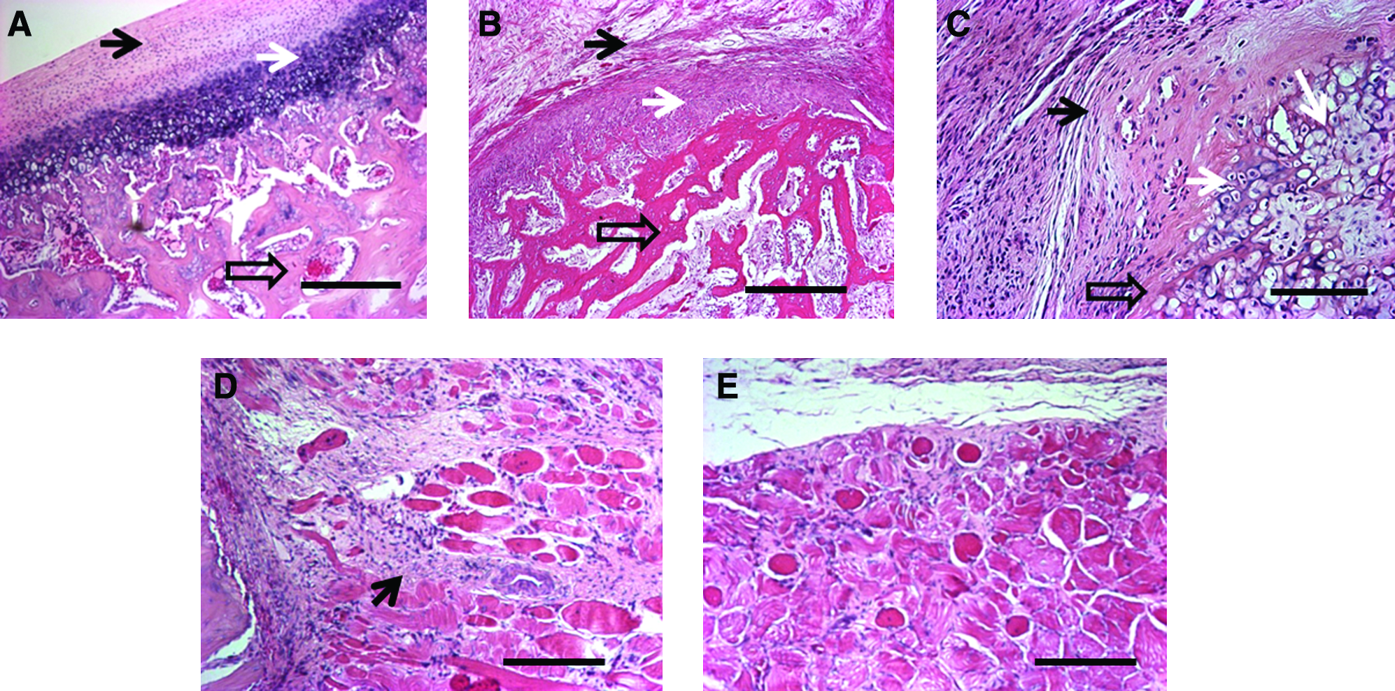
Photomicrographs of the histological examination (H&E staining) of normal condyle (
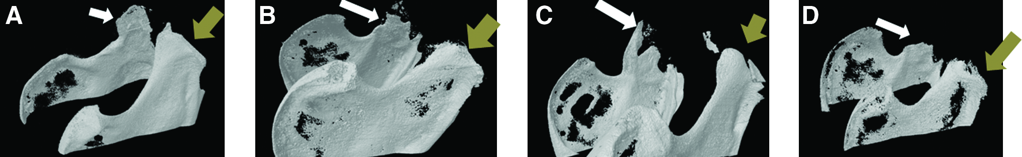
MicroCT scanning of (
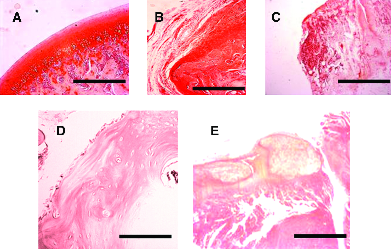
Photomicrographs of Safranin-O-stained histological slides of (
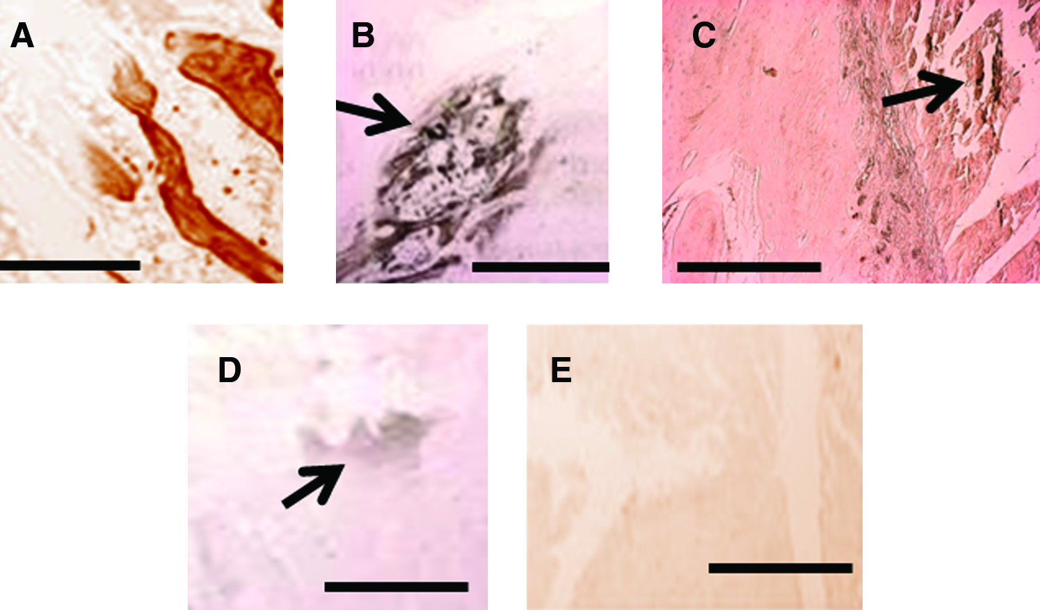
Photomicrographs of von Kossa–stained histological slides of (
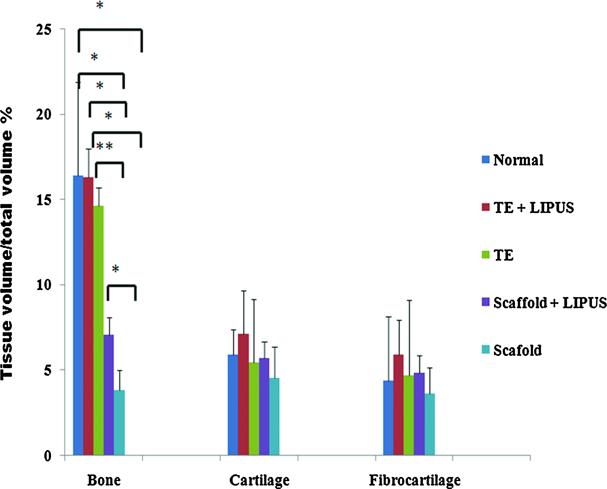
Bone morphometric analysis of bone trabeculae, cartilage, and fibrous tissue surface area in each group. There is a significant difference in BV/TV between normal condyles and groups 3 and 4. Also, there is a significant difference between groups 1 and 3 and between 1 and 4. Moreover, there is a significant difference in BV/TV between groups 2 and 3 and between 2 and 4. However, there was no significant difference between groups 1 and 2 or between normal condyle and either group 1 or 2. Also, there was no significant difference between all the groups and normal condyle in terms of the chondrogenic tissue or fibrocartilaginous tissue area *p < 0.05, **p < 0.01. BV, bone volume; TV, tissue volume. Color images available online at
Histomorphometric measurement (in μm2) of the three main components of the mandibular condyle in each group is given. There is no statistical difference in BV/TV between normal condyle and LIPUS-treated TEMCs. The TEMCs show only decreased BV/TV than those of normal condyles or TEMCs with LIPUS; however, this decreased BV/TV is not statistically significant. There are significant decreases of BV/TV in both extracellular matrix scaffolds with or without LIPUS than TEMCs, TEMCs with LIPUS, or normal condyle. Also, LIPUS-treated scaffolds show statistically significant increased BV/TV than scaffolds without LIPUS. There is no statistical difference in chondrogenic or fibrous tissue morphometric analysis between all groups.
BV, bone volume; TV, tissue volume; TEMCs, tissue-engineered mandibular condyles.
Discussion
This study evaluated the effect of LIPUS on enhancing differentiation of BMSCs into chondrogenic and osteogenic lineages, and assessed the effect of LIPUS on matrix production of the TEMCs in rabbits. The in vitro portion of this study confirmed that LIPUS enhanced BMSC differentiation into chondrogenic and osteogenic lineages. This was demonstrated histochemically (Fig. 1), immunologically (Fig. 2), and with gene expression (Fig. 3 and Table 1) and increased osteopontin and collagen II production by LIPUS (Fig. 3 and Table 1). These results are in agreement with previous reports that showed LIPUS enhanced chondrogenic and osteogenic differentiation of stem cells in vitro.41–43
The in vivo portion of this experiment was a pilot study using rabbit mandibles. The use of the left TMJ as the experimental side and right TMJ as a self-control was planned to allow the rabbits to eat after surgery. There was no concern about the potential effects of the LIPUS on the control (right) side in the LIPUS-treated groups as its effects on these conditions have been shown to be neglible. 49 The results of this experiment demonstrated that LIPUS enhanced chondrogenic and osteogenic matrix production in TEMC but not in empty scaffolds. This was expected based on previous studies that reported increased production of bone and cartilage matrices using LIPUS. Further, it demonstrated that differentiated cells were required for tissue regeneration, suggesting that the effect of LIPUS is on the cells themselves, not the matrix. These results were consistent and confirmed physically by microCT, histologically, and using histomorphometric measures (for bone only).
On the basis of the histological and microCT analysis, LIPUS enhanced the TEMC formation and led to better integration of the chondrogenic and osteogenic portions of the TEMCs. To the best of our knowledge, our current research is the first attempt to replace entirely excised TMJ condyles with TEMCs in vivo. Previous reports have shown that it is possible to tissue engineer articular condyles from BMSCs;21–25 however, those attempts were limited to tissue engineering the mandibular condyle without implanting the TEMC into the TMJ, or they were not replacing the entire TMJ condyle by the TEMC (those attempts were limited to small articular defects). In addition, those studies did not show structural integration of the TEMCs with the native bone of the mandibles. Further, the in vitro part of this research was in agreement with previous reports and supported the successful results of LIPUS-assisted TEMCs in vivo as demonstrated here.
In this experiment, ProDense was used as a bone cement to help initial integration of the implanted TEMC in all groups as it is commonly used in orthopedic surgery to augment bone fracture healing. 58 There was no evidence that the ProDense had any effect on bone regeneration in the TEMCs in this study, as the groups without cells did not show significant bone regeneration and ProDense was used in all groups. That said, the interaction between LIPUS, ECM, and calcium phosphate (ProDense) requires further investigation.
The results of these experiments suggest that the use of ECM matrix along with chondrogenic and osteogenic differentiated BMSCs shows promise for joint tissue engineering of mandibular articular condyles and that ECM without cells is not a practical option for mandibular condyle tissue engineering. Our research describes a new technique of using LIPUS to enhance chondrogenic and osteogenic differentiation of BMSCs in vitro as well as to enhance functional integration of the TEMCs in vivo in rabbits. Future research will be directed to evaluate the mechanical properties of TEMCs using ECM with and without LIPUS application.
Footnotes
Acknowledgments
This research was supported by the University of Alberta Start-up fund to T.E.B., the University of Alberta Fund for Dentistry, and the University of Alberta Hospital Fund grants. The authors would also like to thank Exogen Inc. for their donation of the LIPUS devices.
Disclosure Statement
No competing financial interests exist.