
Abstract
This work presents an integrated microfluidic device on which the proliferation of rabbit chondrocytes was investigated in the presence of insulin-like growth factor-1 (IGF-1), basic fibroblast growth factor (bFGF), and their combinations. The microfluidic device was mainly composed of an upstream concentration gradient generator and a downstream perfusion-based three-dimensional cell culture module. The rabbit articular chondrocytes were cultured for 2 weeks at the different concentrations of growth factors generated by concentration gradient generator. IGF-1, up to 57.14 ng/mL, had the ability to promote the proliferation of chondrocytes in a dose-dependent manner, and there were no further promotions at higher concentrations. bFGF increased chondrocyte proliferation dose dependently up to 5.72 ng/mL, and then the proliferation rate decreased when the concentration was increased. The combination of IGF-1 and bFGF could synergistically promote the proliferation, and the group of 85.73 ng/mL IGF-1 and 1.43 ng/mL bFGF presented an optimal effect (up to 4.76-fold), which had statistically significant differences compared with IGF-1 and bFGF, respectively. Moreover, the proliferation test using the conventional method was performed simultaneously and revealed similar results. The results obtained in this study demonstrated that the microfluidic device is an effective platform for cartilage tissue engineering. With this device, experimental conditions are flexible and can be optimized by changing either the category of growth factors or the concentration of input growth factor. Further, the small number of cells (1–100) required, with which parallel experiments could be performed simultaneously, makes it an attractive platform for the high-through screening at the cellular level in autologous chondrocyte implantation.
Introduction
In vivo, chondrocytes typically reside in the environment with very specific three-dimensional (3D) features. The biological behaviors of cells are sensitive to the presence of cytokines, hormones, biomechanical forces, and other factors. Although the in vitro cell culture technique is widely used in conventional laboratory experiments, it is doubted by some researchers whether chondrocytes grown in vitro are identical to those grown in vivo, as conventional methods provide a static and macro-scale environment that is entirely different from the environments in vivo.8,9 A newly emerging technology known as microfluidics has been demonstrated to be very useful for cell culture. 10 It could supply and transfer media, buffers, and even air, and the waste products from cellular activities are drained by resembling the human circulatory system.11,12 It also creates new opportunities for the spatial and temporal control of cell growth and stimuli by integrating relative functional modules into a chip that carries out 3D culture, medium exchange, as well as cell seeding, observation, and assay in microchannels.13,14 Therefore, microfluidic systems provide an in vivo-like environment for a cell culture as well as a reaction environment for a cell-based assay. Further, it reduces the number of cells required and the need for large volumes of culture medium and costly reagents, which makes microfluidics an attractive platform for the ACI.
In this work, the perfusion-based micro 3D cell culture platform with concentration gradient generator (CGG) was developed by soft lithography of poly (dimethylsiloxane) (PDMS). At the different concentrations of growth factors generated by CGG, rabbit articular chondrocytes were cultured for 2 weeks. The effects of insulin-like growth factor-1 (IGF-1) and basic fibroblast growth factor (bFGF) on the proliferation of chondrocytes were investigated. In addition, conventional experiments in 96-well plates were carried out to demonstrate that the proposed microfluidic platform can be exploited for cartilage tissue engineering at the micro-level.
Materials and Methods
Microfluidic device design and fabrication
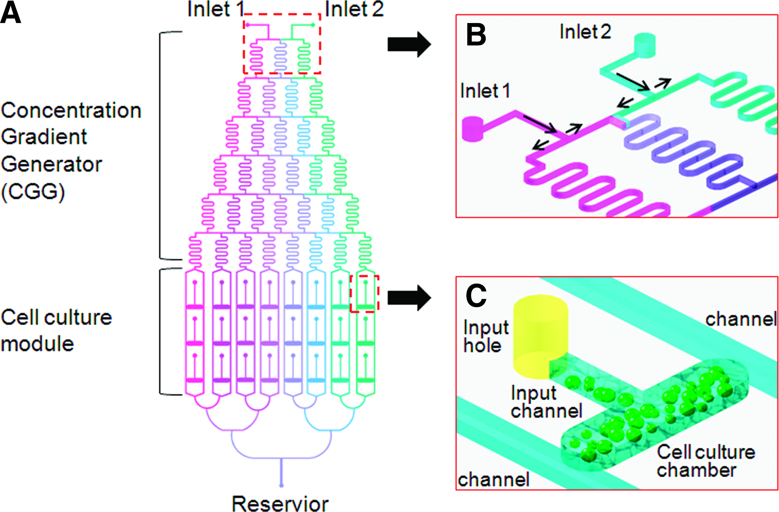
The microfluidic device in our study was mainly composed of an upstream CGG and a downstream cell culture module (Fig. 1A). The design of the CGG was based on the work previously presented by Jeon et al. 15 According to the Reynolds experiment, at small scales (channel diameters of around 100 nanometers to several hundred micrometers), the Reynolds number (which compares the effect of momentum of a fluid to the effect of viscosity) can become very low. A key consequence of this is that fluids, when travel side by side, do not mix necessarily in the traditional sense; molecular transport between them must be carried out by diffusion in the serpentine channels (Fig. 1B). This property is critical in our microfluidic device. As two streams travel down in CGG, they are repeatedly split at the nodes, combined with neighboring streams in laminar fashion, and allowed to mix by diffusion in serpentine channels. Consequently, the solutions are continuously diluted and a series of concentrations are produced in the outlets of CGG. For the current study, if one solution (concentration 0) and the other one (concentration C) were introduced into CGG, the concentrations in the eight outlets are, respectively, 0, 1/7C, 2/7C, 3/7C, 4/7C, 5/7C, 6/7C, and C according to the equation described by Jeon et al.
Microfluidic device design. (
The cell culture module is composed of an array of cell culture chambers that are integrated with the CGG unit. Three cell culture chambers with the same function were connected between two parallel channels of each outlet of CGG. The chambers were miniaturized: 700 μm long, 500 μm wide, and 100 μm height. The volume of each chamber is around 0.4 μL. The chondrocyte–gel mixture could be seeded into a chamber through an input hole and channel. When solutions flow down in the parallel channels, growth factors and other substance would diffuse into the chamber by osmosis till the cell–gel mixture is saturated. Since the concentrations of solutions in the two channels are same, the saturated concentration of the mixture in the chamber is uniform and consistent with that in the parallel channels (Fig. 1C).
All microfluidic devices were fabricated in PDMS using rapid prototyping 16 and soft lithography. 17 Briefly, a high-resolution printer was used to generate a mask (in the form of a transparency) from a computer aided design (CAD) file. The transparency mask (which can have a minimum feature size of about 20 μm, when generated by a 3300 dpi printer) was used in 1:1 contact photolithography with SU-8 photoresist (MicroChem) to generate a negative master, consisting of patterned photoresist on an Si wafer. Positive replicas with embossed channels were fabricated by molding PDMS against the master. The inlets and outlets (1-mm-diameter holes) for the fluids were punched out of the PDMS using a sharpened needle. The surface of the PDMS replica and a clean glass substrate were activated in an air plasma (2 torr, 60 s, 100 W) and brought together immediately after activation. An irreversible seal was formed between the PDMS and the glass substrate; this assembly produced the required systems of microfluidic channels. Polyethylene tubing with outer diameter slightly larger than the inner diameter of the port was inserted into the hole to make the fluidic connections. The pieces of tubing were then connected to a syringe pump (Orion Sage, M362) to complete the fluidic device.
Microfluidic chip operation
A syringe pump was used to drive fluid flows. Culture media with and without growth factors were simultaneously infused into the microfluidic device. As the fluid streams traveled through the network, they were repeatedly split, mixed, and recombined. The concentration gradient of growth factors was established at eight outlets of CGG 30 s later. Solutions carrying different concentrations of growth factor diffused into the chondrocyte–gel mixture when they flow in the parallel channels of each outlet. The device was then kept in an incubator at 37°C, 5% CO2, and 100% humidity. The flow speed was controlled at 0.1 μL/min (Fig. 2).

Microfluidic device operation. A syringe pump is connected to a chip located inside an incubator by polyethylene tubes. It drives the medium into the chip; meanwhile, waste liquid is drained and collected. Color images available online at
CGG performance validation
Since the fluorescence probe, fluorescein isothiocyanate–dextran (FITC-Dextran molecular weight [MW] 20,000 Da; Sigma-Aldrich), has a comparable MW with the IGF-1 (Peprotech; MW 7649 Da) and bFGF (Peprotech; MW 18,000 Da), it was used as an indicator for estimating the gradient produced by CGG. By introducing FITC-Dextran and blank solution into two inlets, respectively, the FITC-Dextran solution was diluted continuously and a series of concentrations of FITC-Dextran were formed in the microchannels and then entered the corresponding downstream cell culture chambers. As the intensity of fluorescence of FITC-Dextran was proportional to the concentration, the intensities of FITC-Dextran at eight outlets of CGG were imaged by confocal laser scanning microscope (Leic) and quantified by Image-Pro Plus software (version 6.0 for Windows XP; Media Cybernetics). Further, the intensities were compared with the theoretical values achieved by the equation and correlation factors were calculated. All the experiments were repeated three times.
Chondrocytes isolation and expansion
Full-thickness articular cartilages were removed from the knees and hip joints of a New Zealand white rabbit, and were subsequently cut into small pieces as described. 18 Chondrocytes were digested and released from cartilage slices using collagenase II (0.25% w/v). The isolated cells were then cultured in Dulbecco's modified Eagle's medium supplemented with 10% fetal calf serum, 100 U/mL penicillin, and 100 μg/mL streptomycin. Cells were incubated at 37°C in a 5% CO2 incubator, and the medium was changed every 4 days. When the cells reached the growth plateau phase, they were harvested by 0.25% trypsin/1.0 mM ethylenediaminetetraacetic acid.
Cell viability assay and the diffusion of FITC-Dextran in the collagen gel
Cell viability in 3D matrix was assessed by applying two specific fluorescence probes, Rhodamine 123 (Rh-123; Sigma Chemicals) and propidium iodide (PI; Molecular Probes). Rh-123 could be sequestered by active mitochondria of living cells, which then emit green fluorescence. PI stained dead cells red. Briefly, we introduced 2 μg/mL Rh-123 into perfusion channels and incubated at 37°C for 40 min, rinsed with phosphate-buffered saline, labeled with 2 μg/mL PI, and imaged immediately. Images were observed using an inverted fluorescent microscope (Olympus IX 71). To observe the diffusion of FITC-Dextran (MW 20,000 Da, similar to those of IGF-1 and bFGF) in the collagen gel, we injected the gel to one of cell culture chambers followed by the perfusion of FITC-Dextran through upstream inlets. The images of FITC-Dextran diffusion in the collagen gel were collected by the inverted fluorescent microscope.
Chondrocytes seeding, culture, and assay
One part of chondrocytes obtained from articular cartilage was embedded in the 0.24% type I collagen gel solution (pH 7; Cell Matrix Type Ia collagen; Koken) at a cell density of 1 × 10⁵/mL at 4°C. This cell–gel mixture was divided into two groups, either of which was seeded into 96-well plates (100 μL/well) or seeded into culture chambers of microfluidic chips by pipetting (approximately 0.4 μL/chamber). The other part of chondrocytes was seeded in 6 cm plate at a density of 1 × 10⁵/mL and cultured for 48 h to compare the morphologies of chondrocytes in 2D and 3D.
The serum-free (SF) medium adapted from Jakob et al. 19 was supplemented with 100 ng/mL IGF-1 and 10 ng/mL bFGF. From the two inlets, (1) 100 ng/mL IGF-1 SF medium and SF medium, (2) 10 ng/mL bFGF SF medium and SF medium, and (3) 100 ng/mL IGF-1 SF medium and 10 ng/mL bFGF SF medium were pumped into three microfluidic chips, respectively. Chondrocytes were cultured at a series of concentrations of IGF-1, bFGF, and their combinations on chips and 96-well plate for 2 weeks, respectively.
Since both PDMS and glass slide are optically transparent, we counted the number of proliferated chondrocytes on chips directly with the inverted microscope after 2 weeks. For the 96-well plate, chondrocytes were released by incubation with 0.25% collagenase (Sigma). Cell suspension was collected, and the cell number was counted on a hemocytometer using the trypan-blue exclusion test.
Statistical analysis
Mean proliferation rate and standard error were calculated. Subsequently, analysis of variance and least-significant difference (LSD) post hoc testing were performed with SPSS for Windows version 14.0 professional (SPSS® Inc.).
Results
CGG performance validation
As streams of the dye travel down the network, they were repeatedly split, combined with neighboring streams in laminar mode at the nodes, and allowed to mix by diffusion in the serpentine channels. A series of solutions with different concentrations of FITC-Dextran were formed at the outlets of CGG (Fig. 3A). The fluorescent intensities of FITC-Dextran in the junctions between the CGG and the cell culture module were quantified, corrected by subtracting the background fluorescence, and compared with the theoretical data. As shown in Figure 3B, there was a good coherence (correlation coefficient = 0.9953) between the experimental and theoretical data.

CGG performance validation. As the fluorescence intensity of FITC-Dextran was proportional to the concentration, the intensities of FITC-Dextran at eight outlets of CGG were imaged by a confocal laser scanning microscope and quantified by Image-Pro Plus software. Further, the intensities were compared with the theoretical values achieved by the equation. (
Cell morphology and viability in 3D matrix
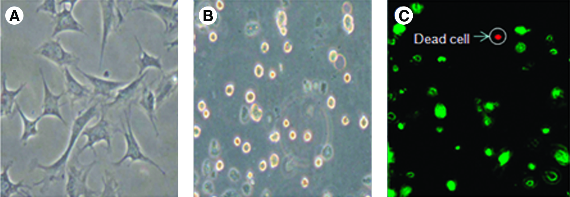
The biological characteristics of chondrocytes were investigated by comparing the morphology features in 2D (6 cm plate) and 3D (embedded in the collagen gel) cultures for 48 h. The cells exhibited fibroblast-like morphology with spindle-shaped appearance on 2D surface (Fig. 4A). However, the morphology was changed from fibroblast-like morphology, as seen in 2D culture, to round in 3D culture (Fig. 4B), which is consistent with the previous studies. 20 In addition, the viability of cells cultured in microfluidic chip was evaluated. After 2 weeks of culture, few red-stained cells (1–3 cells), detected by inverted microscope, were found in every cell culture chamber (around 100 cells). The viability of chondrocytes cultured in the chip was calculated to be above 95% (Fig. 4C).
Cell morphology and viability in three-dimensional matrix. (
The diffusion of FITC-Dextran into the collagen gel
To observe the diffusion of FITC-Dextran (MW 20,000 Da, similar to the MW of IGF-1 and bFGF) in the collagen gel, images of FITC-Dextran diffusion in the collagen gel were collected by the inverted fluorescent microscope. Figure 5A and B depicts the diffusion of FITC-Dextran into the collagen gel in 5 and 60 min, respectively.

The diffusion of FITC-Dextran in the collagen gel. To observe the diffusion of FITC-Dextran (MW 20,000 Da, similar to the MW of IGF-1 and bFGF) in the collagen gel, images of FITC-Dextran diffusion in the collagen gel were collected by the inverted fluorescent microscope (
Chondrocytes proliferation
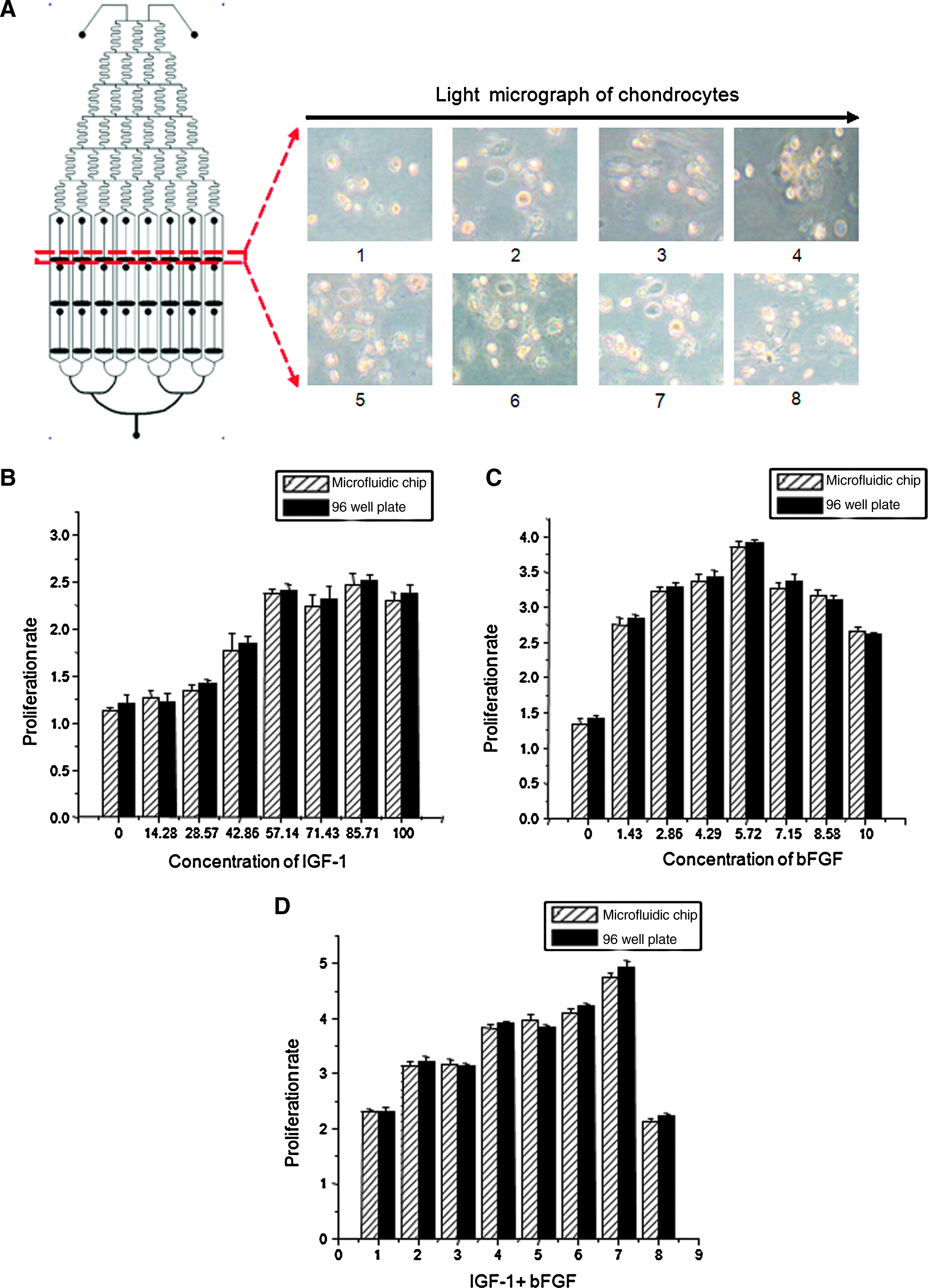
Chondrocytes embedded in the collagen gel maintained their round morphology after 2 weeks at different concentrations of IGF-1, bFGF, and their combinations (Fig. 6A). The dose dependency of chondrocyte proliferation in the presence of IGF-1 was apparent between 0 and 57.14 ng/mL. The proliferation rate reached the peak in the 57.14 ng/mL group (2.38-fold), which has the statistically significant difference with the control group (p < 0.01). There was no statistically significant difference between 57.14 ng/mL group and the groups of higher IGF-1 concentrations (p > 0.05). bFGF increased chondrocyte proliferation dose dependently between 0 and 5.72 ng/mL. The proliferation rate reached the peak in the 5.72 ng/mL group (3.85-fold). When the concentration was increased, the proliferation rate appeared to decrease from 5.72 ng/mL to 10 ng/mL. The eight combinations of IGF-1 and bFGF are the following: 0/10, 14.29/8.57, 28.57/7.14, 42.86/5.72, 57.14/4.29, 71.44/2.86, 85.73/1.43, and 100/0 ng/mL. Among these combinations, the group of 85.73 ng/mL IGF-1 and 1.43 ng/mL bFGF exerted the optimal synergistic effect on chondrocyte proliferation (4.76-fold), which had the statistically significant differences compared with the control group, 57.14 ng/mL IGF-1 group, and 5.72 ng/mL bFGF group (F = 540.129, p < 0.01; see Figure 6B–D). Further, there were no statistically significant differences between the chips and 96-well plate with the proliferation rates in every corresponding group (F = 111.388, p > 0.05).
Chondrocyte proliferation in the presence of IGF-1, bFGF, and their combinations. (
Discussion
Microfluidic device
Microfluidics, also known as “lab-on-a-chip,” is an emerging technology that represents a revolution in laboratory experimentation, bringing the benefits of integration, miniaturization, and automation to many research areas. It is the science and technology of systems that control small (10−9 to 10−18 liters) amounts of fluids in channels with dimensions of tens to hundreds of micrometers. 21
As mentioned before, chondrocytes typically reside in the environment with very specific 3D feature in vivo. The biological behaviors of cells are sensitive to the presence of cytokines, hormones, biomechanical forces, and other factors. The ultimate aim of cartilage tissue engineering is to imitate the environment where chondrocytes live in vivo. In the previous experiments on cartilage tissue engineering, the culture conditions of chondrocytes are poor to mimic the cellular environment in vivo because it is very hard for researchers to take so many factors involved into consideration in one study with traditional methods. However, microfluidics can create a new opportunity for the spatial and temporal control of cell growth and stimuli by integrating various functional units.
In this study, two modules were integrated: CGG and 24 perfusion-based cell culture chambers. With the CGG module, as depicted in Figure 1A and B, eight concentrations of growth factors were generated precisely/rapidly in 30 s (Fig. 3). Several growth factors regulate the metabolism and activation of cartilage cells. To enhance culture conditions and effectiveness for in vitro cartilage engineering, researchers should characterize the interactions and combinations of different growth factors. When this microfluidic platform is used, experimental conditions are flexible and can be optimized by changing either the category of growth factors or the concentration of the input growth factors.
In addition to the CGG design, we have integrated 24 cell chambers to culture cells at different concentrations of growth factors. Chondrocytes were embedded in the collagen gel and seeded in the chambers, and showed good compatibility with our chip since more than 95% of cells remained alive after 2 weeks (Fig. 4). PDMS is biocompatible and highly permeable to CO2 and O2, 22 thereby guaranteeing rapid exchange of these gases between the atmosphere around the chip and the medium in the culture chambers. As a new cell culture platform, microfluidic devices have been successfully used for the long-term culture (up to 4 weeks 23 ) of a variety of cells, including cell lines and primary cells. 24 The devices were also used to test different culture conditions for their ability to drive cell growth and to maintain cell function.
For most conventional experiments, the culture medium was supplied in a noncontinuous manner. For instance, the culture medium is replaced manually and periodically in a common static culture, which not only risks contamination but also, more importantly, results in a fluctuating culture environment. 9 Conversely, continuous medium supply and waste removal in our study not only kept the culture environment stable but also reduced the contamination possibilities of medium change. Further, in the perfusion-based culture pattern, medium in the parallel channels diffused into the cell–gel mixture, and metabolic waste generated by cellular activities was drained simultaneously, imitating the exchange between extracellular conditions and cells in vivo (Fig. 5).
For the current study, the consumption of the medium was less than 5 mL and the consumption of IGF-1 and bFGF was less than 200 ng. What is more important is the reduction in the required number cells. The optimal culture condition of chondrocytes in vitro differs in the cells, which come from either patients with different age and illness or the site and layer where cells are located in vivo. In the ACI, few chondrocytes were harvested and available for the subsequent expansion. It is virtually impossible for researchers to perform many parallel experiments using conventional methods to figure out the optimal condition with a very limited number of chondrocytes. The unique advantage that the number of cells required on microfluidic chip is very small (1–100 cells), with which many parallel experiments could simultaneously be performed, makes it an attractive platform for the high-through screening at the cellular level in ACI.
In the following experiments, we can integrate more relative modules into the microfluidic chip to implement the effects of 3D culture, growth factors, and biomechanic forces on the chondrocytes. In the near future, using the microfluidic platform we will try to explore a deliberate menu of multiple factors that can substantially proliferate chondrocytes while keeping good morphology in vitro. This menu is unique for individuals and could meet the need of expansion in vitro in different phases. On the basis of seeded cells of high quality, ACI promises a good prognosis.
Effects of IGF-1 and bFGF on chondrocytes proliferation
For the purpose of supplying sufficient numbers of chondrocytes, researchers seeded the chondrocytes into plates for monolayer culturing. Although a large number of cells were gained in a short time by this method, chondrocytes lost their typical round morphology, forming a spindle fibroblast-like shape, and cartilaginous protein synthesis was decreased. This is a common phenomenon known as de-differentiation. 25 The prevention of de-differentiation or the preservation of chondrocyte quality during proliferation culture is a vital step for the enhancement of regenerative cartilage medicine. Although the expansion of chondrocytes in the collagen gel (3D) could maintain the chondrocyte phenotype during cultivation, unfortunately, this method showed a much slower proliferation speed compared with the monolayer culture. 20 It is necessary to accelerate the speed of proliferation in 3D culture.
IGF-1 is a circulating cytokine that reaches articular cartilage through the synovial fluid. It is a single polypeptide with protein sequencing similar to insulin. IGF-1 was found to stimulate the biosynthetic activity and proliferation of articular chondrocytes in a dose-dependent manner. 26 bFGF was originally isolated and identified from bovine brain and pituitary based on its stimulatory activity on fibroblast proliferation. 27 Many studies have implied a potent anabolic effect of bFGF on cartilage homeostasis and suggested its use for cartilage regeneration and repair.28,29 In bovine adult articular cartilage, bFGF has been associated with a modest stimulation of proteoglycan (PG) synthesis and cell proliferation. 30 However, most studies about the effect of bFGF and IGF-1 on proliferation of chondrocytes were examined not in a 3D culture, but in a monolayer culture. In the previous studies, the effect of growth factors on chondrocytes proliferation was completed using a wide gap of growth factor concentration (e.g., 0, 50, and 100 ng/mL). In comparison, our platform was able to generate detailed concentrations of growth factors in a rapid/automated fashion. The concentration gradient of IGF-1 is 0, 14.29, 28.57, 42.86, 57.14, 71.44, 85.73, and 100 ng/mL, and the concentration gradient of bFGF is 0, 1.43, 2.86, 4.29, 5.72, 7.14, 8.57, and 10 ng/mL with an increase by 1/7 of highest concentration, respectively.
In the present study, it was shown that IGF-1, up to 57.14 ng/mL, promoted the proliferation of cultured articular chondrocytes in a dose-dependent manner and no further promotion happened at higher concentrations. Rechler and Nissley 31 suggested that in IM-9 lymphocytes, human skin fibroblasts, and BC3H-1 mouse muscle cells, the limitation of proliferation at higher IGF-1 concentrations was caused by the downregulation of IGF-1 receptors. Chiyokura 32 reported that although the number of IGF-1 receptors was not exactly known, expression of IGF-1 receptors on articular chondrocytes was detected continuously and did not seem to decrease significantly with time during the culture period. From these results, it appeared that IGF-1 receptors were saturated with IGF-1 at higher IGF-1 concentrations and the possibility of downregulation in the cultured articular chondrocytes was also indicated. Further studies are required.
bFGF increased the chondrocyte proliferation dose dependently up to 5.72 ng/mL. When the concentration was increased, the proliferation rate appeared to descend, which indicates that higher concentrations of bFGF inhibit chondrocyte proliferation. The proliferation inhibition in our study is similar to that in the previous work.33,34 This inhibition seems due to the change of extracellular signaling mediated by bFGF. Extracellular signals from bFGF to the cells are transduced through one of the four structurally related high-affinity receptors (FGF receptors 1–4). bFGF binding to the FGF receptor 1 (FGFR1) has been demonstrated to increase proliferation of growth plate chondrocytes, whereas bFGF binding to the FGF receptor 3 (FGFR3) inhibits the proliferation and promotes differentiation.35,36 In human articular chondrocytes, Muddasani et al. 37 suggested that FGFR1 is the major FGF receptor that is responsible for the bFGF-mediated biological consequences, such as cellular proliferation and production of matrix metalloproteinase-13 in vitro.
For this study, FGFR1 was highly expressed and the binding of bFGF and FGFR1 resulted in receptor dimerization, which, in turn, activated multiple downstream signaling cascades in cells to promote the proliferation when the concentration of bFGF was between 0 and 5.72 ng/mL. As bFGF increased from 5 to 10 ng/mL, more FGFR3 may be activated and suppressed the chondrocyte proliferation. 38
Among the combinations of IGF-1 and bFGF, the group of 85.73 ng/mL IGF-1 and 1.43 ng/mL bFGF had the optimal effect on the chondrocyte proliferation (4.76-fold). Previous studies disagree on the notion that IGF-1 and bFGF interact. In adult articular chondrocytes, IGF-I and bFGF have been reported to have 39 and have not 33 a synergistic effect on DNA synthesis. Variations in the observed responsiveness of chondrocytes to bFGF and IGF-1 may have been due to the differences in the species and age of the donor animals, the initial seeding intensity, the method of culture (2D or 3D), the biological substance used, the duration of cultures, and so on.
The mechanism underlying this interaction is not clear. However, because IGF-1 and bFGF produce their cellular effects through separate surface-membrane receptors, the data could be explained by the interaction between postreceptor pathways. Further investigations are needed to define the interactions of these growth factors and to determine their application in chondrocytes expansion in vitro.
Conclusions
The current study indicated that both IGF-1 and bFGF promoted the proliferation of rabbit articular chondrocytes in 3D culture while maintaining their morphology. The combination of IGF-1 and bFGF could synergistically promote chondrocyte proliferation. The data obtained revealed that our microfluidic device is a candidate for cartilage tissue engineering. The abilities of miniaturization and integration of valuable functional units make microfluidic technology an attractive platform for the high-through screening at the cellular level in ACI.
Footnotes
Acknowledgments
The authors are grateful to Dr. Mais Jebrail and Dr. Hassan Dib for their help in revising and editing the article.
Disclosure Statement
No competing financial interests exist.