
Abstract
Electrospun collagen scaffolds must be crosslinked to improve stability. Chemical crosslinking methods are often associated with cytotoxicity and can require lengthy rinsing procedures to remove the crosslinker. Physical crosslinking using dehydrothermal (DHT) treatment is utilized to stabilize fibrous collagen sponges; however, little is known regarding the effect of DHT crosslinking on electrospun collagen. To investigate the efficacy of DHT crosslinking, soluble type I collagen was electrospun and exposed to DHT crosslinking, chemical crosslinking with N-(3-dimethylaminopropyl)-N′-ethylcarbodiimide hydrochloride (EDC; 5 mM), and DHT+EDC. DHT crosslinking produced no change in scaffold fiber diameter or interfiber distance and reduced scaffold degradation. Strength was significantly improved by DHT (139.0 ± 34.9 kPa) compared to control but was weaker than EDC or DHT+EDC (222.7 ± 58.4, 353.3 ± 19.0 kPa, respectively). Fourier transform infrared spectroscopy (FTIR) indicated increased amide bond formation with DHT compared to control but a lower amide bond density than EDC or DHT+EDC. After crosslinking, sterilization, and rinsing (a total of 50 h for DHT, 98 h for EDC, and 122 h for DHT+EDC), fibroblasts adhered and proliferated on all scaffolds; however, cell metabolism was 12% less on DHT scaffolds. These data indicate that DHT crosslinking can be utilized to stabilize electrospun collagen scaffolds; however, a tradeoff exists between scaffold stability/strength and rapid processing.
Introduction
Physical and chemical crosslinking are often performed to stabilize protein scaffolds for use in aqueous environments. Crosslinking collagen using dehydrothermal (DHT) treatment is a common method of physical crosslinking. Numerous studies have been performed that utilize DHT stabilization of collagenous natural materials and collagen scaffolds with the use of DHT crosslinking.11–18 During DHT crosslinking, ester and amide bonds form,12,19 leading to significant increases in the strength of collagen compared to noncrosslinked collagen materials 20 and reduced degradation rates upon exposure to collagenase. 21 Fibrous, insoluble collagen crosslinked using DHT crosslinking has also been shown to support cell proliferation and tissue formation.17,20,22 Thus, electrospun collagen could significantly benefit from DHT crosslinking methods that can be performed while the scaffold is dry.
Chemical crosslinking is the most widely used method of crosslinking collagenous tissue engineering scaffolds. Several different chemicals have been used, including glutaraldehyde (GA),3,23,24 hexamethylene diisocyanate,15,25–27 and N-(3-dimethylaminopropyl)-N′-ethylcarbodiimide hydrochloride (EDC).22,25,28 Despite the broad utilization of GA as a crosslinking agent, it has long been associated with scaffold failure due to cytotoxicity and calcification. 29 Hexamethylene diisocyanate has also been shown to cause cytotoxicity due to un-reacted pendant molecules.15,30 Crosslinking of electrospun collagen with EDC reduced scaffold degradation,11,31 maintained cell viability, 10 and has been used successfully to crosslink scaffolds used for bioengineered skin, 22 bone,25,32 and cartilage. 33 Collagen crosslinked with EDC has also been shown to have an increased strength and denaturation temperature compared to similar crosslinking agents 27 due to the formation of amide and ester bond crosslinks. 34 EDC crosslinking has been shown to effectively crosslink electrospun collagen scaffolds but has been associated with cytotoxicity at concentrations >10 mM 35 and often requires a lengthy rinsing procedure, up to 4 days, to ensure no crosslinker remains within the scaffold.22,36,37 Thus, it would be beneficial to directly compare efficacy of DHT crosslinking versus EDC crosslinking to determine if the use of chemical crosslinking can be avoided to avoid possible degradation during processing and cytotoxicity and to reduce processing times.
The goal of this study was to evaluate the efficacy of DHT crosslinking of electrospun type I collagen compared to standard chemical crosslinking alone and in conjunction with standard chemical crosslinking. Additionally, the utility of these crosslinked scaffolds for dermal tissue engineering was examined. Collagen scaffolds were prepared by electrospinning soluble type I collagen. Scaffolds were then dehydrothermally (DHT) crosslinked, chemically crosslinked, or both DHT and chemically crosslinked. As-spun scaffolds served as a control. The effects of crosslinking on scaffold architecture, stability, mechanical properties, and cytotoxicity were investigated.
Materials and Methods
Electrospinning solutions
Electrospinning solutions were prepared by mixing acid-soluble collagen from bovine hide (Kensey Nash; Exton) with 1,1,1,3,3,3-hexafluoro-2-propanol (Sigma-Aldrich Co.) at a concentration of 10 wt./vol.%. The solutions were mixed for 2 days before electrospinning.
Collagen scaffolds
Collagen-1,1,1,3,3,3-hexafluoro-2-propanol solutions were electrospun (4 mL/h, 30 kV) onto a grounded, Al-foil-covered brass plate positioned 20 cm away. As-spun scaffolds were then placed into a vacuum-sealed desiccator or dehydrothermally crosslinked (DHT) at 140°C for 24 h. A subset of these scaffolds were crosslinked in an ethanol and EDC (Sigma-Aldrich Co.) solution (5 mM EDC) for 24 h. Four different scaffolds were prepared: as-spun collagen (control), collagen with DHT (DHT), collagen with EDC crosslinking (EDC), and collagen with DHT and EDC crosslinking (DHT+EDC).
Scanning electron microscopy
Scaffold morphology was qualitatively examined using scanning electron microscopy (SEM; FEI Sirion). Small, circular punches (6 mm in diameter) from each dry scaffold were collected, prepared for SEM, and imaged with 5 kV accelerating voltage. From the collected digital images, average fiber diameter for each scaffold type was calculated. Fiber diameter (>100) was measured from six independent samples from each group using Image J software and reported as average fiber diameter ± standard deviation (SD). To quantitatively assess the pore size in these scaffolds, the free space between fibers or interfiber distance was calculated. Interfiber distances (>20/sample), measured perpendicularly from one fiber to its nearest neighbor in the same plane, were calculated from six samples per group. Mean interfiber distance ± standard deviation was reported. Scaffold morphology (overall structure, fiber diameter, and interfiber distance) was also examined after incubation in HEPES buffered saline (HBS) for a total of 7 days to evaluate if significant alterations in scaffold morphology would occur due to hydration. Hydrated scaffolds were prepared for SEM by rinsing in phosphate-buffered saline (PBS), dehydrating in a series of alcohol, and chemical drying with decreasing solutions of ethanol and hexamethyldisilazane (HMDS; Ted Pella Inc.). 37 Dry samples were then prepared for and imaged using SEM with average fiber diameter and interfiber distance quantified as above.
Transmission electron microscopy
To examine the microstructure of electrospun collagen, the scaffolds were crosslinked using the above methods (control, DHT, EDC, and DHT+EDC), rinsed two times with milli-Q water for 1 h, and postfixed with 1% osmium tetroxide for 1 h. Next, the collagen scaffolds were rinsed three times with deionized water for 5 min each rinse, exposed to a graded ethanol series (50%, 70%, 80%, 95%, 100%, and 100% ethanol for 10 min), exposed to 100% HMDS three times for 10 min, and dried overnight in 100% HMDS. Then the samples were trimmed and embedded in epoxy resin followed by sectioning on a Leica EM UC6 ultramicrotome cutting at 100 nm. Sections were examined with a Tecnai G2 Spirit transmission electron microscopy (TEM) operated at 100 kV.
Scaffold stability
Scaffold resistance to degradation was quantified through exposure to bacterial collagenase type I (Worthington Biochemica Corp.). Dry scaffolds were punched into 18-mm-diameter circles (n = 6 per group), weighed, sterilized in 70% EtOH, and incubated for 3 days in medium with 1 U/mL collagenase per mg of collagen. Aliquots from the medium were removed at 1, 3, 6, 24, and 72 h and analyzed for total collagen using a hydroxyproline assay. 35 As these scaffolds were fabricated from collagen that contains unknown quantities of lipid and carbohydrates (<2%), the standard curve was formed using known amounts of the same collagen raw material used for the electrospun mats. The concentration of collagen in the samples was determined using the standard curve within the linear region. Data were presented as average percent of scaffold degraded + SD.
Tensile testing
Collagen scaffold mechanics were quantified via uniaxial tensile testing (n = 6 per group; TestResources 100R). Scaffolds were hydrated with HBS and punched into dogbone specimens with a gauge length of 20 mm and width of 4 mm. The specimens were strained to failure at a rate of 2 mm/s. If samples failed outside of the gauge length, data were discarded. Ultimate tensile strength (UTS), linear stiffness, and elongation at failure were calculated and reported as mean ± SD.
Fourier transform infrared spectroscopy
Infrared spectra of the electrospun collagen scaffolds were obtained using the attenuated total reflectance mode of a Nicolet Nexus 670 benchtop Fourier transform infrared spectrometer with a Continuum microIR microscope (Thermo Scientific). A germanium crystal was used for the attenuated total reflectance analysis. Spectra were obtained between the wave numbers of 600–4000 cm−1 and were normalized to the methyl peak at 2950 cm−1. The extent of crosslinking was assessed between samples by comparing the peak height at 1546 cm−1, which indicates the formation of amide bonds and at 1100 cm−1 indicative of ester bonds.
Cellular interaction: Viability and penetration
The toxicity of the crosslinking procedure was determined by assessing the viability of fibroblasts cultured on the scaffold for 7 days. All scaffolds were crosslinked following protocol described above (control, DHT, EDC, DHT+EDC). Following the crosslinking step, if any, scaffolds were sterilized for 24 h in 70% ethanol, rinsed 2× in PBS for 24 h, rinsed in HBS 5× 10 min, and then rinsed in culture medium 2× for 30 min in preparation for cell inoculation. This rinsing protocol has been previously utilized to ensure that no cytotoxicity is observed. 37 Scaffolds were then placed onto sterile polypropylene meshes (N-terface; Winfield Labs), which were then set on top of inoculation sponges (Hydrasorb; Carwild Inc.). Inoculation sponges were hydrated but not saturated and aided in inoculation by preventing inoculum from leaking over the sides of the scaffold and promoted cellular infiltration through the wicking action of the sponge. Human primary dermal fibroblasts were inoculated into the scaffolds (1 × 10⁶ cells/cm2) and incubated on the inoculation sponge for 2 h. Subsequently, scaffolds were removed from the inoculation sponges and maintained submerged in the culture medium. Fibroblasts were cultured for a total of 7 days with 4 mm punch biopsies removed from the scaffold at 1, 3, 5, and 7 days (n = 6 per group, n = 6 per time point). Cell metabolism on the scaffolds was assessed using a modified MTT assay following a protocol previously described. 37 Briefly, biopsy punches were incubated for 3 h in 0.5 mL of sterile MTT solution (0.5 mg MTT/mL PBS) at 37°C and 5% CO2. The MTT solution was then removed and 0.5 mL methoxyethanol (Fisher Scientific) was added to each well. Plates were gently agitated for 3 h after which the absorbance of the solution was measured at 590 nm using microplate reader (Gemini XPS; MolecularDevices). Average absorbance values ± SD were reported.
To determine cell penetration depth into scaffolds, biopsies from the fibroblasts inoculated scaffolds were collected at day 7, embedded, and cryosectioned (8-μm-thick sections). Cryosections were then immunostained with propidium iodide (cell nuclei) and rabbit anti-collagen type I (Santa Cruz Biotechnology). Sections were digitally imaged using the laser scanning confocal microscope (Zeiss LSM 510 Meta). Average cell penetration depth in each section was measured using the cell penetration depth of at least 10 cells. Average cell penetration within each group was calculated using the average penetration depth from five sections, each from a different sample within the same group.
Statistical analyses
All data were analyzed using SigmaStat 3.10 (Systat Software Inc). Student's t-tests were performed to determine statistical significance with p-values <0.05 considered statistically significant.
Results
Scaffold structure
Collagen scaffolds were comprised of randomly oriented ribbon-like fibers 1.59 ± 0.60 μm in diameter (Figs. 1A and 2D). Exposure to DHT crosslinking maintained the as-spun fiber morphology with no change in fiber diameter or interfiber distance (Figs. 1 and 2D, E). After incubation in HBS for 7 days, the control group completely degraded while the DHT, EDC, and DHT+EDC groups remained intact and were characterized by larger fiber diameters (Fig. 2A–C). Fibers crosslinked with EDC, only experienced the greatest swelling after hydration with a 50% increase in fiber diameter, whereas the fibers with DHT crosslinking increased by 15% on average (Fig. 2D). In addition to causing fiber swelling, exposure to aqueous media also decreased the free space between fibers or interfiber distance (Fig. 2E). The EDC and DHT groups had a significant reduction in interfiber distance after soaking in HBS for 7 days (p < 0.01) but were not statistically different than one another. The DHT+EDC group maintained the greatest interfiber diameter and had significantly larger pores than the DHT group (Fig. 2E; p < 0.05). TEM confirmed the fiber morphology observed in SEM; however, no samples displayed the 67 nm banding, which is a characteristic of native collagen as seen previously with electrospun collagen 38 (Fig. 3). Control scaffolds degraded during processing; thus, no TEM imaging could be conducted on these samples.
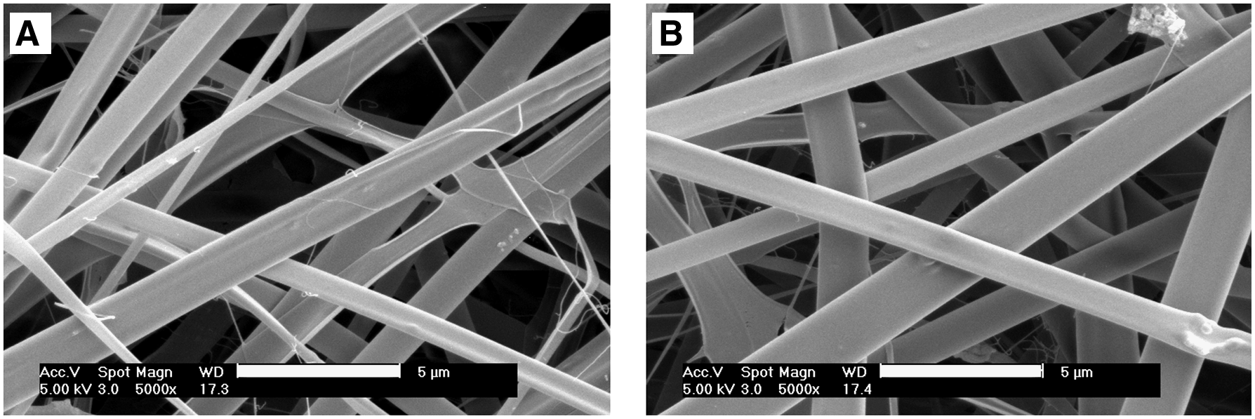
Scanning electron micrographs of electrospun collagen scaffolds. (
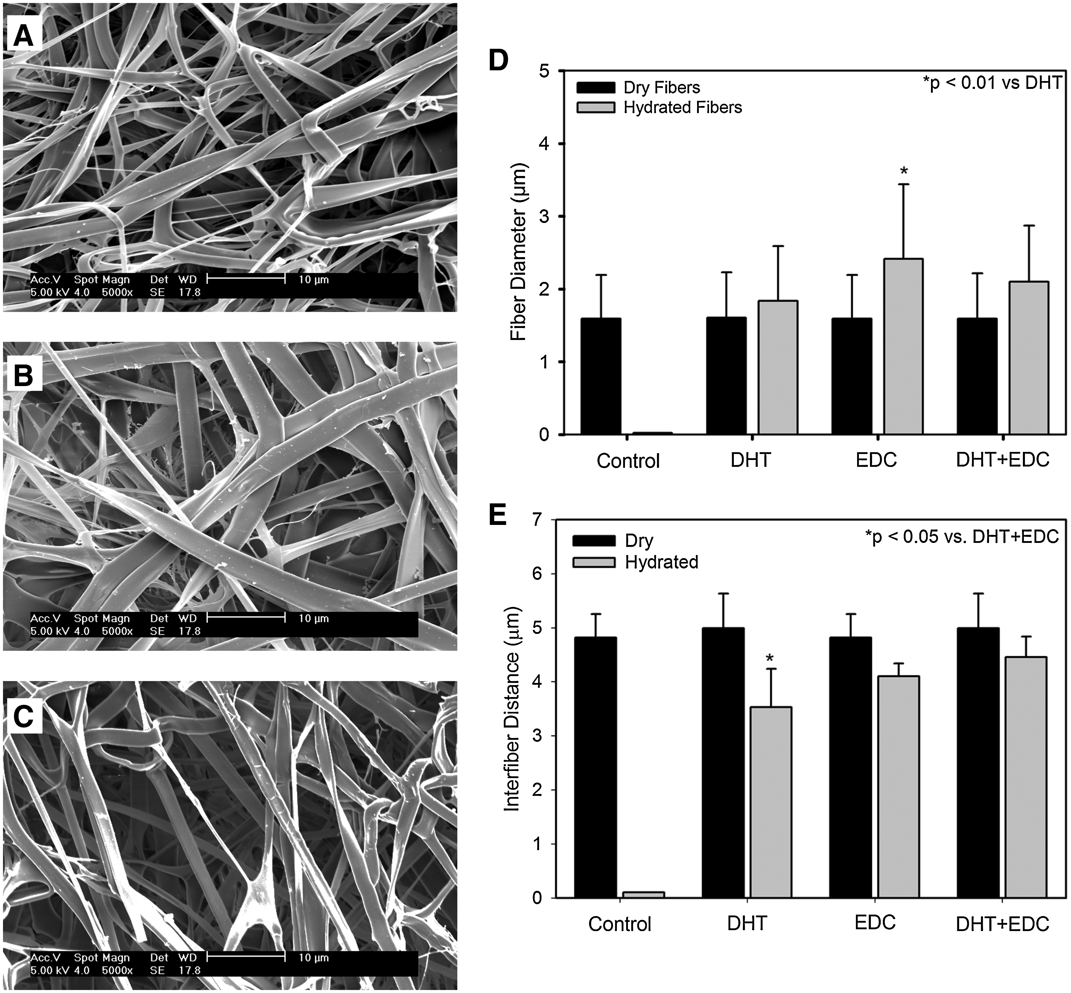
Scanning electron micrographs of (
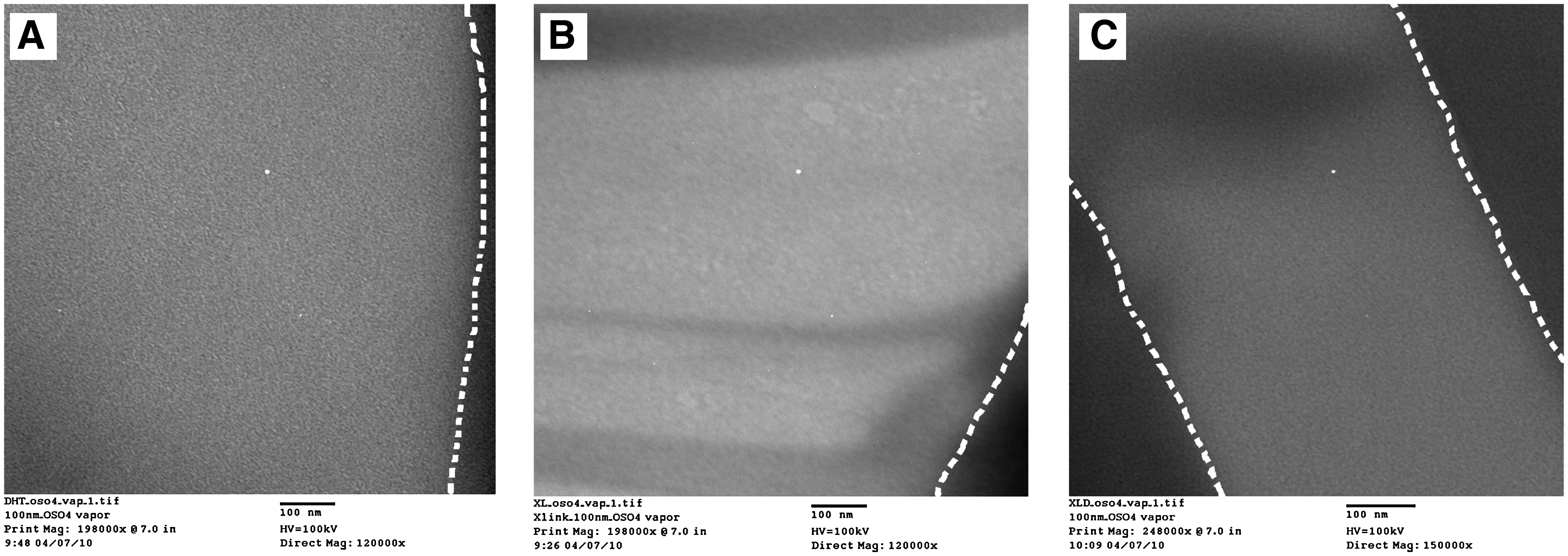
Transmission electron micrographs of OsO4-stained electrospun collagen fibers crosslinked using (
Scaffold stability
As the stability of a scaffold is important for manipulation during cell inoculation and subsequent tissue engineering operations, the degradation rate of scaffolds is extremely important. All scaffolds exposed to bacterial collagenase showed signs of degradation (Fig. 4). Control samples rapidly lost mechanical integrity and fragments after ∼3 h exposure (data not shown). A hydroxyproline assay was used to quantify the amount of degraded collagen found within the medium and confirmed the visual observation that noncrosslinked control group had the most rapid rate of degradation (Fig. 4). After 24 h of collagenase exposure, the control group was 89.95% ± 10.15% degraded whereas the DHT and EDC-crosslinked samples were 69.07% ± 14.15% and 62.37% ± 12.22% degraded, respectively. The DHT+EDC sample was significantly less degraded than all other groups, with the DHT and EDC groups significantly less degraded than control but not statistically different from one another (Fig. 4). After 72 h of exposure, no control scaffolds were visible; however, the DHT+EDC scaffolds maintained their shape and integrity with 45.05% ± 7.34% of the scaffold degraded (Fig. 4).

Resistance to degradation of collagen scaffolds as a function of crosslinking method when exposed to the medium containing 1 U/mL collagenase. The percentage of collagen degraded after 1, 3, 6, 24, and 72 h in solution is shown. a,c,d,e,fp < 0.05 versus all; bp < 0.001 versus DHT, EDC, and DHT+EDC; gp < 0.05 versus DHT and control; and hp < 0.05 versus control and DHT+EDC.
Scaffold strength
In addition to maintaining biostability, a scaffold must also maintain sufficient strength for easy manipulation in vitro, during surgical application and to reduce failure in vivo. The UTS of the scaffolds was highly dependant on crosslinking method. The control scaffold degraded and lost all mechanical integrity after hydration; thus, their mechanical properties could not be quantified. Collagen scaffolds crosslinked with DHT significantly improved strength over the control scaffolds but were exhibited at 60% of the strength of the EDC-crosslinked scaffolds (Fig. 5A). Utilizing both crosslinking methods (DHT+EDC) resulted in a significant increase in strength (p < 0.001) compared to the processes alone. Stiffness followed a similar trend with the DHT+EDC and EDC exhibiting the largest stiffness and DHT alone the smallest (Fig. 5B). No significant difference between the EDC and DHT+EDC groups was observed (Fig. 5B).
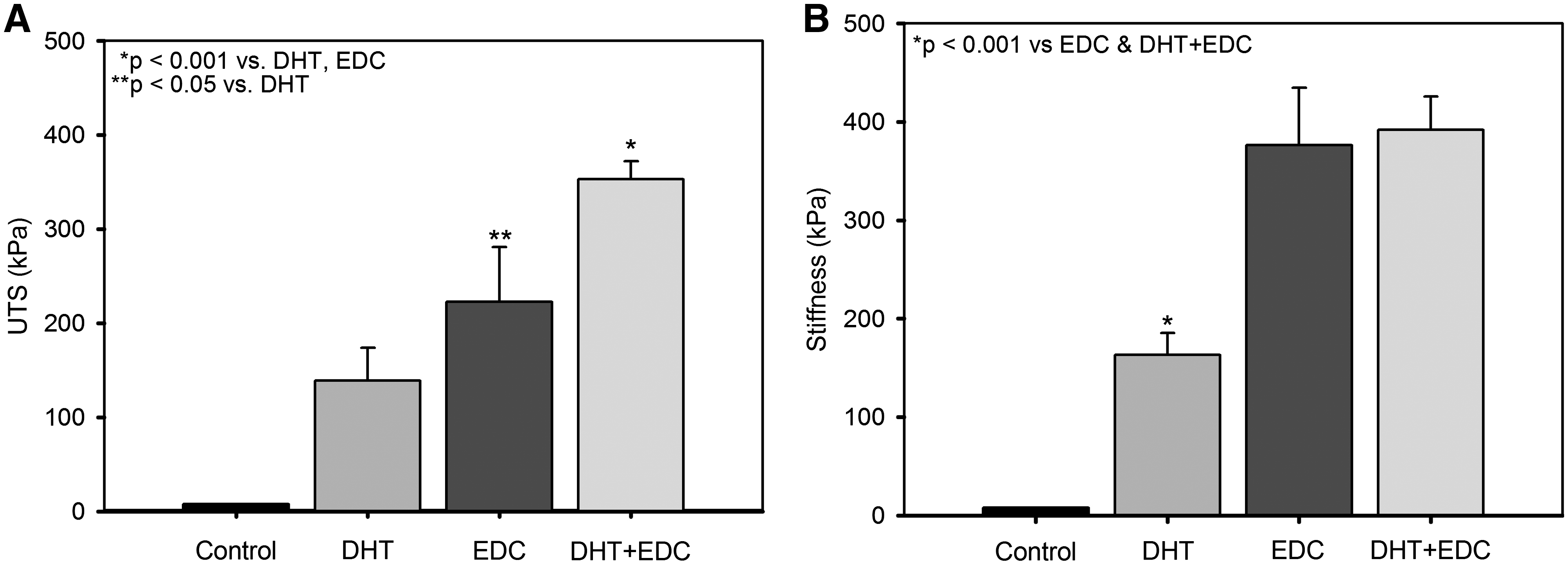
Ultimate tensile strength (UTS) of scaffolds (
Fourier transform infrared spectroscopy
The spectra obtained from the FTIR scans show a definitive increase in peak height at the 1546 cm−1 wavenumber, which is a characteristic of amide bonds,12,28 between the control samples and crosslinked samples (Fig. 6). The observed peak height increase indicates an increase in the number of amide bonds and crosslinks formed during the reaction. 12 Peak height at 1546 cm−1 indicated that the EDC samples possessed a higher amide bond density than controls but a similar crosslink density to the DHT+EDC-crosslinked sample (Fig. 6). For the DHT samples, peak heights at 1546 cm−1 were smaller than the EDC and DHT+EDC (Fig. 6). An analysis of ester bond formation at wave number 1100 cm−1 was attempted; however, the peaks were too small to observe any significant trends.
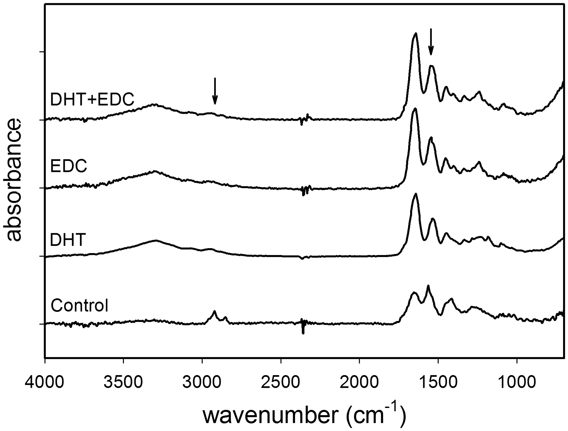
Fourier transform infrared (FTIR) spectra of control and crosslinked electrospun collagen scaffolds. Arrows indicate the normalization peak at 2950 cm−1 and amide peak at 1546 cm−1.
Cell adhesion and viability
Primary human dermal fibroblast viability within these scaffolds was assessed using an MTT assay. The control scaffold degraded during the sterilization and rinsing process; thus, no data on cellular behavior within the scaffolds could be obtained. Significantly reduced cell viability was seen at all time points in the DHT group (Fig. 7). At day 7, the viability of fibroblasts on the DHT scaffolds was on average 13% lower than the viability of the EDC scaffolds and 4% lower than the DHT+EDC group. The EDC and DHT+EDC groups were not statistically different from one another at days 3, 5 and 7 (Fig. 7).
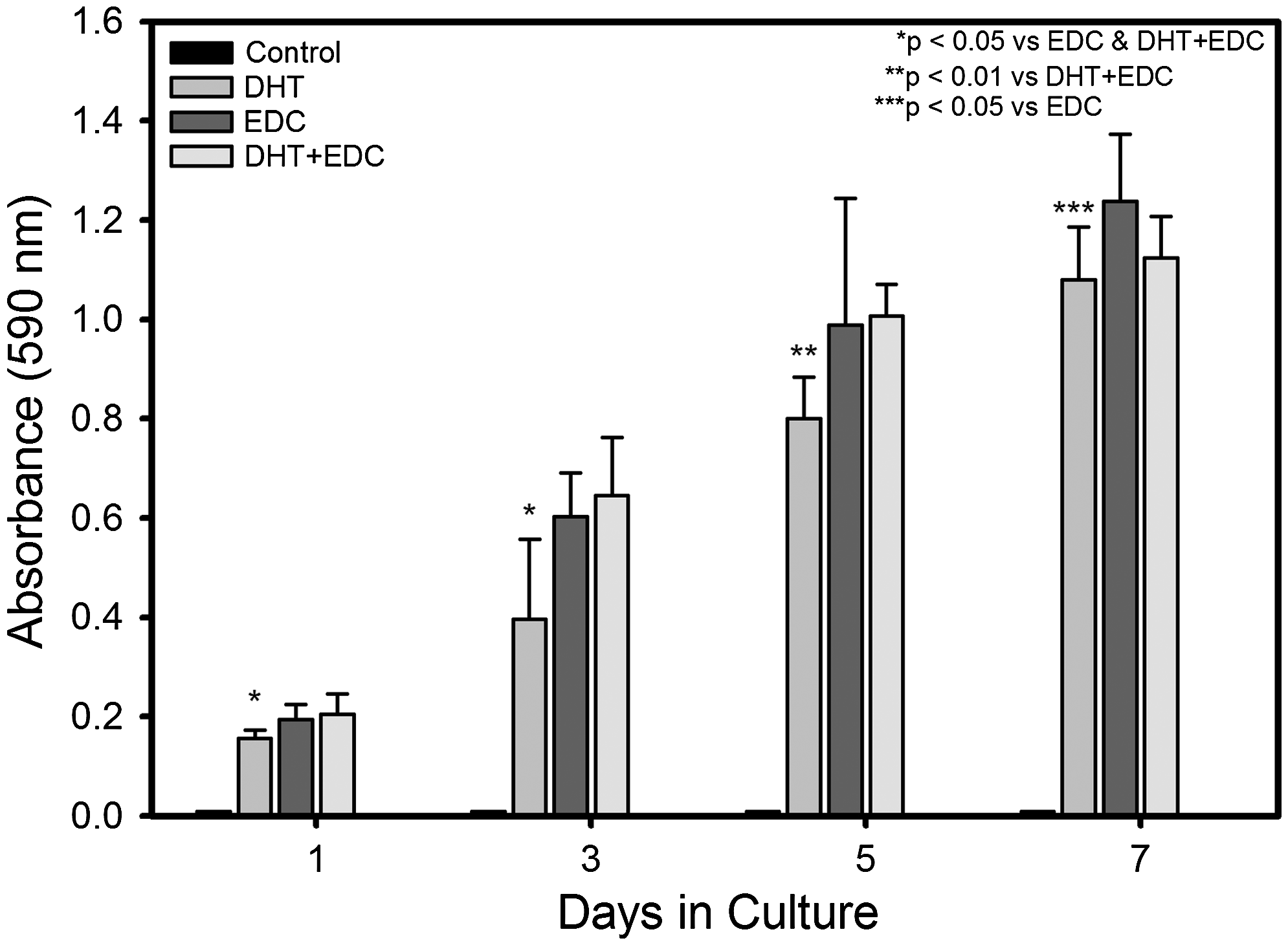
Metabolic activity assay (MTT) of fibroblasts cultured on respective scaffolds for 1, 3, 5, and 7 days in culture. Control scaffold degraded during the sterilization and rinsing process; thus, no cellular data could be obtained.
Cellular penetration into the scaffolds was altered by scaffold crosslinking. Cells infiltrated over half of the DHT and DHT+EDC scaffolds (59.9% ± 6.2% and 56.7% ± 8.8%, respectively) at culture day 7, and they reach the upper half (49.9% ± 4.8%) of the EDC scaffold (Fig. 8). The differences are not significantly different for the DHT and DHT+EDC scaffolds or the EDC and DHT+EDC scaffolds; however, the penetration of cells into the EDC and DHT was significantly different (p < 0.05).
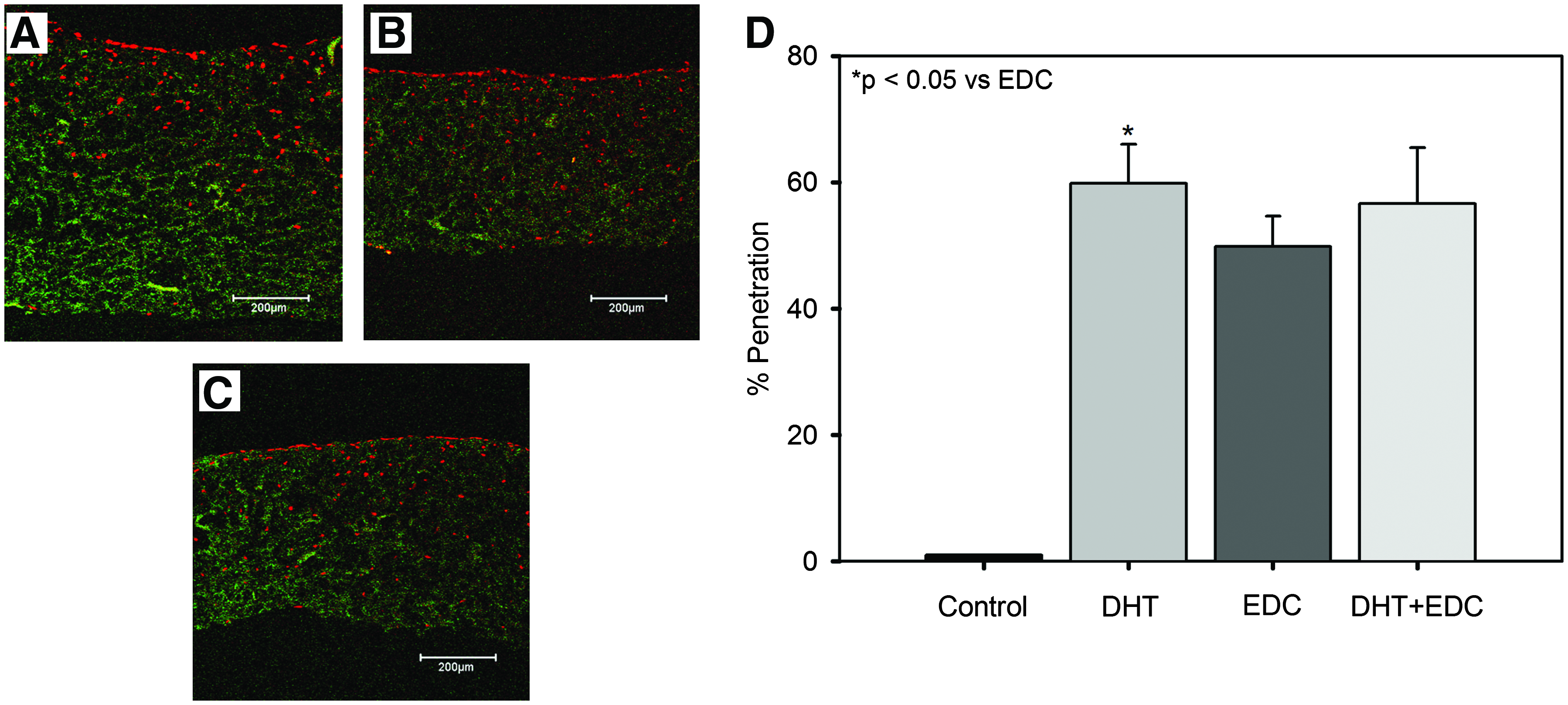
Immunostained sections of human dermal fibroblasts cultured on (
Discussion
Fiber morphology
As-spun and DHT-crosslinked samples showed no significant difference in morphology, fiber diameter, or interfiber distance (Figs. 1 and 2). In contrast, EDC-crosslinked fibers exhibited the largest increase in fiber diameter, whereas the DHT-crosslinked fibers swelled to a minor degree. It is not uncommon for electrospun proteins to swell after hydration.9,37 This phenomenon has been previously observed in GA-vapor-crosslinked gelatin nanofibers. 9 In general, swelling of protein-based scaffolds and protein fibers has been inversely correlated with crosslink density, showing a decrease in swelling ratio with increased crosslinker concentration.9,39 DHT crosslinking has been previously shown to generate lower crosslink densities compared with chemically crosslinked collagen, 25 yet our results indicate that the DHT sample had the least amount of fiber swelling. During DHT crosslinking, ester and amide bonds are formed reducing the amount of the more hydrophilic free carboxyl, amine, and hydroxyl moieties and leading to more hydrophobic materials as has been previously reported in gelatin scaffolds. 40 Collagen films treated with DHT have also been reported to have slightly greater hydrophobicity than their nontreated counterparts. 41 In contrast, crosslinking with EDC was not shown to influence collagen film contact angle greatly. 41 The reduced fiber swelling in the DHT-crosslinked scaffolds may be a result of increased scaffold hydrophobicity reducing the interaction of the aqueous medium with the fibers.
As DHT crosslinking has been shown to denature collagen molecules,20,25 it was important to investigate the ultrastructure of the electrospun scaffolds to determine if they exhibited cross-banding and if that cross-banding was eliminated during processing. TEM analysis of the scaffolds indicated that no banding was seen in any of the scaffolds irrespective of the crosslinking method performed (Fig. 3). This is in contrast to previous reports where banding was observed in electrospun type I collagen. 38 Collagen fibrils within the noncrosslinked, control scaffolds could not be observed, as they degraded during processing. It is possible that the initial raw material processing denatures the collagen before it is electrospun, as the source of type I collagen was different between these two studies. It is also possible that the collagen solubilization process also causes denaturation. 42 The lack of collagen banding in all samples in addition to no significant changes in denaturation temperature as measured by differential scanning calorimetry (data not shown) suggests that the collagen was denatured during initial raw material processing, during electrospinning, or both. Thus, DHT processing of electrospun collagen does not likely further denature the collagen significantly.
Scaffold stability and mechanical properties
The crosslinking method had a strong influence on the physical properties of the scaffolds. Without crosslinking, collagen fibers were not able to resist degradation in the presence of an aqueous medium (Fig. 4) and provided no mechanical integrity. The DHT and EDC scaffolds exhibited similar degradation rates with slightly reduced average degradation in EDC scaffolds, whereas the combination of DHT+EDC was the most stable with ∼55% of the scaffold remaining after 72 h of collagenase exposure (Fig. 4). A greater crosslink density in the EDC samples may have been responsible for its improved biostability compared to DHT alone. FTIR spectrum indicated an increase in amide bond formation as a result of exposure to EDC crosslinking (Fig. 6). DHT crosslinking increased the number of amide bonds formed compared to control scaffolds but were reduced compared to EDC (Fig. 6). Previous studies would support the hypothesis that EDC crosslinking is more efficient than DHT crosslinking. 11 Although the combination of DHT and EDC crosslinking did not appear to significantly improve FTIR peak height compared to EDC alone, the degradation rate of the DHT+EDC samples was significantly lower than DHT or EDC (Fig. 3). This may be due to a combination of increased amide and ester bond formation as a result of the crosslinking procedures and increased hydrophobicity as a result of the DHT processing. Previous reports have indicated a stronger increase in hydrophobicity of collagen films after exposure to DHT and EDC. 41
Mechanical properties of the electrospun collagen scaffolds were improved by all crosslinking methods; however, DHT crosslinking was significantly weaker and less stiff than the EDC and DHT+EDC scaffolds (Fig. 5A). UTS was significantly improved by using both methods in conjunction with one another, whereas no statistical difference in stiffness was observed (Fig. 5B). As previously discussed, DHT crosslinking generates a lower crosslink density as suggested by the degradation rate data, FTIR spectrum, and prior reports 25 as is likely the cause of the reduced mechanical properties compared to the EDC-crosslinked samples.
Cell–scaffold interaction
All scaffolds that were stable in aqueous medium were capable of supporting fibroblast attachment and growth. For DHT samples, the scaffolds were electrospun, dehydrothermally crosslinked for 24 h, sterilized for 24 h, and then inoculated after ∼2 h of rinsing (50 total hours). In contrast, the DHT+EDC samples were electrospun, dehydrothermally crosslinked for 24 h, chemically crosslinked in EDC for 24 h, sterilized for 24 h, rinsed in PBS for 48 h, and then rinsed with HBS for 2 h before inoculation (122 total hours or 2.4× greater processing time). At days 3, 5 and 7, fibroblast metabolism on the EDC and EDC+DHT scaffolds was not statistically different from one another (Fig. 7), and on average these scaffolds promoted increased cellular metabolism compared to the DHT group alone. DHT crosslinking has been shown to reduce the wetting ability of the scaffolds 41 and may lead to lower seeding efficiency compared to the EDC crosslinking alone and, as a result, lower cell number. Additionally, cell penetration into electrospun collagen scaffolds was dependant on crosslinking method. Fibroblast migration into scaffolds was similar for all groups but was greater for the scaffolds treated with DHT in the EDC-crosslinked and uncrosslinked condition. The reduced number of crosslinks might allow the fibroblasts to degrade the matrix and migrate within the scaffold with greater ease than a highly crosslinked scaffold.
Conclusion
These data indicate that physical crosslinking using DHT treatment improves mechanical strength and biostability compared to noncrosslinked controls. However, DHT crosslinking appears to produce a lower crosslink density than EDC-crosslinked scaffolds resulting in more rapid degradation rates and reduced strength compared to chemical crosslinking. Dermal fibroblasts adhere and grow on DHT-crosslinked scaffolds; however, data suggest that the seeding efficiency may be less than that of EDC-crosslinked scaffolds, leading to a 12% reduction in cell viability after 7 days in culture. A combination of both processing methods (DHT+EDC) produces scaffolds with the slowest degradation rate and greatest strength, but this method requires 122 h of postspinning processing in contrast to a total of 50 h for DHT. DHT crosslinking can clearly be utilized to stabilize electrospun collagen scaffolds; however, one must determine the ideal balance of mechanical properties and processing times for their specific application.
Footnotes
Acknowledgments
The authors thank Campus Microscopy and Imaging Facility at OSU for the use of the confocal microscope and the transmission electron microscope, and the Campus Electron Optics Facility for the use of the scanning electron microscope. The authors would also like to thank Dr. Mark Ruegsegger for assistance with FTIR analysis and Jessica Wolever for assistance with the cellular studies.
Disclosure Statement
No conflicts of interest exist.