
Abstract
Both physical and chemical crosslinking methods have been shown to be effective in improving the biological stability and mechanical properties of porous collagen scaffolds. However, the wetting of the collagen fibril surface by a culture medium is reduced and it is difficult for the medium to diffuse into the 3D structure of a porous collagen scaffold. This article reports a strategy for the surface processing of crosslinked collagen scaffolds by an integrated ultraviolet/ozone perfuse processing technique. Ultraviolet/ozone perfuse processing improved surface wettability for both the exterior and interior surfaces of the porous 3D collagen scaffold. This leads to a significant improvement in the scaffolds ability to take up water without compromising the bulk biological stability and mechanical properties. In vitro evaluation using mesenchymal stem cell demonstrated that surface processing enhanced cell colonization of the scaffolds, cells could migrate deep into the structure of the scaffolds, and significantly higher levels of cell proliferation were achieved. In contrast, the cells were unable to migrate deep into the scaffolds, and most of the cells that survived were observed only in the top seeding layer resulting in a low level of cell activity in the unprocessed scaffolds.
Introduction
Both synthetic, such as polylactic acid (PLA), polyglycolic acid, and polyesters,5,6 and naturally occurring polymers, such as polysaccharides (starch, chitosan, cellulose, etc.) and proteins (collagen, elastin, alginate, etc.), have been suggested as scaffolds7,8 to provide a structure into which biological cells and molecules can be placed such that they undergo the appropriate stimuli to enable them to produce viable tissue. 9
One of the major shortcomings of these synthetic polymers is their reduced functionality in vitro, compared with natural biomaterials. For example, most polymer surfaces are hydrophobic and these are not appropriate substrates for cell attachment, ingrowth, and proliferation.10–12 In vitro evaluation commonly reveals that the cells are only able to migrate and survive close to the surface to within a critical depth, which is cell dependent. 8 Methods to enhance the key properties, for both interior and exterior surfaces, that can promote cell growth within the scaffold and induce the formation of an ECM from cells that come into contact with the polymers as 3D scaffolds are of interest in this regard.10,11,13,14
Collagen is a significant constituent of the natural ECM.15,16 Type I collagen provides a structural framework for connective tissue and plays a central role in the temporal cascade of events leading to the formation of new bone. Scaffolds made from collagen have been used in a variety of applications due to a number of useful properties such as haemostasis and low antigenicity.17,18 In its physiological functional form, collagen I occurs in the solid state as fibers joined in a continuum throughout the human body. It can modulate cell proliferation, migration, differentiation, and specific gene expression. 19 When it is used as scaffold material, crosslinking treatment is an effective method to improve its biological stability and mechanical properties. 20 The crosslinking can also suppress the immunogenicity of the artificial implants. Two crosslinking treatment methods, physical and chemical, are frequently used in practice to improve the properties of porous collagen scaffolds. However, accompanying the improvement in biological stability and mechanical property, either physical or chemical crosslinking treatments results in reduced surface wettability and the collagen fibril surface becoming hydrophobic in nature. 10 The wetting of the collagen fibril surface by a culture medium is reduced and it is difficult for the medium to diffuse into the 3D structure of porous collagen scaffold. Hence, this is not a fully appropriate substrate for cell attachment, ingrowth, and proliferation.10,14,21 Low temperature plasma surface processing technology has been used for collagen/chitosan composite scaffold surface modification. The surface wettability of the scaffold was improved, and it subsequently prompted cell attachment and cell proliferation, as observed in vitro evaluation. 10 However, the major limitation of the conventional scaffold surface modification using plasma technique for tissue engineering is the depth of penetration of the active species into the porous structure.
Ultraviolet (UV) light at 184.9 and 253.7 nm wavelengths are known to excite molecular oxygen to form ozone and to photosensitize polymer surface. UVO treatment leads to the incorporation of oxygen-containing functional groups such as carboxyl and hydroxyl groups onto substrate materials. 22 These functional groups specifically interact with the receptors on cells through attractions between opposite charge (ionic bonding).13,23 It is known that attachment of cell binding proteins, such as fibronectin, increases with improved hydrophilicity at the substrate.24,25 Similarly, the deposition of secreted fibronectin by adhesion and spreading cells is also associated with hydrophilic surface. 25 UV and ozone have often been used for biomaterial surface modification and can improve the surface wettability and subsequently promote cell adhesion performance. However, few investigations use UVO-integrated method for the 3D porous scaffold surface processing.26,27
Surface engineering techniques, either direct or indirect, provide routes for scaffold surface modification. This article reports the strategy for the surface processing of crosslinked collagen scaffolds by UVO perfuse processing technique to achieve an improved surface wettability both for exterior and interior surface of the porous 3D collagen scaffold. The in vitro performance in terms of cells' colonization of the scaffolds and proliferation behavior using human bone marrow mesenchymal stem cells (MSCs) is reported.
Materials and Methods
Scaffold fabrication and crosslinking treatment
Bovine Achilles' tendon collagen type I (Sigma-Aldrich) was used for scaffold fabrication. A collagen dispersion of 1% was achieved by adding the respective mass of collagen I in 0.05 M acetic acid solution (pH 3.2) and homogenizing on ice. The mixture was then degassed in a bell jar and stored at 4°C before use. The collagen scaffold was made by casting the collagen dispersion into a polytetrafluoroethylene mold 10 mm diameter and 2 mm height (cast on polytetrafluoroethylene sheet to obtain thin collagen sheet for surface wettability measurements), and then freezing at −80°C for 24 h followed by freeze-drying to obtain the porous scaffold. The dried scaffolds were further subjected to dehydrothermal treatment (DHT) at 105°C under a vacuum pressure of <10 Pa for 72 h before additional chemical crosslinking treatment. The process of crosslinking treatment has been described previously.28,29 In brief, the collagen scaffolds were submerged/immersed and incubated in 50 mM MES (2-[morpholino]ethanesulfonic acid) solution (pH 5.5) containing 2.5 M lysine for 1 h at room temperature. Then, 1 mL MES (Sigma-Aldrich) solution containing 40 mM 1-ethyl-3-(3-dimethylaminopropyl)carbodiimide (EDC; Sigma-Aldrich) and 20 mM N-hydroxysuccinimide (NHS; Sigma-Aldrich) was added. After incubation for 24 h, the scaffolds were removed from the solution, washed with distilled water, and freeze-dried to obtain porous scaffolds.
Scaffold surface processing
The surface processing was carried out in a purpose-built UV-integrated ozone reactor (UVO reactor). In this reactor, a tubular glass chamber (with internal diameter of 10 mm and 100 mm long) was placed into a compartment containing a high-intensity low-pressure mercury vapor grid lamp. An earlier study demonstrated that UV-assisted ozone surface processing has a combination of effects on polymer surface processing. 30 For scaffold surface processing, the samples were packed into the tubular glass chamber, the chamber was evacuated, and the pressure of the chamber was stabilized to 100 Pa. Ozone gas (from an ozone generator) was introduced into the chamber to perfuse through the porous scaffolds for a predetermined period, 50 s in this study. After ozone perfusion, the samples were washed in sterilized water before in vitro evaluation.
Mechanical property testing and surface characterization
Dynamic mechanical properties of the scaffolds were analyzed using a TRITEC200B dynamic mechanical analyzer (Triton Technology). The dynamic shear stress scan was performed on scaffolds in wet conditions at 37°C. The sample disc (8 mm diameter and 4 mm thick) was loaded into a sample holder, and then dynamic shear stress was applied to the sample to a displacement of 0.1 mm at frequency of 1 Hz. The changes in strain, phase angle, tan δ, and modulus, related to the viscoelastic behavior of the scaffolds, were monitored during the test.
A surface chemistry analysis was performed in a Kratos Axis HSi X-ray photoelectron spectrometer equipped with a five-channel detector and a monochromated Al Kα X-ray source with an energy of 1486.6 eV operating at 150 W. A low-energy electron flood gun was used to compensate for surface charging on the insulating films. The carbon 1s signal from each film was corrected for surface charging to the hydrocarbon signal at 285.0 eV.
Surface wettability was evaluated using an FTÅ125 Dynamic Contact Angle Analyser (First Ten Ångstroms). A sessile drop of ∼20 μL water was advanced toward the surface at a set rate. As the drop was released from the needle, a sequence of digital images was taken automatically by the camera and then analyzed to give an advancing contact angle. The known weight of scaffold was soaked in phosphate-buffered saline (PBS) at 37°C for 24 h, and then the scaffold was blotted using a lint-free paper to remove excess PBS, and weighed. The water intake capability was defined as the percentage of water intake to the initial weight of the scaffold.
The pore size and size distribution of the samples were analyzed by a high-resolution micro X-ray computed tomography (micro-CT) system (μCT 40; Scanco Medical) operated at a voltage of 55 kV and a current of 145 mA. Samples were scanned at 8 μm volume pixel (voxel) resolution with an integration time of 300 ms to produce 3D reconstructed images.
In vitro cell attachment and proliferation evaluations
The in vitro biodegradation of the scaffolds was evaluated using a collagenase assay, as reported elsewhere. 28 In brief, scaffolds of about 5 mg dry weight were incubated in 1 mL 0.1 M Tris-HCl (pH 7.4) solution containing 0.05 M CaCl2 for 1 h at 37°C. Subsequently, 200 U bacterial collagenase (Clostridium histolyticum; Sigma) in 1 mL 0.1 M Tris-HCl was added. The reaction was stopped at given time intervals by addition of 0.2 mL 0.25 M EDTA and cooling the mixture on ice. The mixtures were filtered through the filter paper, and the filter cake was air-dried and weighed. The degree of biodegradation is defined as the percentage of weight loss compared to the initial weight of the scaffold.
In vitro evaluations of scaffolds were performed using green fluorescent protein–labeled human bone marrow MSCs. Disc-shaped scaffolds (10 mm diameter and 3 mm thick) were sterilized in sterile-filtered 70% ethanol, thoroughly washed in PBS, and then equilibrated in tissue culture media. Scaffolds were placed on sterile filter paper to remove excess media, and then placed on a nontreated 12-well Nunc polystyrene plate and seeded with MSCs at 5 × 10⁵ cells per scaffold. Seeded scaffolds were left for 2 h to allow the cells to adhere to the scaffold, and then 2 mL tissue culture medium was added to each well. Seeded and control (nonseeded) scaffolds were cultured under static conditions for up to 7 days. Cell viability/proliferation was assessed using an alamarBlue metabolic assay at days 1, 4, and 7. The alamarBlue working solution was measured at an excitation wavelength of 560 nm and an emission wavelength of 590 nm using a fluorescence plate reader and compared to a standard curve. The differences in metabolic activity are indicative of differences in live cell numbers, and cell metabolic activity multiplication was expressed as percentage of metabolic activity, as measured by alamarBlue assay, to initial metabolic activity.
Results
Microstructure
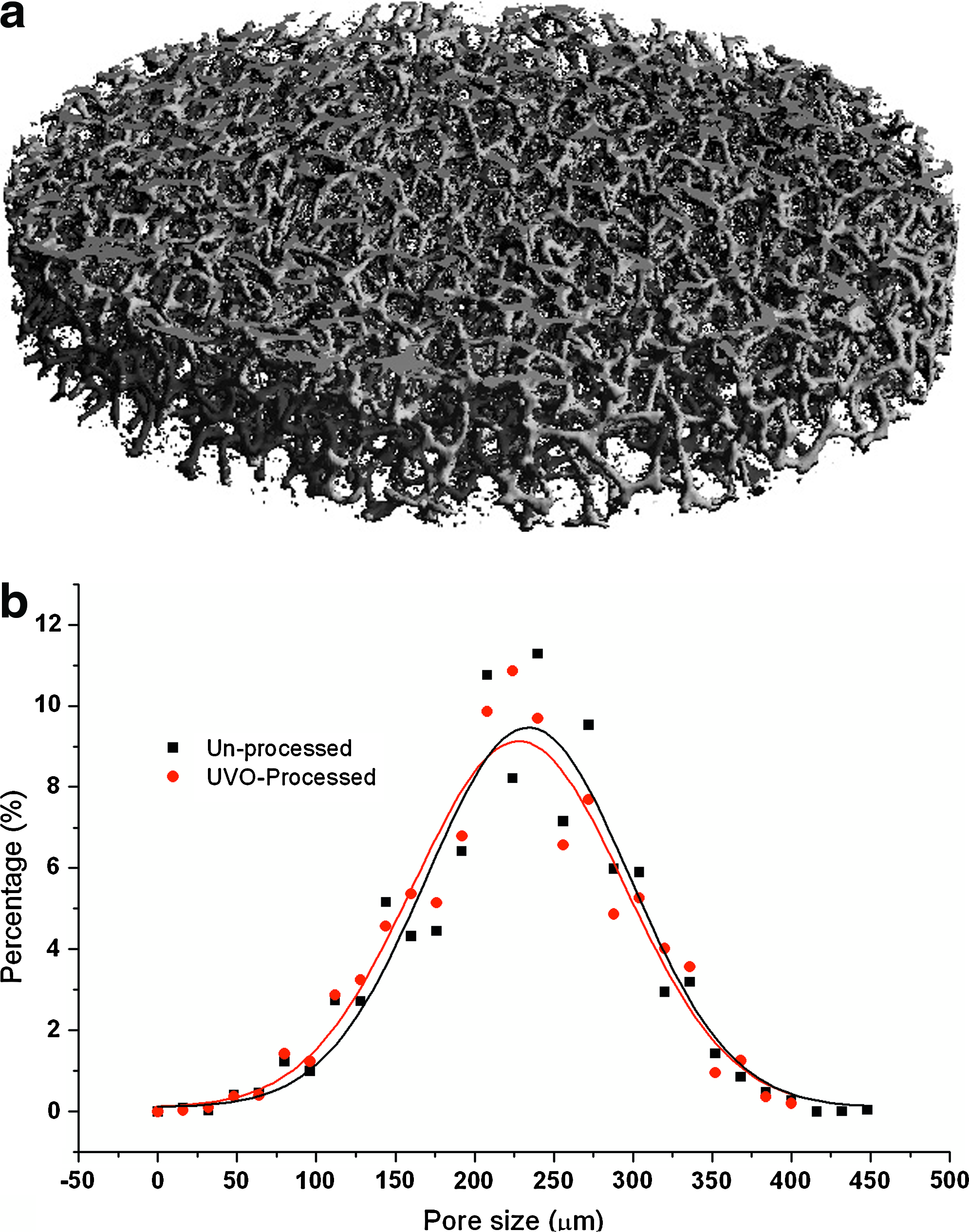
It is relatively straightforward to fabricate collagen-based scaffolds using our technique. The architecture of the scaffolds such as sample dimensions and pore size can be readily controlled. Micro-CT examination revealed that the collagen scaffold obtained from the 1% collagen dispersion featured a porous architecture with interconnected open pores, as shown in Figure 1a. The interconnected pores could improve the mass transportation of nutrients and oxygen, as well as allowing removal of cell metabolic waste during the in vitro culturing process. The pores, as shown in Figure 1a, which were formed during freezing of the collagen dispersion, encourage collagen to aggregate in the interstitial spaces and to create an interconnected network of collagen fibrils. Hence, the pore size can be adjusted by altering concentration of the collagen dispersion, the freezing rate, and the pH, as these factors are known to affect both nucleation and growth rates of the ice crystals. 31
(
The pore size and pore size distribution, as obtained from micro-CT examinations, within the scaffold are shown in Figure 1b. This figure illustrates that the pore size has a wide distribution. The resultant scaffolds, both unprocessed and UVO-processed scaffolds, had a broad range of pore sizes ranging from 20 to 350 μm. F-test confirmed that there is no significant difference for pore size distribution, at 5% significant level, between unprocessed and UVO-processed scaffold (F = 1.1 < Fcritical = 1.96). Previous experiments 28 observed that the lower dispersion concentration condition produced larger pores than at higher concentration case. The 1% scaffolds have a peak pore size range of 100–150 μm for both unprocessed and surface-processed scaffolds. Detailed analysis revealed that pores in the size range of 50–200 μm represent 72% of total pores. Small pores (<20 μm) and large pores (>250 μm) together only represent about 6% of total pores in the scaffolds studied.
Dynamic mechanical properties
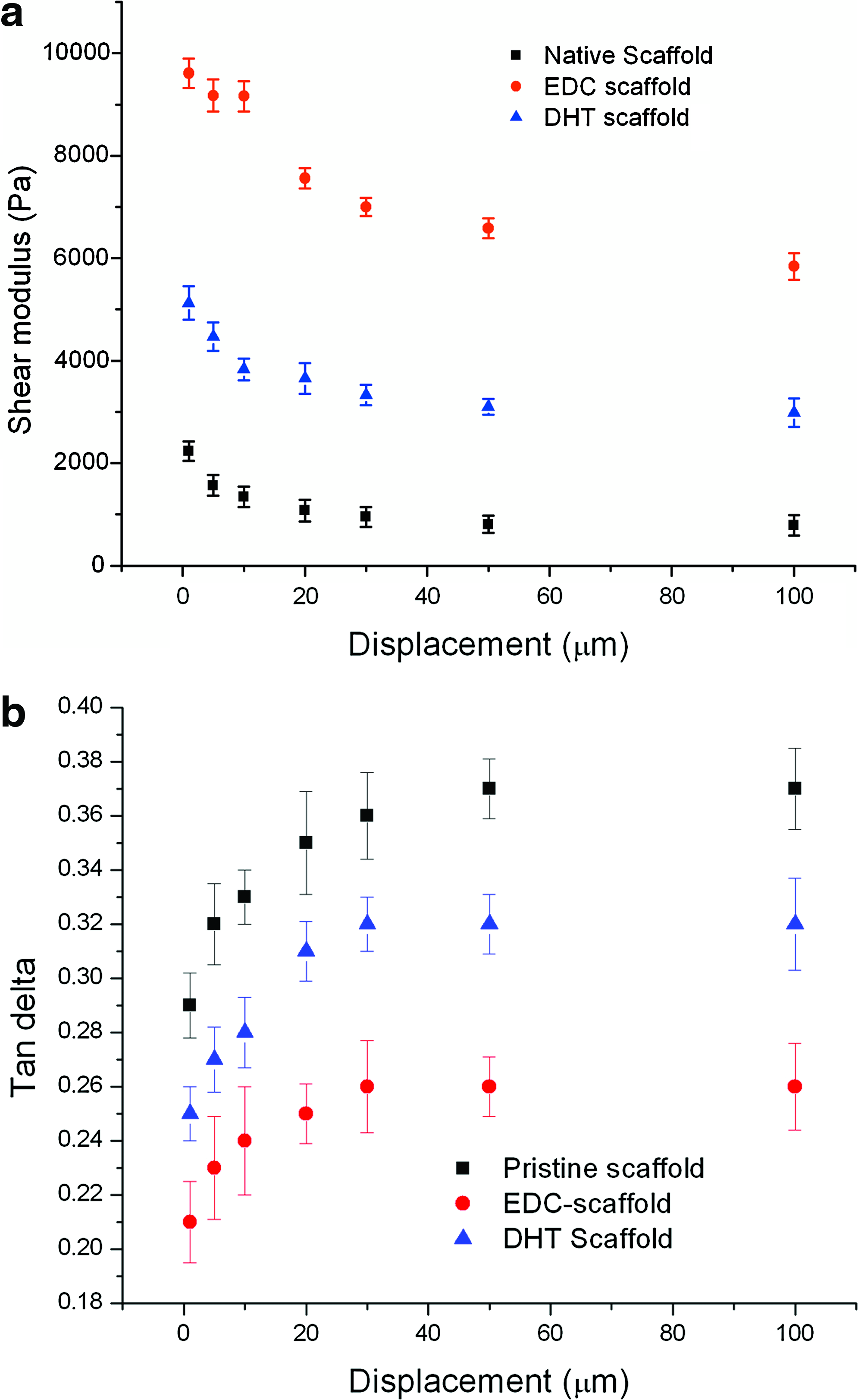
The dynamic mechanical testing of the pristine collagen scaffold and crosslinking-treated scaffolds (both DHT and EDC crosslinking treated) are shown in Figure 2. These data demonstrate that the mechanical property (in shear modulus) of scaffolds varied with the shear displacement. The shear modulus of the collagen scaffolds reduced with displacement under dynamic shear stress for all samples. Two discrete stages were identifiable: an initial higher rate of decrease followed by a steady decrease leading to a plateau. A 21% decrease and 29% decrease in shear modulus, when the scaffold was shear displaced by 20 μm, were observed for EDC- and DHT-treated scaffold, respectively, whereas a 52% decrease was observed for pristine scaffold in the initial stage of shear displacement. Beyond this initial displacement, the shear modulus tended to level off for all samples. The variation of tan δ of the scaffolds with the shear displacement is shown in Figure 2b. The tan δ values for all three scaffolds increased with the displacement. Similar to the trend of shear modulus, the variation of tan δ in association with shear displacement also exhibited two stages for all three scaffolds, that is, an initial high rate increase stage followed by a plateau.
Dynamic mechanical analysis (DMA) testing of scaffold in shear mode. (
It was revealed from the shear modulus–displacement curves that both DHT and EDC treatment methods enhance the mechanical property and that the scaffolds become more rigid after crosslinking treatment. In contrast, the pristine scaffolds exhibited a more soft and flexible behavior, as demonstrated by tan δ measurements (Fig. 2b). The pristine scaffold exhibited higher tan δ value than did both DHT- and EDC-crosslinking-treated samples. In other words, it demonstrates more viscous, less elastic behavior than treated samples. In comparison to DHT-treated scaffolds, the EDC-method-treated samples demonstrated a sustained increase of mechanical property. They exhibited a much higher shear modulus and lower tan δ value at each displacement points than that of the DHT-treated scaffold. The shear modulus was increased to about 6 and 3 kPa, from 0.8 kPa for pristine scaffold at displacement of 0.1 mm, for EDC- and DHT-crosslinked scaffolds, respectively. Correspondingly, the tan δ-value reduced to 0.26 and 0.32, from 0.37 for pristine scaffold, as a result of EDC and DHT crosslinking treatment, respectively.
Surface chemistry, wettability, and water intake capability
High-resolution X-ray photoelectron spectroscopy (XPS) was used to study the chemcal changes of scaffolds before and after surface processing. Figure 3a shows the survey scan recorded by XPS on unprocessed and surface-processed scaffolds. It reveals the typical C1s, N1s, and O1s peaks of collagen. Quantification analysis of the spectra, as listed in Table 1, revealed that surface processing by UVO increased the surface oxygen concentration significantly. About 4% more oxygen was incorporated onto the scaffold surface. As a result, the O/C ratio was increased to 22.3% from the original 18.4%.
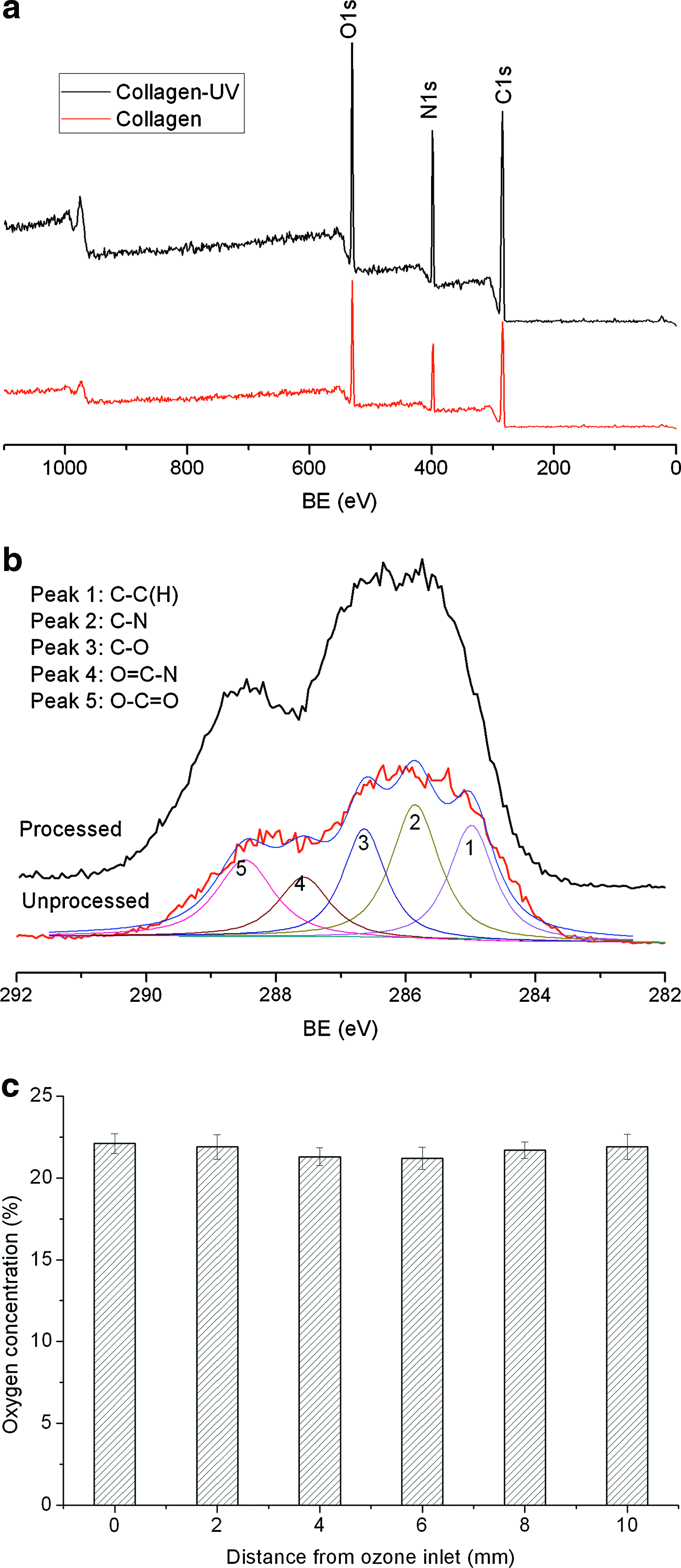
X-ray photoelectron spectroscopy (XPS) analysis of scaffolds: (
The high-resolution C1s spectrum of scaffolds (Fig. 3b) can be deconvoluted to reveal five main peaks. The peak centered at 284.9 eV is assigned to a carbon atom bound to carbon and hydrogen [C–(C, H)]. The peak at about 286.7 eV is attributed to carbon singly bound to oxygen alcohol functional group (C–O). On the left, a component at 288.7 eV is assigned to carbon atom involved in an ester or carboxyl functional group (O–C=O). Two peaks arising from the amide, located at 285.9 eV and 287.8 eV, are assigned to carbon singly bound to nitrogen (C–N) and of carbon involved in an amide bond (O=C–N), respectively. Quantification analysis (Table 1) demonstrated that, after UVO processing, there was a significant increase in the relative concentration of the oxygen-containing functional groups, especially component O–C=O, which exhibited a 7.6% increase compared with the unprocessed sample. The uniformity of UVO processing was assessed by analyzing the distribution of oxygen concentration across scaffold. Figure 3c shows the variation of oxygen concentration with the depth as determined by XPS examination by examining a sequence of slices taken from the original 10-mm-long sample. It reveals that perfusion of ozone through the scaffold, adopted in this study, rendered a uniform oxygen incorporation through the scaffold.
The dynamic contact angle measurements using the sessile drop technique were carried out to study the surface wettability changes of collagen films upon exposure to UVO processing; the water contact angles on different collagen films were shown in Figure 4. Both DHT and EDC crosslinking methods resulted in increased water contact angle. The water contact angle for DHT- and EDC-crosslinked collagen film increased to 74° and 76° from 51° for pristine collagen, respectively. This demonstrated that the crosslinking treatment decreased the surface wettability significantly. The water contact angles for all samples, including pristine and crosslinked collagen films, reduced to about 40° after exposure to UVO for a short period of 100 s, demonstrating that surface processing is an effective method to improve the surface wettability of collagen. This was attributed to the oxygen incorporation onto the surface by UVO exposure, as revealed by XPS analysis (Table 1).
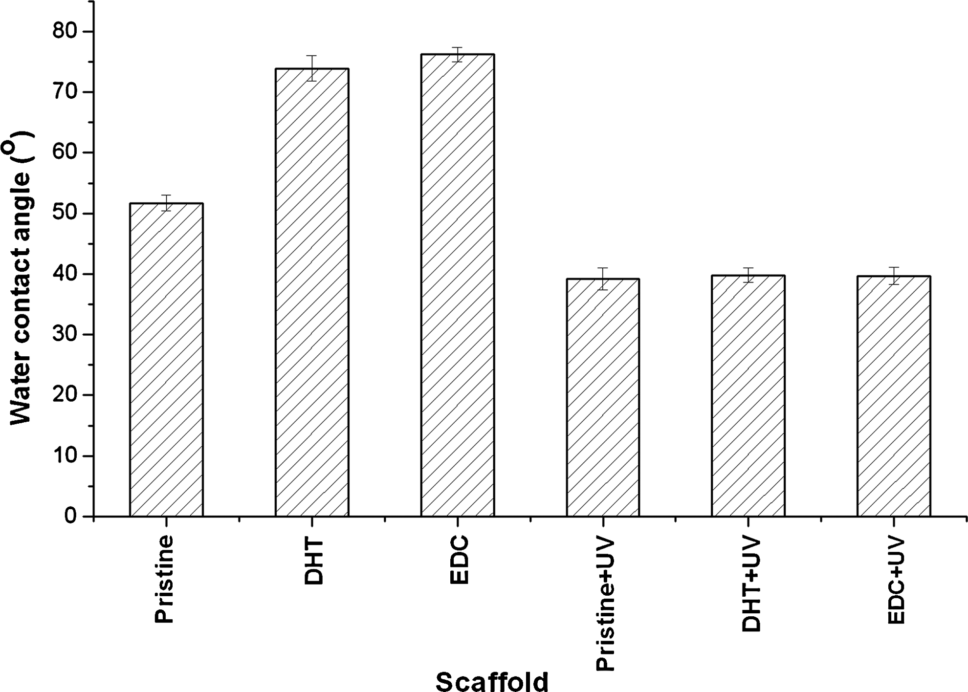
Water contact angle measurements indicated that both DHT and EDC treatment reduced surface wettability, whereas surface processing by UVO can improve the surface wettability significantly. DHT, dehydrothermal treatment; EDC, 1-ethyl-3-[3-dimethylaminopropyl]carbodiimide.
The study also revealed that the EDC collagen was very sensitive to the UVO processing. The surface oxygen increased rapidly with exposure to UVO and the saturation level (22.5%) was achieved at treatment times of 50 s (Fig. 5), which corresponds to a steady-state between oxygen incorporation by photo-oxidation and the loss of volatile photolysis products from the collagen surface. As a result of surface oxygen incorporation, the surface wettability improved as evidenced by the water contact angle measurements. The water contact angle reduced to about 39°, from its original 76°, at 50 s exposure. With extended exposure, the water contact angle on EDC collagen surface leveled off, as demonstrated in Figure 5.
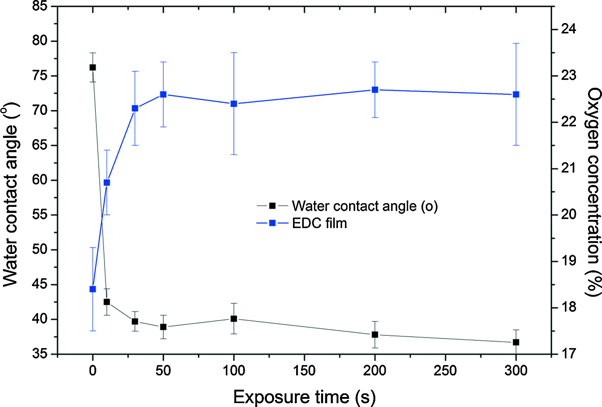
Variation of water contact angle and oxygen incorporation on EDC-treated collagen films with UVO exposure duration. Color images available online at
The aging effect on the surface wettability was evaluated for EDC collagen films that had been exposed to UVO for 50 s, by measuring the water contact angle daily for 2 weeks. The variations of water contact angle with aging period (in air) are shown in Figure 6. It is apparent that water contact angle recovers, that is, surface wettability decay, quickly within first 2 days. The water contact angle recovered to about 44° at day 2 of aging. After 2 days of aging, the surface wettability decay process slowed down and leveled off after 4 days. The water contact angle recovered to about 46° after 2 weeks of aging. However, this value remains much lower than the unprocessed samples.
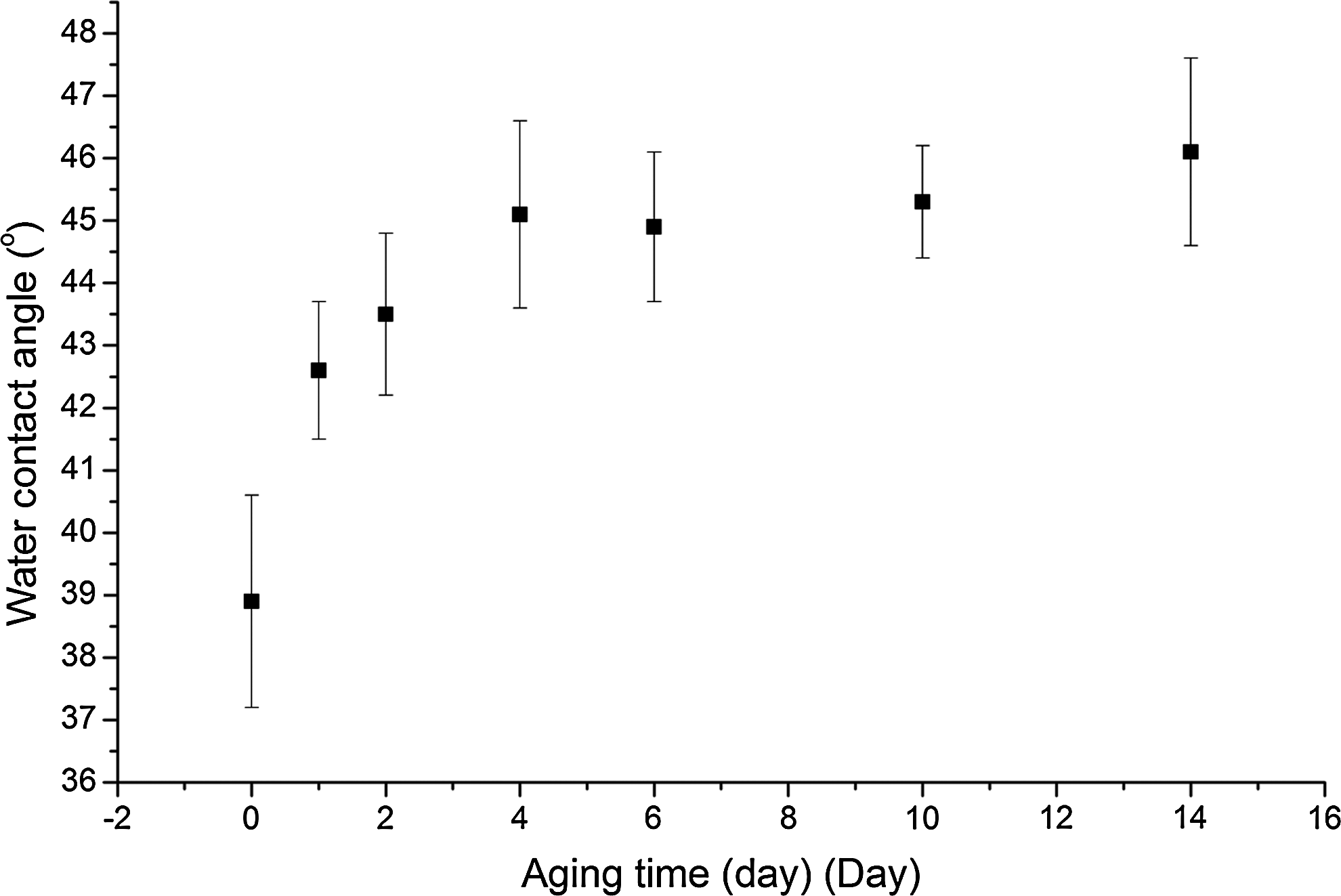
The aging effect on the water contact angle of surface-processed EDC collagen film.
When a water droplet contacted with the crosslinked porous collagen scaffold, it did not diffuse into the structure but remained on the top surface of the scaffold. In contrast, the water droplet immediately diffused into porous structure of processed scaffold as observed during the experiment. This was attributed to the fact that the processed scaffold has improved surface wettability, and water could wet the surface and thus diffuse into the structure immediately. When dropped into PBS, the processed scaffold immediately sank into the PBS solution to the bottom, whereas unprocessed scaffold floated on the surface; thus, a mesh has to be used to force the scaffold to immerse into PBS for water intake capability measurement.
Biodegradation behavior of the scaffold
Scaffolds treated by DHT and EDC crosslinking method should have an increased level of crosslinkage, and it is assumed that this would have an effect on the biodegradation behavior of collagen scaffolds. The in vitro biodegradation behavior of the scaffolds was evaluated using a collagenase assay; the degradation profiles of pristine scaffold and crosslinked scaffolds are shown in Figure 7. Plots demonstrate that all the samples undergo biodegradation with collagenase, the degree of degradation increasing with time for both pristine and crosslinked samples. However, biodegradation rate varies with crosslinking treatment methods. The pristine scaffolds exhibited a rapid degradation, with about 80% of the scaffold degraded in 30 min of incubation, and it takes 1 h to degrade completely. Both DHT and EDC method improved the biodegradation stability. However, DHT-treated samples demonstrated a much higher degradation rate than did the EDC-treated samples at each time point. The degree of degradation reached 60% after only 30 min of degradation time for DHT samples, and 90% of degradation after 1 h of incubation. By comparison, EDC-treated samples exhibited only 33% degradation after 2 h of collagenase incubation. The biodegradation profile of surface-processed EDC-treated scaffold is also shown in Figure 7. It was revealed that UVO processing had a limited effect on the biodegradation degree, causing the scaffold to biodegrade slightly faster, about 2% greater than the unprocessed scaffold, at each time point.
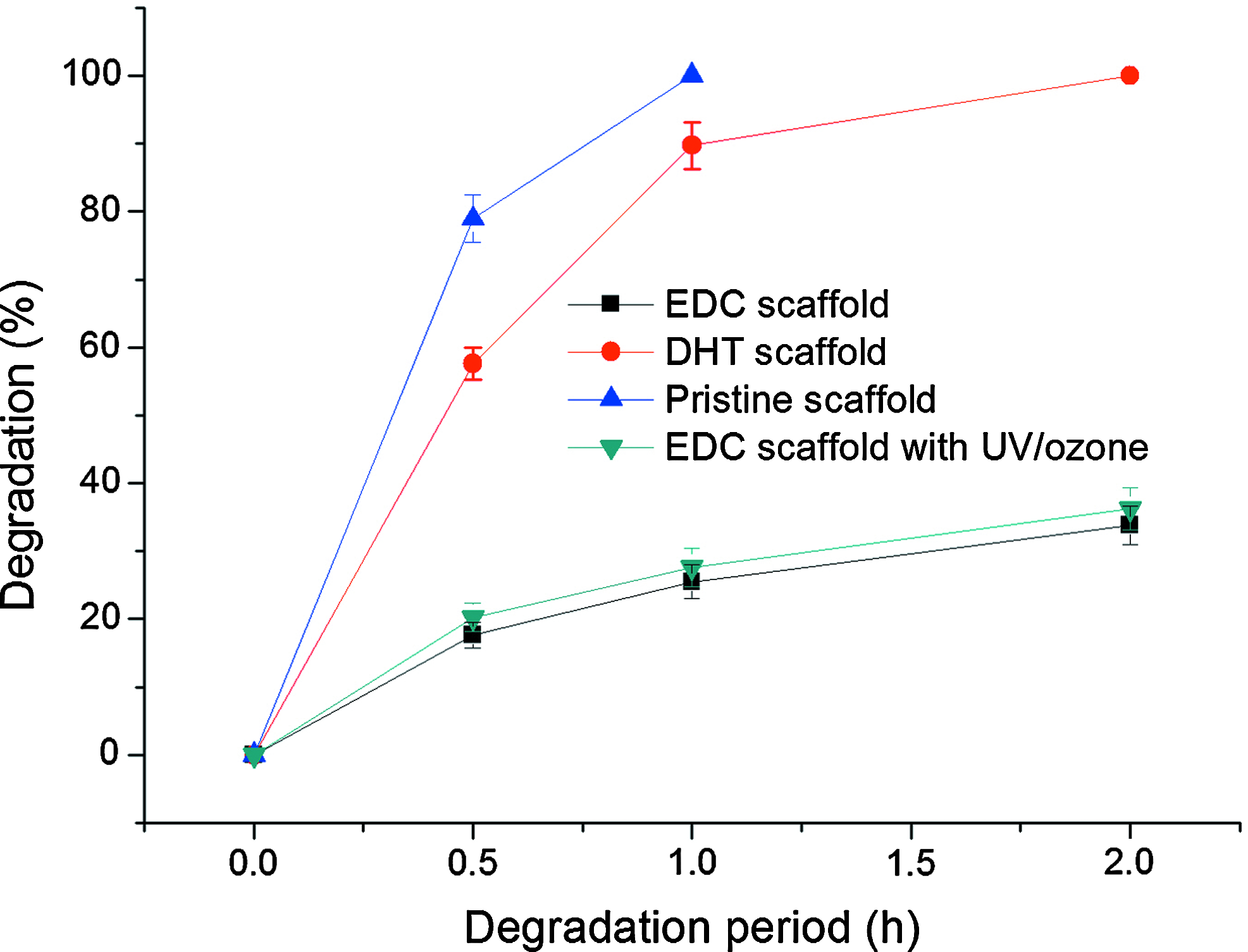
Effect of crosslinking treatment on biodegradation of collagen scaffolds. The EDC crosslinking treatment reduced biodegradation rate of collagen scaffold significantly. Color images available online at
Cell colonization and proliferation performance
Surface-processed and unprocessed EDC- and DHT-treated scaffolds were used to evaluate the effect of surface processing on cell colonization and proliferation. In this study, the green fluorescent protein–labeled human MSCs (hMSCs) were seeded onto scaffolds (n = 6) at a seeding density of 5 × 10⁵ cells per scaffold and statically cultured for 7 days.
The metabolic activity was assessed using an alamarBlue assay over the evaluation period. Figure 8 shows cell proliferation data from these experiments. It was demonstrated that the scaffolds exposed to UVO showed significantly higher total metabolic activity than unprocessed scaffolds, for both EDC- and DHT-treated scaffolds, at each time point (p < 0.05). A sustained increase in metabolic activity of cells was revealed for surface-processed scaffolds over the 7-day culturing period. The metabolic activity of cells had increased from the initial value to 230% ± 25.3% and 170% ± 11.6% (mean ± standard deviation) at the end of 7 days culture on surface-processed EDC- and DHT-treated scaffolds, respectively. In contrast, reduced metabolic activity was demonstrated for both surface unprocessed scaffolds over the period. The total cellular activity at day 7 represent about only 60% of initial cell metabolic activity. The alamarBlue assay reveals the total cellular activity on the scaffold and is unable to demonstrate differences in regional variations in cell migration. The distribution of cells within the scaffolds at 7 days of culturing was examined using a fluorescence microscope and is shown in Figure 9. The in vitro evaluation revealed a poor cell migration within non-surface-processed scaffolds, with most viable cells limited to the top seeding layer, about 0.7 mm thick. Few viable cells were observed in the deep region (Fig. 9a). In contrast, the surface processing enhanced the cell migration within the scaffold and persisted even deep inside the scaffold. The viable cells were observed throughout scaffolds after 1 week of culture, as revealed in Figure 9b. The higher magnification observation (Fig. 9c, d) revealed the cells attached to the collagen fiber and formed a 3D structure. This observation, together with alamarBlue assay results, suggests that surface processing by exposure of the scaffold to UVO improves the surface wettability and water intake ability, and that this also contributed to the enhanced cell colonization and proliferation performance.

AlamarBlue activity of the processed and unprocessed scaffolds (EDC crosslinked, seeding density = 5 × 10⁵ cells per scaffold) (n = 6). Color images available online at
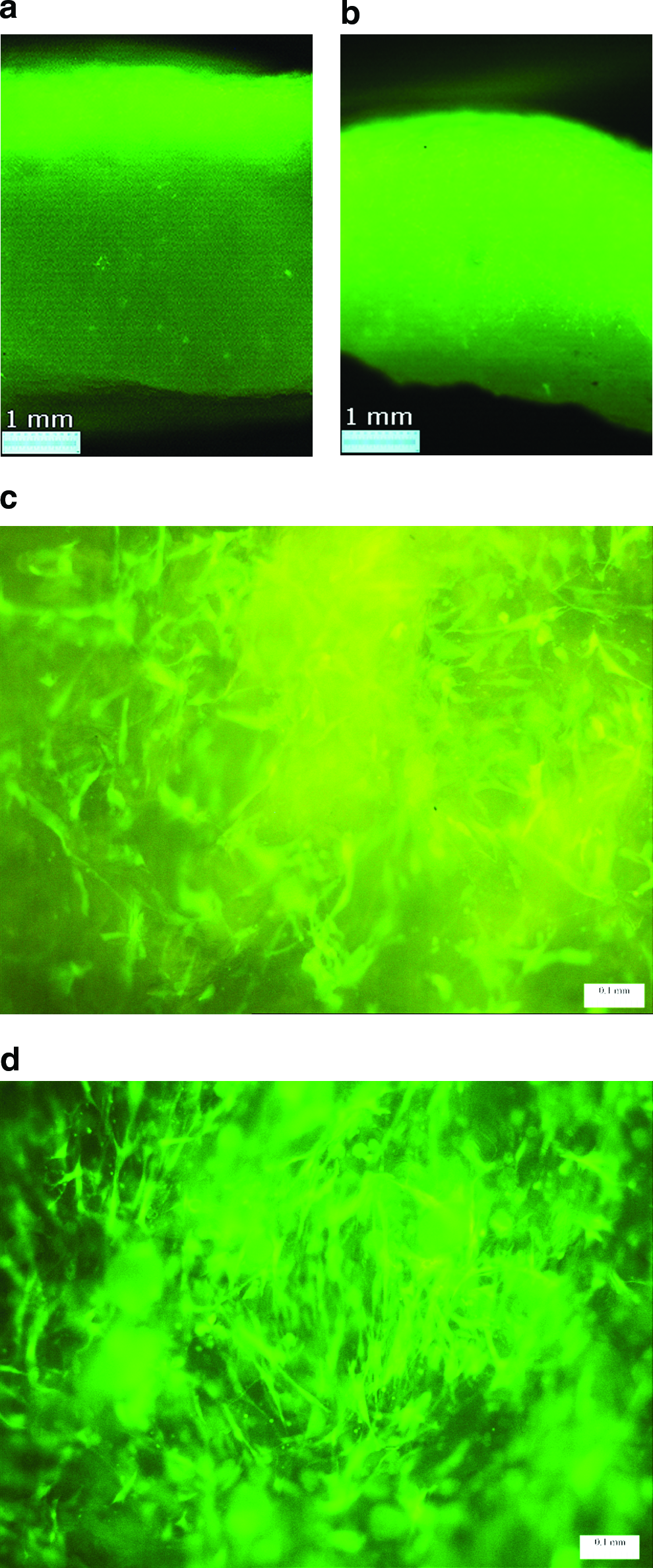
Fluorescence microscopy of green fluorescent protein–labeled human mesenchymal stem cells cultured on EDC-treated scaffolds for 1 week in vitro. (
Discussion
We have demonstrated that crosslinking treatment of collagen scaffolds improves mechanical integrity and reduces biodegradation rate significantly. However, the crosslinking treatment results in a hydrophobic nature of collagen. The collagen surface already contained oxygen, but we have demonstrated that surface processing of crosslinking-treated collagen scaffolds by exposure to UVO leads to a further increase in surface oxygen concentration and improves the surface wettability of the collagen scaffolds.
Surface properties are crucial in controlling interactions between cells and a substrate. Although surface properties are often derived from the bulk properties of materials, the bulk materials do not entirely define them, because 3D matrices are coated with proteins almost immediately after implantation in the body or immersion in culture media. Surface chemistry and topography determine the identity, quantity, and conformational change of these adsorbed proteins.24,32
Cell adhesion to the substrate will influence the cell's capacity to proliferate and to differentiate, and its colonization of the scaffold upon contact. The delivery of a precise number of cells into a 3D matrix with a homogeneous distribution is critical because variation in cell numbers hampers systematic comparison of scaffold-to-scaffold experimental results, and the migration of viable cells within the scaffolds is necessary for the long-term regeneration of viable tissue. 33 To obtain a satisfactory cell colonization, the ideal scaffold material must degrade in a controllable way, maintain its integrity, and possess surface chemistry that promotes cell attachment and proliferation. Shen et al. 34 observed that fibroblast growth factor beta could not be immobilized on the internal section of the poly(lactic-co-glycolic acid) (PLGA) scaffold and cells were prevented from migration into the internal structure of the scaffolds due to the surface hydrophobicity nature. They also observed that surface processing by CO2 plasma improved the surface wettability, which facilitated cell migration into the scaffold structure with the culture medium and resulted in an enhanced cellular performance.
It is the surface of a scaffold that first comes into contact with cells containing the medium; hence, the initial response of cells to scaffold greatly depends on the surface properties. The wetting and adsorption capabilities of scaffolds by the culture medium play an important role during in vitro culture. They allow the cells not only to attach but also to migrate inside and colonize the scaffold to form a 3D structure. The water intake capability was measured using PBS as the percentage of PBS intake to the initial weight of the scaffold, and the results are listed in Table 2. As expected, the crosslinked scaffolds, especially the EDC-crosslinked scaffold, exhibited much lower water intake capability compared to the pristine scaffold. This was attributed to the decreased surface wettability as a result of crosslinking treatment. However, all samples exhibited an enhanced water intake capability after UVO exposure. The DHT and EDC scaffolds exhibited a water intake capability of 8.1 and 8.6 g of water per gram of scaffold, respectively, after surface processing. This represents a 2.2- and 4.8-fold increase in water intake capability compared to unprocessed DHT- and EDC-crosslinked scaffolds, respectively. A 1.3-fold increase was also observed for the pristine scaffold after exposure to UVO. These results, together with water contact angle measurements, also verify that the reported processing allows for successful through thickness surface (both interior and exterior surface) modifications of porous scaffolds. Thus, cell colonization of the scaffolds is enhanced and the cells proliferated to a greater extent in surface-processed scaffolds.
DHT, dehydrothermal treatment; EDC, 1-ethyl-3-(3-dimethylaminopropyl)carbodiimide.
The present investigation supports the literature reports that the surface modification, by using plasma technologies, enhanced the in vitro performance of 3D scaffolds for tissue engineering.11,35 However, the major limitation of the conventional scaffold surface modification using plasma technique for tissue engineering is the depth of penetration of the active species into the porous structure. By comparison, the method reported in this investigation shows that both the exterior and interior surfaces of the 3D scaffold could be modified. Oxygen was uniformly incorporated onto the collagen fiber surface, as determined by XPS analysis, through the thick sample (10 mm thick), as observed in Figure 3c. The reported method renders scaffold an appropriate surface for cell attachment, ingrowth, and proliferation, and promotes cell growth within the scaffold.
Although we have used only collagen scaffolds produced from type I collagen for the experiments described here, we believe that the surface-processing technique reported in this study could be extended to other polymer scaffolds such as PLA/PLGA. surface modification to tuning the surface wettability and to improve the cell attachment and enhance the cellular performance without compromising their mechanical integrity.
Conclusions
In this study a UVO-processing protocol was developed to modify both interior and exterior surface properties of porous collagen scaffolds for tissue engineering applications. We have demonstrated that crosslinking treatment enhances the mechanical properties of the collagen scaffolds and biodegradation stability significantly. However, the crosslinking treatment reduces the surface wettability of collagen scaffolds and water intake ability significantly, whereas surface processing using UVO reactor significantly improves their surface wettability and water intake ability, a factor that could be attributed to the incorporation of oxygen and oxygen-containing functional groups onto the surface. In vitro evaluation demonstrated that hMSCs are unable to migrate into the interior of the crosslinked scaffold but not surface-processed scaffolds; in contrast, surface processing enhanced hMSC migration deep into the structure of the scaffold, leading to uniform colonization and proliferation therein. We believe that the enhanced cell colonization and cellular response to crosslinked collagen scaffolds by UVO processing, observed in this study, could be applied to other synthetic polymeric scaffolds such as PLA or PLGA scaffolds.
Footnotes
Acknowledgments
This work was financially supported by the Northern Research Partnership Program (NRP). NRP is a partnership of the Robert Gordon University, University of Dundee, and University of Aberdeen.
Disclosure Statement
No competing financial interests exist.