
Abstract
To improve the mechanical properties of polymers used in bone repair, it has been suggested to incorporate single-walled carbon nanotubes (CNTs). However, concern exists about the biosafety of the CNTs in vivo. Therefore, the aim of this study was to develop a magnetic resonance imaging technique to examine the distribution pattern of CNTs after release from a degrading poly(lactic-co-glycolic acid) (PLGA) scaffold in vivo. Five rats received a PLGA scaffold with incorporated gadolinium-labeled single-walled CNTs (“gadonanotubes”) subcutaneously. The rats were analyzed up to 5 weeks, subsequently euthanized, followed by histological evaluation of the explanted scaffolds with their surrounding tissue. A significant increase in intensity of the scaffold surrounding tissue was shown in the time period around 3 weeks, as compared to internal control areas. The intensity declined soon thereafter. This is suggested to be caused by the release of gadonanotubes from the degrading scaffold into the surrounding tissue. Histological imaging showed encapsulation by connective fibrous tissue and some mild inflammation around the scaffolds. In conclusion, magnetic resonance imaging is an excellent technique to study the biological fate of gadonanotubes. However, to formulate solid conclusions on the distribution pattern of gadonanotubes in vivo the experimental setup requires further optimization.
Introduction
Materials and Methods
Synthesis of gadonanotubes
SWCNTs (Sigma Aldrich) were cut into ultra short (US) tubes by a chemical cutting method. 18 Briefly, SWCNTs were fluorinated for 2 h at 50°C under 1% gaseous fluoride diluted in helium. This step was followed by pyrolysis at 1000°C for 1 h under an inert atmosphere what lead to spontaneous cutting of the micron-long SWCNT into US-SWCNTs. Besides cutting the tubes, this method also purified the US-tubes. In the next step, the US-tubes were filled with Gadolinium (Gd3+) by a solution-phase filling protocol. 14 This method involved stirring of the US-tubes with anhydrous GdCl3 (Sigma Aldrich) (1:1 ratio by weight) in deionized high-performance liquid chromatography grade water, followed by sonication for 1 h. Then, the solution was left undisturbed overnight upon the Gd3+-loaded US-tubes flocculated from the solution. The supernatant was decanted off and next the sample was washed with fresh deionized high-performance liquid chromatography-grade water and batch sonicated to remove any unabsorbed GdCl3. This flocculation and washing procedure was repeated three times. The sample was then air-dried and an inductively coupled plasma spectrometry analysis was performed to obtain the Gd3+ content. Finally, the filled US-tubes were thermally annealed to seal the carbon nanotube ends, thereby preventing leakage of the encapsulated Gd3+.
Preparation of gadonanotubes incorporated PLGA scaffolds
Disc-shaped scaffolds were prepared by dispersing 2 wt% gadonanotubes in low-molecular-weight (average molecular mass of 5 kDa) PLGA (50:50 lactic-to glycolic-acid copolymer; Sigma Aldrich) polymer using an established procedure. 3 Briefly, gadonanotubes were dispersed in chloroform by high shear mixing for 5 min and sonication for 15 min and then added into a PLGA/chloroform solution. This procedure was followed by 15 min of sonication, rotary evaporation, and vacuum drying. Finally, the composite material was cast into molds that produced solid discs of 6 mm in diameter and 1 mm in height. Besides the composite scaffolds, also one plain PLGA scaffold was produced by the same process to serve as a reference sample to asses for artifacts. The discs were sterilized by ultraviolet (UV) irradiation for 2 h.
In vitro phantom MR study
A concentration series of gadonanotubes ranging from 0.0002 to 20 wt% dispersed in agar were prepared in eppendorf tubes. Subsequently, spin echo MR measurements were performed with varying repetition time (TR) or echo time (TE) to characterize the MR relaxation behavior of the gadonanotubes at different concentrations. Applied parameters for these images were a TR ranging from 50 to 5000 ms with a fixed TE of 13 ms, and a TE ranging from 13 to 200 ms with a fixed TR of 2000 ms. From these images the signal intensities of all samples were measured using software on the MRI scanner—Image Display (MR Solutions). Here, a region of interest (ROI) was drawn in which the intensity was measured. From these data, signal intensity–time curves were plotted and T1 and T2 values were calculated by fitting the curves using the following formulas:
Mt = M0 [1 − e(−(Ta + Tau)/T1)] and Mt = M0 × e−TE/T2, where Mt = nuclear spin magnetization at time t M0 = nuclear spin magnetization at t = 0 Ta = aquisition time Tau = delay time between two excitation pulses minus Ta (varied) T1 = spin-lattice relaxation time TE = echo time (varied) T2 = spin-spin relaxation time
In vivo MRI
Essentially, a similar experimental approach was used as described previously for dispersed gadonanotubes. 15 In the current study, six male Wistar rats, weighing ∼250 g, were used. National guidelines for the care and use of laboratory animals were observed and the study was performed under Ethics Committee number DEC-2007-096. Five rats received a PLGA/gadonanotubes scaffold subcutaneously and the sixth rat received the plain reference PLGA scaffold. For this purpose, the rats were placed under general anesthesia (isoflurane/O2, N2O) and the dorsal skin was shaved, washed, and disinfected with iodine. One full-thickness longitudinal incision of 1 cm was made through the skin on the right side of the spine. Subcutaneous pockets were created by blunt dissection with scissors and a PLGA/gadonanotube or PLGA scaffold was inserted. Finally, the wound was closed. All rats were analyzed on day 1 after implant placement. Although, MRI did not allow to analyze all animals simultaneously. Still, each rat was regularly measured at intervals of ∼1 week (±2 days) up to 5 weeks.
For each MR examination, the rats were placed in a custom-build probe on top of a radiofrequency microstrip coil, 19 consisting of a copper strip taped onto a 7-mm-thick Teflon layer. The probe with the rat was then placed inside a 7-tesla MR magnet (Magnex), with a horizontal bore of 200 mm, which was interfaced to a spectrometer (SMIS; MR Solutions). Then, T1-weighted coronal multi-slice MR images (14 in total) with an image matrix size of 256 × 256 and a slice thickness of 1 mm were obtained. A TE of 15 ms was used, while the TR was about 1500 ms. The exact time was set by triggering the imaging on the breathing of the rats, which was, by inhalation anesthesia, ∼38–44/min. From the images obtained in this way, signal intensities were measured in an ROI and in an internal control region in muscle tissue. After the final MRI measurement the animals were euthanized and the scaffolds with surrounding tissue were harvested and examined histologically.
Histological analysis
The retrieved scaffolds with their surrounding tissue were fixed in 10% formalin, dehydrated in a series of ethanol, and embedded in polymethylmethacrylate. Polymerization sections of 10 μm in thickness were prepared using a modified diamond-blade sawing microtome technique (Leica Microsystems GmbH), stained with methylene blue and hematoxylin–eosin, and examined with a light microscope (Leica Microsystems B.V.). 20
Statistical analysis
All intensity measurements were performed in duplicate on a total of five rats. For statistical analysis the ratio of the ROI versus the internal control region was calculated (n = 5 for both groups). The mean of these data was used in an unpaired two-tailed t-test comparing to a hypothetical mean of 1.0. Data were tested on a normal distribution and a p-value of 0.05 or less was considered to indicate significance. Calculations were performed in InStat software (v 3.05 Graphpad).
Results
In vitro phantom MR study
Series of images with different TR and TE were acquired from a range of dilutions of gadonanotubes dispersed in agar to obtain T1 and T2 values as a function of the concentrations of gadonanotubes. For each concentration signal intensitiy curve (Fig. 1), T1 and T2 values were generated (Table 1). These results indicate that, with the acquisition parameters used in the in vivo MRI of the study, very high concentrations of gadonanotubes will result in a negative contrast (T2 weighted), whereas low concentrations (less than about 2 wt%) will result in a positive contrast (T1 weighted).
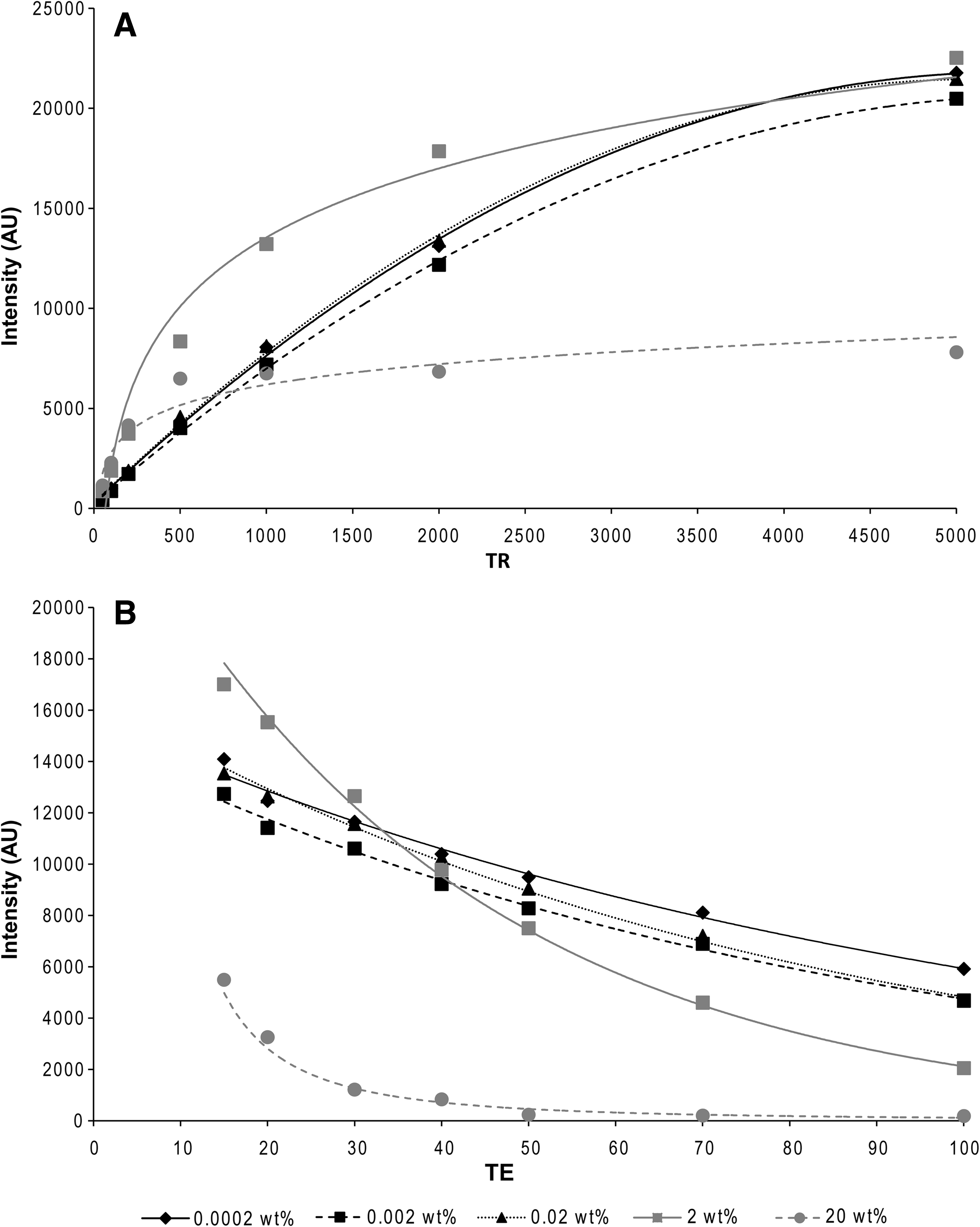
Signal intensity–TE and TR curves for a concentration series of gadonanotubes dispersed in agar. For the T1 curve a fixed TE of 13 ms was used (
Values were calculated from the signal intensity–time curves.
SD, standard deviation; T1, spin-lattice relaxation time; T2, spin-spin relaxation time.
In vivo MRI
All rats were analyzed at intervals of ∼1 week (±2 days) up to 5 weeks. MR images were created from the signals of water protons. In Figure 2, MR images of a rat with a PLGA/gadonanotube scaffold and of a reference rat with a plain PLGA scaffold are depicted. Images were made in the coronal plane of the rats; the spine of the rats appears slightly curved due to small differences in positioning of the rats. With MRI at TR of 1500 ms and TE of 15 ms, tissues are visible in gray tones, whereas the water-impermeable scaffolds appear as black disks due to the solid structure of the disks compared to tissue. In time, the size of the scaffolds decreased, as well as the negative contrast of the disk, due to the degradation and water uptake of the polymeric scaffolds. Around the scaffolds several bright spots were visible, considered susceptibility artifacts caused by the water to solid hydrophobic scaffold transition. These have no relation to the amount of gadolinium at the site, since the reference rat that received the pure PLGA scaffold showed the same artifacts. The MR images suggested that the scaffolds adapted a bent shape, starting after ∼1 week. In the five rats that received a PLGA/gadonanotubes scaffold, the signal intensity of the scaffold surrounding tissue significantly increased (T1 weighted) compared to the control areas around week 3 (Fig. 3). This indicates the release of gadonanotubes from the degrading scaffold into the surrounding tissue. A week later, the first elevated signal intensity was declined again to its original value. However, no increase in MR signal intensity was observed in body organs (like kidney, brain, or liver), not even when the rats were repositioned over the radiofrequency coil to obtain more optimal images of these sites.
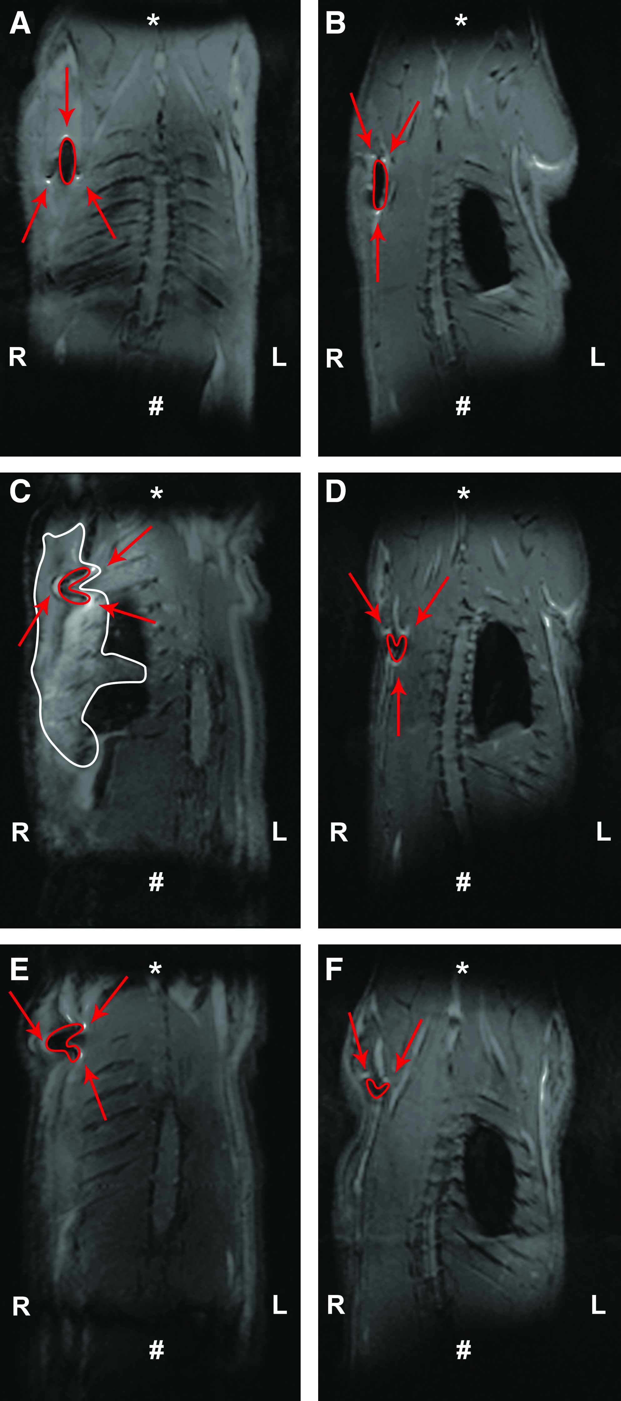
Coronal magnetic resonance images of a rat that received a PLGA/gadonanotubes scaffold (
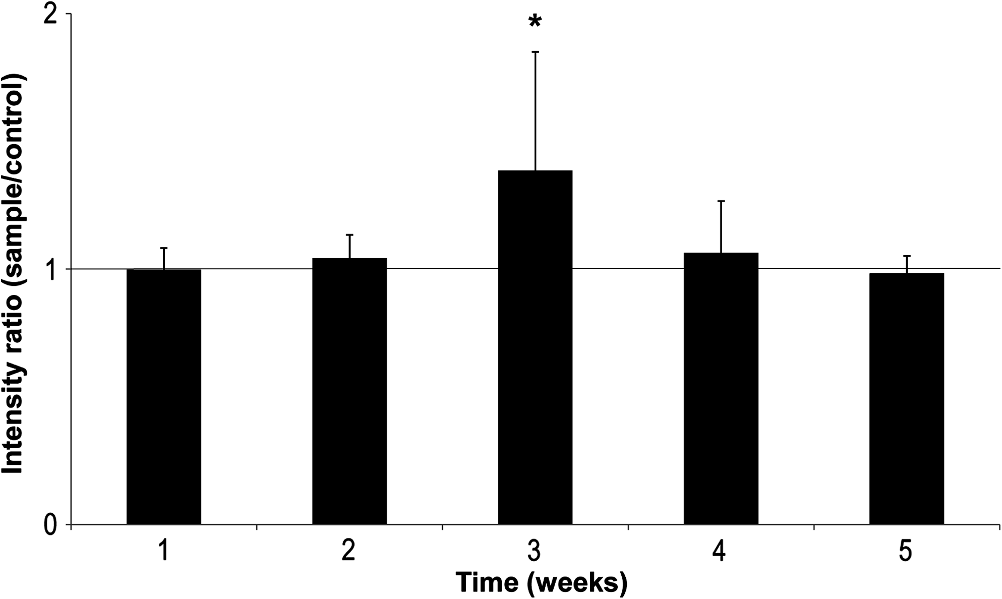
The average ratio of the sample values and their internal control values in the time intervals for a total of five rats. *The average ratio is significantly increased around 3 weeks compared to a mean of 1.0 (p ≤ 0.05).
Histological analysis
PLGA/gadonanotube scaffolds were retrieved without complications. Upon visual inspection, all scaffolds displayed heavy degradation, but maintained their shape (Fig. 4). No reference PLGA scaffold could be explanted, as this scaffold was undetectable after 5 weeks, due to complete degradation. As a consequence, histological comparison of the tissue response between PLGA/gadonanotubes scaffolds and pure PLGA scaffolds was not possible.

(
The light microscopical examination of the PLGA/gadonanotubes scaffold sections showed encapsulation of all scaffolds by a vascularized connective fibrous tissue capsule of ∼20 cell layers in thickness. The capsule tissue was fibrous but immature, as characterized by the presence of many fibroblasts, inflammatory cells, newly formed blood vessels, and little collagen. There was no apparent difference in thickness or vascularization of the fibrous tissue between the different explanted scaffolds. Within the scaffold material, some aggregated gadonanotubes were visible as black spots. Due to degradation, the scaffolds were fragmented and each fragment was separately encapsulated by fibrous tissue. The interstitium in between these fragments showed ingrowth of immature fibrous tissue as well. At the capsule–scaffold interface scattered foci of inflammatory cells were present. However, no giant cells, correlated to a foreign body giant response, were observed.
Discussion
The aim of this study was to examine the distribution pattern of CNTs in an in vivo situation after release from a degrading scaffold by the means of MRI. As described before, gadonanotubes function as an excellent contrast agent for MRI in in vitro and in vivo applications, even at very low gadolinium concentrations.14–16 Based on in vitro testing, it is assumed that the gadolinium inside the gadonanotubes remains stable under physiological conditions. 21 In a previous study, subcutaneously injecting a gadonanotubes solution (10 μL containing 3 × 10⁵ mmol gadolinium) in rats showed a negative contrast on the MR image as a result of high T2 relaxivity values. The injected solution was shown to be maintained in the vicinity of the injection site, but this was only assessed up to 24 h. 15
The current study demonstrates that CNTs are released from a degrading PLGA scaffold into the surrounding tissue, and demonstrates the possibility to monitor this process by MRI. An increase in signal intensity in the tissue surrounding the scaffold was observed around week 3. This increase disappeared soon thereafter. These observations are evidently the result of released, gadonanotubes from the degrading PLGA scaffold. In contrast to the previously described in vivo study, where the rats received an injection with gadonanotubes, the monitored signal increased. 15 This difference in signal evoked by the gadonanotubes is clearly explained by the in vitro phantom study results; that is, high concentrations of gadolinium result in a strong T2 effect on imaging, giving a negative contrast, whereas lower concentrations result in a T1 effect, giving a positive contrast. Comparison of the signal intensity of the measured rats that displayed an increase in signal intensity with the in vitro T1 and T2 measurements indicated that the concentration of gadonanotubes that is present in the tissue is very low. A more quantitative assessment of the gadonanotube level would require the in vivo measurement of T1 and T2.
Although we aimed to follow the fate of the released CNTs, no changes in MR signal intensity were found in organs where the gadonanotubes might possibly aggregate, like kidneys, brain, spleen, and liver. However, this is not necessarily indicating that the gadonanotubes are systematically cleared from the body. It is still possible that very low quantities of gadonanotubes below the detection limits of the MR measurement have been present. It is too soon to claim that no CNTs aggregate in any of the organs, and the experimental setup has to be optimized (by higher dosage, higher field strength, and/or dedicated coils) to make valid statements for this research question. Since our composite was developed for hard tissue regeneration, it would be of great interest to asses different (bone) implantation sites. With the acquired knowledge from this study, in future studies it would be beneficial to increase the number of measurements, especially around the release period of week 3, for instance, with other dedicated nano detection techniques such as high-resolution transmission electron microscopy. Moreover, it has been reported that carbon nanotubes present in blood are cleared quickly from the body trough the urinal and fecal excretion.12,13 Therefore, as an interesting positive control group a renal deficient model like the model described by Jost et al. could give some valuable new insights. 22 In this model, renally impaired rats (ZSF1 rats) were used to determine the kidney retention of different contrast media. ZSF1 rats have a three- to eightfold decrease in creatinine clearance compared to commonly used rat lines like SHHF, SHR, and WKY. This model would be ideal as a positive control to observe agglomeration of the gadonanotubes and it also provides a possibility to examine potential kidney damage caused by retention of the nanotubes.
In our histological evaluation degradation of the composite scaffolds could not be compared to the plain PLGA scaffold, as its retrieval was impossible due to complete degradation at the time of explantation. This was unexpected, although a fast degradation of the scaffolds was desired. Specifically, a low-molecular-weight PLGA was selected, known to degrade almost completely in 40 days. The degradation of such PLGA depends on many factors, like pH, degree of swelling, ionic strength, ion concentration, temperature, and buffering capacity.17,23,24 These factors cannot be completely controlled in an in vivo study and inter-animal differences will always occur. Besides these factors, the applied UV sterilization treatment is also known to reduce polymer chain length, thus increasing the degradation rate of PLGA. One hour of UV radiation was shown to already affect the degradation of PLGA nanofiber scaffolds. 25 In our study all CNT containing scaffolds could be retrieved, although often fragmented. The difference in degradation of both scaffold types was unexpected and suggests a slowing effect of the incorporated gadonanotubes on the degradation rate. Unfortunately, no definite explanation can be given, as no material with a comparable composition has been described before. Also, literature is not consistent on the influence of CNTs on the degradation rate of polymeric scaffolds. One study reported an increase in degradation rate after the incorporation of multiwalled carbon nanotubes in poly(L-lactic acid), 26 whereas two other studies reported no effects of incorporated nonfunctionalized SWCNTs in poly(propylene fumarate) (PPF) and PLGA scaffolds, respectively, on the degradation rate.27,28
Another technical note is that the MR images suggested that the scaffolds adapt to a bent shape in time. In contrast, explantation of the scaffolds showed that the scaffolds were not bent. The apparent folding in the MR images was probably caused by an irregular uptake of water in the scaffold as a result of which some of the areas of the scaffold were brighter than others in combination with some susceptibility effects. This implies that this experimental setup is not applicable to measure the effects of the gadonanotubes within the scaffold; however, this was not the intended goal of this study.
Histological analysis showed the scaffolds to be surrounded by fibrous tissue, which is a natural occurrence after subcutaneous implantation of foreign body materials. 29 As described before, no comparison between the PLGA/gadonanotube scaffolds and the reference plain PLGA scaffold was possible due to complete degradation of the control. Therefore, it is recommended for future research to bring the histological analysis time point forward and also to expand it with a few time points around 3 weeks, as it is known now that the release occurs around week 3. In addition, histological examination of organs like the kidneys, brain, spleen, and liver at the 3-week time point might prove to be valuable.
Detailed examination of the light microscopical sections showed the presence of foci of inflammatory cells at the scaffold–fibrous capsule interface, which were evidently attracted to remove the degradation products of the PLGA and possibly to remove the CNTs. However, no severe inflammatory reaction with the formation of giant multinucleated cells was observed with these quantities of PLGA and gadonanotubes.
When comparing our data to earlier studies, designed to obtain information on the distribution of CNTs in vivo, the results are still debatable. Two approaches, with radiolabeled SWCNTs or multiwalled CNTs injected either intravenously or intraperitoneally in rats or mice, reported rapid clearance rates within 24 h of the CNTs through the renal excretion route, without significant retention in any of the secondary organs, like liver or spleen after 24 h.12,13 This would corroborate our current findings. However, another study reported injected iodine-125–labeled SWCNTs in mice to be distributed throughout the whole body (except the brain) and to accumulate in bone. 11 It has to be emphasized that in these CNT distribution studies, body exposure to and transportation of CNTs was significantly enhanced compared to the current study, due to direct injection into the bloodstream or peritoneal cavity.
Before the first distribution studies even started many toxicity studies were already performed and toxicity in the pulmonary area and bloodstream was reported.8,30–32 However, thorough purification of toxic residual metal catalysts33–35 as well as functionalization of the CNTs6,8,36 diminished toxicity to a great extent and thus it remains unclear up to what level such early information is still dependable. Additionally, in all these studies high concentrations of CNTs were used. In contrast, a recent rabbit implantation study with degradable PPF scaffolds containing US-SWCNTs reported a significant improvement in bone-healing capacity of the PPF/US-SWCNT composite, without any toxic effect of the released US-SWCNTs. 27 Taking these reports into consideration, there is still a high interest in CNT application in tissue engineering constructs. Therefore, the further development of proper analysis techniques is still desirable.
Conclusions
We conclude that MRI detection forms an excellent tool to follow the release of gadolinium-labeled nanoparticles from scaffold materials. On basis of our preliminary results, gadonanotubes were released from degrading PLGA scaffolds into the scaffold surrounding tissue around 3 weeks, followed by a rapid clearance from the site. However, the experimental setup requires optimization before more detailed conclusions on the final biological fate of gadonanotubes can be made. Such an effort will be essential before further explorations of CNTs in biomaterial and tissue engineering science can be considered.
Footnotes
Acknowledgments
This study was supported by the EU FP6 Nanobiocom project (grant #STRP516943) and the travel grant EMIL-LSHC-CT-2004-503569. We thank Valerio Zerbi for his valuable support on the relaxation time calculations.
Disclosure Statement
No competing financial interests exist.