
Abstract
Spatially controlled coculture in three-dimensional environments that appropriately mimic in vivo tissue architecture is a highly desirable goal in basic scientific studies of stem cell physiological processes (e.g., proliferation, matrix production, and tissue repair) and in enhancing the development of novel stem-cell-based clinical therapies for a variety of ailments. This study describes a novel fabrication system for photopatterning and assembling cell-laden oligo(polyethylene glycol)-fumarate:poly(ethylene glycol)-diacrylate hydrogels with high spatial fidelity and thickness using a controlled, inert nitrogen environment without the need for expensive precision equipment. Cross-linking was performed using Irgacure-2959 photoinitiator and 365-nm light (∼7 mW/cm2) to form gels ranging from 0.9 to 3 mm in width. Employing a nitrogen environment increased gel thickness up to 240%, generating gels >1 mm thick before swelling. This technique was further applied for spatially controlled patterning of primary tendon/ligament fibroblasts and marrow stromal cells in a single 1.5-mm-thick laminated hydrogel construct. Cells encapsulated using this technique maintained viability over 14 days in culture. This system potentially enables better understanding of paracrine effects on a range of stem cell functions and therefore may be useful as an in vitro model system for a wide array of regenerative medicine applications.
Introduction
Toward this end, the use of 3D hydrogel biomaterials as cell carriers has enabled researchers to address many complex questions regarding the role of specific niche components and architecture in regulating the dynamic responses of stem cells to well-defined model microenvironments.5,6 Of these, synthetic poly(ethylene glycol) (PEG)-based hydrogels, such as oligo(poly(ethylene glycol) fumarate) (OPF), are widely utilized for their cytocompatibility, intrinsic resistance to protein adsorption and cell adhesion, polymer network configurations and hydration state that mimic mechanical and molecular transport properties of native ECM, and chemical versatility that allows tethering of bioactive molecules.7–9 Importantly for the prospect of coculturing multiple diverse cell types, robust and mechanically stable interfaces can be created by laminating several OPF-based hydrogels together. 7 To further control the microscale architecture of hydrogels with different cells or ligands, novel patterning techniques have been adapted for their fabrication. In particular, photopatternable polymers, 10 in combination with patterning techniques for cell encapsulation,11,12 enable precise definitions of ECM density and type, as well as cellular location, proximity, and density to facilitate study of specific cell–microenvironment interactions. Recently, microfluidic devices, traditionally used for fluid handling at the microscale and miniaturized high-throughput assays, have been utilized for rapid fabrication of photopatterned, cell-laden hydrogel microstructures on the order of 100 μm.13,14 However, to date, precision systems for photopatterning hydrogels have not been developed for long-term (∼weeks) coculture of cells in constructs of tissue-scale thickness (>1 mm thick).
In response, we describe a novel, facile photolithographic patterning scheme for generating and assembling thick (>1 mm), spatially controlled hydrogel constructs with high fidelity and minimal alteration in standard photocross-linking chemistry. Our system was readily calibrated to predict gel size before and after equilibrium swelling. Cell-laden gels containing spatially patterned primary isolates of tendon/ligament fibroblasts and marrow stromal cells (MSCs) were successfully laminated together into a single 1.5-mm-thick construct as a coculture model for understanding stem cell interactions with injured tendon/ligament tissue. The patterning technique developed in this proof-of-concept study helps maximize diffusion between cell types while maintaining spatial segregation. Importantly, viability for primary-isolated cells is maintained in these constructs over culture times relevant for tracking biological phenomena (up to 14 days). Accordingly, this work represents a simple enabling platform that facilitates development of in vitro biological model systems for informing future stem-cell-based clinical therapies.
Materials and Methods
Polymer synthesis and characterization
OPF (Mn 16,952 ± 131 Da, polydispersity index [P.I.] 4.92 ± 1.03) and PEG diacrylate (PEG-DA; Mn 3759 ± 18, P.I. 1.07 ± 0.001) were synthesized, purified, and stored as previously described.15,16 OPF and PEG-DA were characterized via a gel permeation chromatography to determine molecular weight and polydispersity. 17
Device fabrication
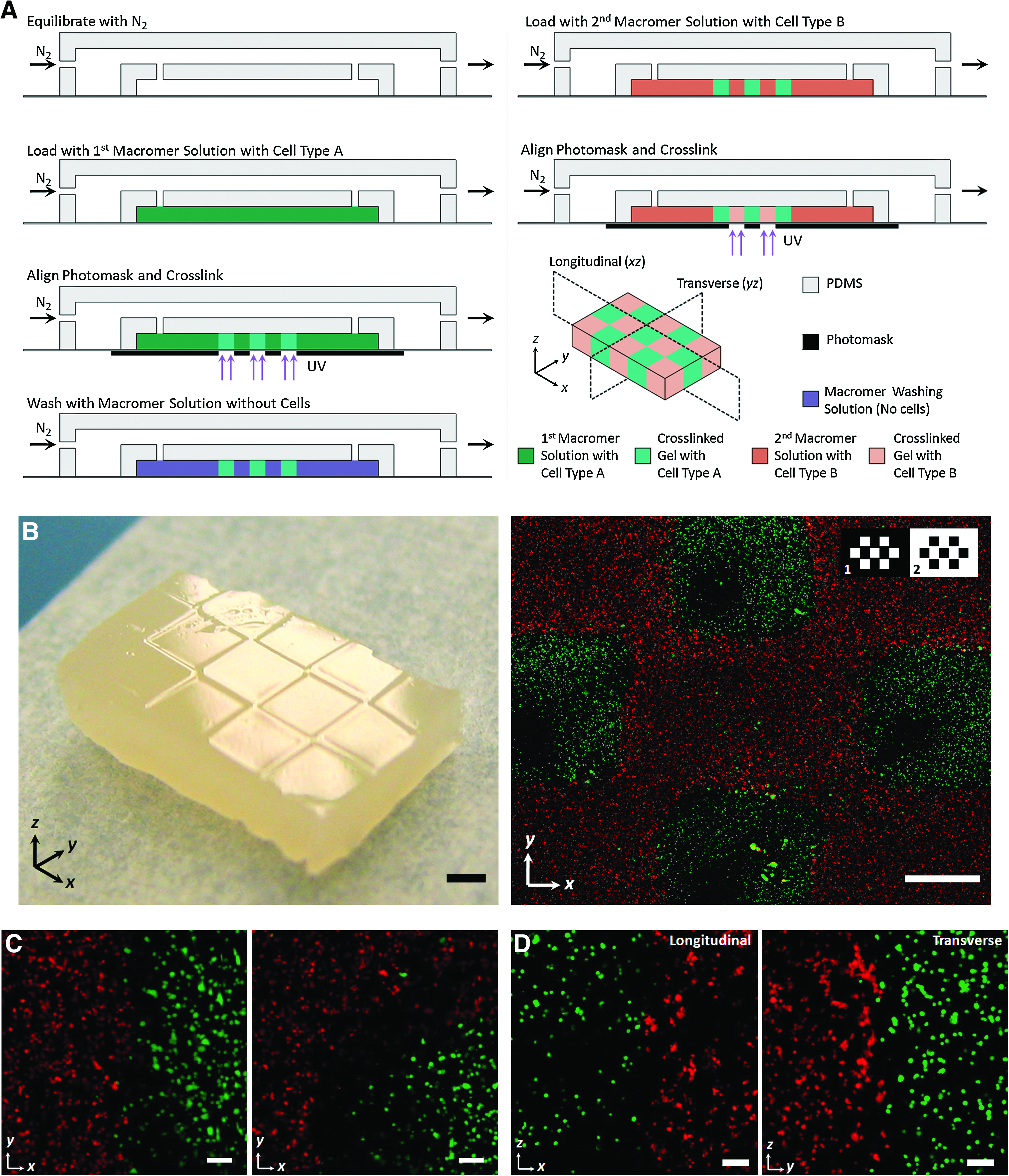
Photopatterning experiments were performed in a microfluidic device fabricated from polydimethylsiloxane (PDMS, Dow Corning Sylgard 184; Essex-Brownwell, Inc.) using replica molding. 18 Devices consisted of a 2-mm-thick rectangular chamber with three inlet and three outlet channels for efficient delivery and removal of macromer solution (Fig. 1A). Briefly, a poly(urethane) master was fabricated using established techniques. 19 We fabricated PDMS devices by curing the device layer (10:1 base:curing agent ratio) over the master at 70°C for 2 h, peeling the PDMS off the mold and cutting individual devices to size, and subsequently bonding each to a separate cover glass using oxygen plasma treatment. 20 Medical-grade platinum-cured silicone microtubing (BB518-12; Scientific Commodities) was used for fluidic connections and was connected to the device channels via type 304 90°-angled stainless steel tubes (21 gauge; Small Parts). Luer lock dispensing needles (21 gauge; McMaster-Carr) were attached to the opposite ends of the tubing for eventual connection to syringes containing macromer solution. A contact-bonded, overlaying PDMS enclosure was fabricated using a different poly(urethane) mold to contain a nitrogen (N2) atmosphere for the device.

Oligo(poly(ethylene glycol) fumarate) hydrogels can be photolithographically patterned into a variety of three-dimensional shapes in a controllable, high fidelity manner at the millimeter scale. (
Calibration of photopatterning method
Photomasks containing polygonal features ranging from 0.9 to 3 mm were used to pattern hydrogels from macromer solutions containing OPF and PEG-DA a 50:50 in ratio by weight with 75% (w/w) initial water content and 0.05% (w/w) D2959 photoinitiator (Ciba) in phosphate-buffered saline (PBS; Invitrogen). Devices were either equilibrated with an N2 atmosphere or left in ambient air before loading the polymer solution (Fig. 1). For devices equilibrated with N2, gas was initially delivered for a minimum of 30 min to the interior of the device via the inlet ports and subsequently delivered within a PDMS enclosure during cross-linking (Fig. 1B, D). The photomask was aligned and the polymer solution injected and allowed to cross-link under exposure to ∼10.5 mW/cm2 of 365 nm light (as measured before passing through the cover glass and mask; ∼7 mW/cm2 of light passes through the glass and mask layers to reach the polymer solution) for 12 or 20 min (Fig. 1B). Hydrogel dimensions immediately after cross-linking and after reaching equilibrium swelling (n = 3) were measured using a stereomicroscope (MZ16F; Leica) and ImageJ software (version 1.43n; NIH).
Cell harvest and isolation
Fibroblasts were isolated from the digested cruciate ligaments and patellar tendons of immature bovine knee joints (Research 87) as previously described 17 and cryopreserved in liquid N2 in Dulbecco's Modified Eagle Medium (DMEM) containing 20% fetal bovine serum (FBS; Hyclone) and 10% dimethyl sulfoxide (Sigma–Aldrich) for storage until use in cell culture experiments. MSCs were isolated from the femora and tibiae of immature bovine hindlimbs (Research 87) similarly to methods of Connelly et al. 21 After initial plating and expansion, cells were subsequently lifted using 0.05% Trypsin/0.53 mM ethylenediaminetetraacetic acid (Mediatech), resuspended in DMEM containing 20% FBS, 10% dimethyl sulfoxide, and 1% antibiotic/antimycotic solution (A/A; Mediatech), and cryopreserved in liquid N2 until further use in cell culture experiments.
Cell patterning and coculture
Before encapsulation, tendon/ligament fibroblasts were thawed and plated at 2 × 10⁶ cells/flask in a growth medium containing DMEM, 10% FBS, 1% nonessential amino acids (Mediatech), 1% HEPES (Mediatech), 1% A/A, and 50 μg/mL ascorbate (Sigma–Aldrich), with medium changes every 2 days. MSCs were thawed and plated at 1 × 10⁶ cells/flask in a growth medium containing DMEM, 10% FBS, 1% A/A, and 1 ng/mL basic fibroblast growth factor (bFGF; Peprotech), with medium changes every 2 days. Cells were grown to near confluency and lifted using 0.05% Trypsin/0.53 mM ethylenediaminetetraacetic acid at passage 2 for encapsulation experiments. Cell populations were distinguished from each other during coculture experiments by differentially staining fibroblasts and MSCs with 10 μM CellTracker Orange CMRA and CellTracker Green CMFDA reagents (Invitrogen), respectively, per manufacturer's recommendations at 1 day before encapsulation.
These cells were subsequently patterned into 3 × 5 arrays of 1.5-mm squares with alternating cell types using sequential photocross-linking steps inside microfluidic devices (Fig. 3A). Completely assembled devices were sterilized using an autoclave before use. Sterilized devices were equilibrated with an N2 atmosphere for a minimum of 30 min before loading the polymer solution. Macromer solutions containing OPF and PEG-DA in a 1:1 ratio were dissolved in PBS at 90% (w/w) initial water content and filter sterilized using 13-mm-diameter syringe filters (0.2 μm pore size; Fisher Scientific). Sterile photoinitiator (0.05% w/w D2959 in PBS) was subsequently mixed into the macromer solution. Cells were resuspended in macromer solution at a concentration of 10 × 106 cells/mL and filtered through nylon mesh with 80-μm pores to dissociate or remove any remaining large aggregates of cells. The first suspension containing one cell type was delivered into the device and patterned into 1.5-mm cubic hydrogel blocks using 365-nm UV light for 12 min (Fig. 3A). The remaining uncross-linked cell solution was washed out of the device using macromer solution containing no cells. A second suspension containing another cell type was delivered into the device and laminated to existing blocks using the same cross-linking parameters through the use of a second photomask. Cells patterned during the first round of cross-linking were protected from a second dose of UV light by overlying dark areas present on the second photomask. Alignment marks were included on the masks and device to allow for registration of laminated gels. Constructs were extracted from devices using a scalpel and placed in six-well tissue culture plates with 5 mL of DMEM containing 10% FBS, 1% nonessential amino acids, 1% HEPES, 1% A/A, 50 μg/mL ascorbate, and 1 ng/mL bFGF.
Image analysis of cell patterning
Image analysis was performed to reveal interfaces between different cell populations after the gel constructs reached equilibrium swelling (∼24 h). Gels containing stained cells were rinsed for 45 min in sterile PBS to remove media and imaged at 5 × and 10 × magnification on a laser-scanning confocal microscope (LSM 510/NLO; Carl Zeiss). A total of 15 overlapping image stacks were acquired for each gel throughout its entire thickness (∼2 mm) at 10-μm intervals. Images were analyzed using ImageJ software. The separate slices of each z-series were examined to verify the absence of an overlap between green- and red-stained cell populations. The images were then processed to provide single images demonstrating a nonoverlapping interface between adjacent cell populations. To accomplish this, the green and red channels were merged for each image slice in the z-series, and then the entire z-series was projected onto a single plane using a standard-deviation-based algorithm. Separate projected images were then stitched together to provide an overall view of the entire construct.
Cell viability assessment
A separate set of studies was conducted to assess the effects of this photopatterning technique on cell viability in OPF:PEG-DA gels over a 14-day period. A series of 3 × 5 hydrogel arrays were fabricated using same methodology as described above and contained homogeneous populations of either fibroblasts or MSCs. Constructs were cultured for various time periods in a medium appropriate for the specific cell type as detailed above, with medium changes every 2 days.
LIVE/DEAD assay
Hydrogel constructs (n = 2) were analyzed on days 1, 7, and 14 using a LIVE/DEAD assay (Invitrogen) as a qualitative indicator of cell viability. Constructs were rinsed in sterile PBS at 37°C and subsequently incubated in staining solution (1 μM calcein AM, and 1 μM ethidium homodimer-1 in sterile PBS) for 30 min at 37°C. After a second PBS rinse to remove excess dye, stained constructs were imaged with confocal microscopy. For each construct, four to five images were collected from different sections of the gel (stack depth = 0–800 μm; 10-μm intervals).
PicoGreen assay
Hydrogel constructs (n = 4) were collected on days 1, 7, and 14; homogenized with a pellet grinder; mixed with 750 μL of dH2O; and subjected to three cycles of freeze/thawing at −80°C and ultrasonication at room temperature to promote cell lysis and DNA release. DNA content was quantitatively assessed as a measure of cell content over time with a plate reader (SpectraMax M2e; Molecular Devices) using the Quant-iT PicoGreen dsDNA Assay kit (Invitrogen) per manufacturer's instructions.
Statistical analysis
All measurements were compared using analysis of variance and Tukey's post hoc test (p ≤ 0.05) performed with Minitab (version 15.1.30.0; Minitab) or SYSTAT (version 12.00.08; SYSTAT) software packages. Results are reported as mean ± standard deviation.
Results
Characterization of patterning fidelity and calibration of gel size
We show that 3D gels with a variety of shapes could be easily and reproducibly patterned using inexpensive, easily fabricated, disposable microfluidic devices (Fig. 1A). Feature shapes in the xy plane roughly resembled those of the applied photomask for straight edges as well as concave and convex corners and arcs (Fig. 1C, top view). When cross-linked under ambient conditions, these gels exhibited somewhat sloped side profiles and shallow thicknesses <1 mm despite relatively long cross-linking times (20 min), indicating incomplete cross-linking of the hydrogel throughout its entire depth (Fig. 1C, side view). Alternatively, efforts to pattern hydrogels in devices equilibrated in an atmosphere of N2 gas (Fig. 1D) yielded improved results: shape features such as edges and corners were more sharply defined; overall hydrogel thickness was visibly greater, exceeding 1 mm for multiple feature types; and side faces of the gels were noticeably straighter and less sloped for the same cross-linking time of 20 min (Fig. 1E).
This photopatterning technique was readily characterized and calibrated by photocross-linking hydrogel blocks using masks with square sizes ranging from 0.9 to 3 mm and measuring gel dimensions before and after swelling. Gels cross-linked under N2 had widths that more closely adhered to the size of features designed into photomask, in sharp contrast to gels cross-linked in ambient air, which were consistently lower than the mask size (Fig. 2A). For smaller feature sizes (<2 mm), hydrogels patterned under N2 exhibited widths significantly greater than those cross-linked in ambient air. Even more pronounced are the significant differences in initial gel thickness observed between gels cross-linked in these two environments. For large features approaching 3 mm in width, gels photocross-linked in ambient air barely approached 1 mm in thickness (Fig. 2B, white bars). Conversely, gel thickness exceeded 1 mm for all mask sizes tested using our N2 atmosphere system, surpassing 1.5 mm in thickness for larger gel widths (Fig. 2B, gray bars). As a consequence of this novel cross-linking environment, larger aspect ratios (thickness:width) could be achieved: 0.49–1.19 under N2 versus 0.33–0.51 under ambient conditions.

Patterning fidelity of oligo(poly(ethylene glycol) fumarate) hydrogels is enhanced under an N2 atmosphere, enabling fabrication of constructs with highly tunable aspect ratios. (
Lamination of multiple gels containing different cell types
Monolithic, laminated hydrogel modules containing segregated cell types were generated through serial photopatterning within the same microfluidic device as described in the methods and depicted in Figure 3A. Using this procedure facilitated the creation of a templated 3 × 5 array pattern of adjacent gels that were well-aligned and remained laminated together after reaching equilibrium swelling within 24 h (Fig. 3B, left). Differential staining of MSCs and fibroblasts encapsulated in alternating blocks revealed excellent patterning fidelity and segregation of cell populations throughout the entire 2-mm thickness of the gel as demonstrated through confocal microscopy image stacks projected onto a single plane (Fig. 3B, C). Well-defined, high-fidelity interfaces including corners and straight edges existed between the two encapsulated cell populations, and there was negligible intermixing within the thick gels (Fig. 3C). The uniformity of this pattern throughout the entire depth of the gel array was verified by longitudinally or transversely sectioning the construct and imaging these cross sections with confocal microscopy (Fig. 3D), revealing a consistently straight interface.
Spatially controlled, tissue scale coculture of multiple cell types can be accomplished through serial photocross-linking and lamination of hydrogels into templated patterns. (
Cell viability during long-term culture
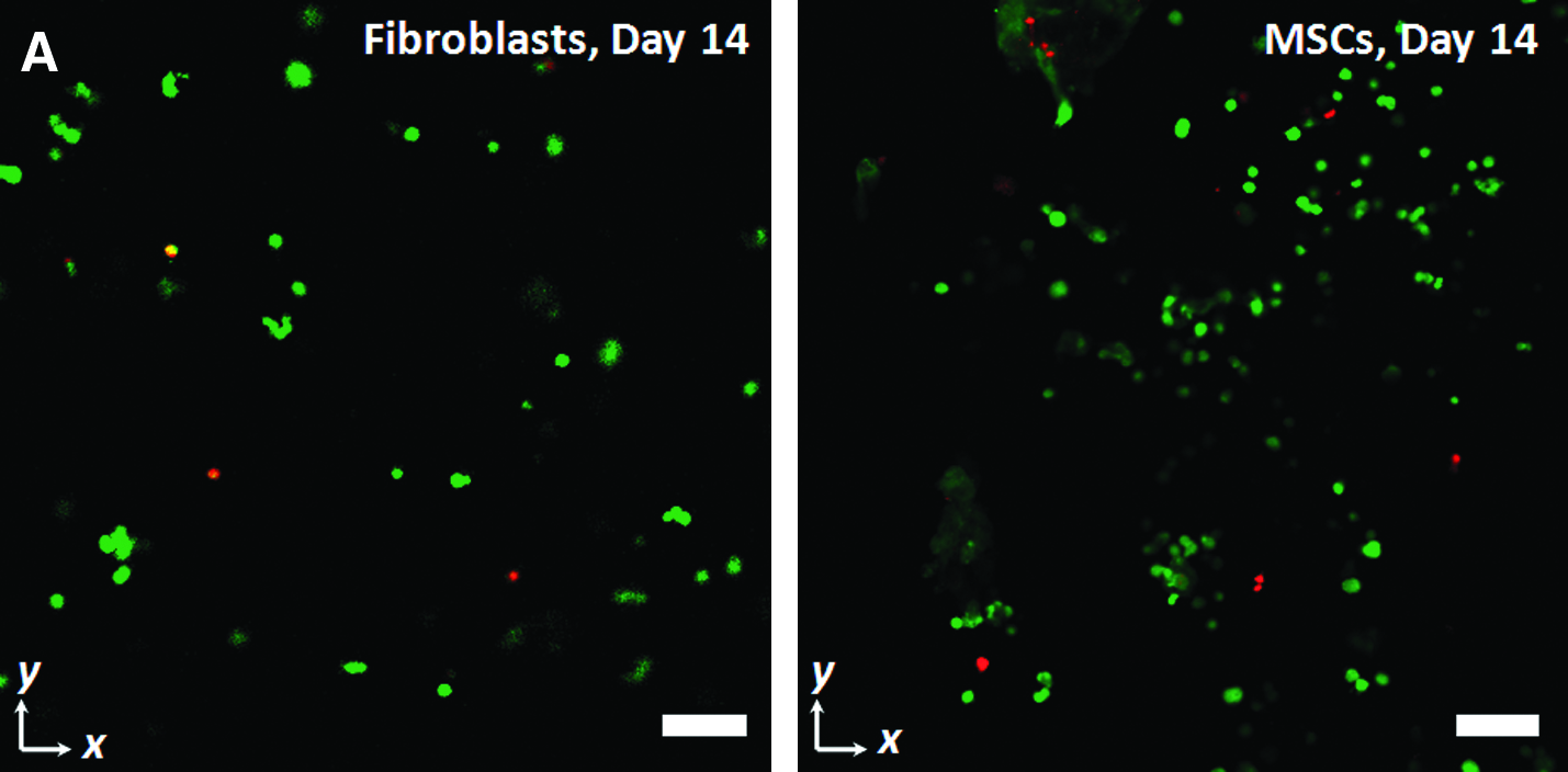
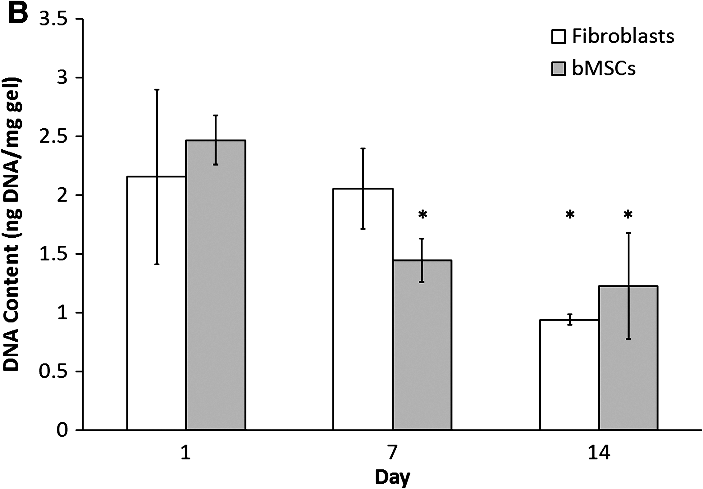
Cell viability was qualitatively and quantitatively assessed for 3 × 5 hydrogel array constructs containing homogenous cell populations (either MSCs or fibroblasts only) after their extraction from microfluidic devices and culture over 2 weeks in their respective medium. LIVE/DEAD assay of intact gels on days 1, 7, and 14 consistently revealed predominately live cells throughout the entire gel thickness when imaged with confocal microscopy (primary bovine tendon/ligament fibroblasts [Fig. 4A, left]; primary bovine MSCs [Fig. 4A, right]). A separate set of samples was analyzed for DNA content as an indicator of cell number over the 2-week culture period (Fig. 4B). Relative to day 1, gels containing fibroblasts exhibited a small yet significant decrease in DNA content at day 14, whereas gels with MSCs showed a slight significant decrease at day 7. No difference was observed between MSCs on day 14 versus day 7.
Primary tendon/ligament fibroblasts and marrow stromal cells remain viable during long-term culture after photopatterning. (
Discussion
This work presents a novel photolithographic technique for spatially controlling hydrogel network formation that facilitates patterning of multiple cell types into 3D hydrogel constructs of >1 mm thick. Shape (Fig. 1) and size (Fig. 2) of hydrogel features within each construct may be tuned and controlled through simple alterations in the photomask and implementation of an N2 atmosphere during the photocross-linking procedure. The success and versatility of this technique in improving patterning fidelity and gel size most likely derives from limiting the presence of oxygen free radicals that hinder the cross-linking reaction by quenching activated photoinitiator or terminating polymer free radicals prematurely.13,22,23 The diffusion of oxygen through the PDMS interface into the cross-linking area could lead to less robust cross-linking; a smaller gel also has an increased surface-area-to-volume ratio, making it more vulnerable to such surface-dependent effects. Purging the unreacted polymer solution with N2 may further improve gelation, but this may also decrease overall cell viability and thus was not explored in this work.
Under reduced oxygen, hydrogels can be consistently photopatterned with this system to thicknesses approaching 2 mm with shape features that accurately reflect the photomask (Figs. 1 and 2). Gel thickness in this environment is thus primarily limited by the concentration and molar absorptivity of the polymer solution, the kinetic efficiency of the free radical initiation and propagation reactions, and the length of the polymer chains and their cross-linkers. 24 Previous efforts demonstrated enhanced patterning fidelity at the microscale by altering the cross-linking chemistry through the use of higher concentrations of photoinitiators, the addition of short cross-linkers, and the use of shorter polymer chains in an effort to induce cross-linking on much shorter timescales for much smaller gels.13,25,26 While each of these enhancements results in improved cross-linking and fidelity of patterned hydrogels, they may be delivered at the expense of cell viability, especially for culturing primary cell types over long periods. Free radical photoinitiators and short cross-linkers are cytotoxic at high concentrations, 27 and the resulting low network mesh size may impose harmful constraints on encapsulated cells due to reduced water content and more limited diffusion of macromolecules. 28 Without altering any of these chemical parameters and instead cross-linking under an N2 atmosphere, we simultaneously avoid these potential detriments and potentially reduce the presence of cytotoxic oxygen free radicals. 29
In addition to patterning of individual gels, this facile photolithographic scheme may be sequentially employed in the generation of multiple laminated, spatially defined hydrogel domains that consistently remain adherent at their interface despite the internal stresses generated while the gels reach equilibrium swelling (Fig. 3A). This serial cross-linking process may be performed multiple times in situ within the same microfluidic device and enables the spatially controlled segregation of multiple cell types in the same laminated hydrogel construct with high fidelity and negligible overlap as demonstrated by confocal microscopy (Fig. 3B–D). Consequently, these templated hydrogel constructs enable tissue-scale coculture between two or more cell types in defined spatial locales and orientations.
Additionally, cell viability for two different types of primary cell isolates (tendon/ligament fibroblasts and MSCs) is largely preserved for at least 2 weeks of culture in the laminated constructs developed in this study (Fig. 4). This phenomenon occurs despite the presence of UV light, the presence of free radicals during cross-linking, and the low oxygen concentration present during cross-linking, all of which could have been potentially harmful to nonimmortalized cell lines. Slight declines were observed in DNA content over time for both cell types after 2 weeks in culture (Fig. 4B), similar to previous observations with cells encapsulated in nonpatterned OPF:PEG-DA gels cross-linked in ambient air. 30 Since remaining cells appeared predominately viable (Fig. 4A), this response may be attributable to the specific cell source studied, the seeding density, or the production of matrix by cells over the culture period that may impair recovery of DNA from the sample.9,30 Future modifications to provide a more optimal microenvironment, such as adhesion or degradation sites, may further enhance cellularity during long-term culture in OPF hydrogels.
Conclusion
In this study, we focused on design, characterization, and preliminary in vitro evaluation of a novel tissue-scale, hydrogel-based scaffold for long-term, 3D coculture of multiple primary cell types with excellent spatial control. Hydrogels were successfully photopatterned into well-defined shapes at 1–2-mm thicknesses using a modified photolithographic process in simple, inexpensive microfluidic devices equilibrated in an N2 atmosphere to enhance cross-linking. Shape fidelity was maintained throughout the entire thickness of the construct, and this system was easily calibrated to allow for the production of hydrogels with tunable sizes and shapes depending on user specifications. Separate hydrogel modules were successfully laminated together with robust, well-defined interfaces, and this process enabled encapsulation and spatially controlled orientation of multiple cell types in monolithic arrays. Cell viability of sensitive primary cell isolates, namely, tendon/ligament fibroblasts and MSCs, was successfully demonstrated for up to 2 weeks in culture in gels photopatterned using this process. The system developed here establishes a proof of concept for examining MSC-based therapies for tendon/ligament tissue regenerative medicine. In the future, this system may be extended to a variety of stem cell types to inform basic science studies of interactions between multiple cell types in stem-cell-mediated healing, as well as to improve design of a wide range of cell-based regenerative medicine therapies.
Footnotes
Acknowledgments
The authors acknowledge funding from the Aircast Foundation and the NIH (1R21EB009153). The authors also thank Nathaniel C. Bloodworth for experimental assistance.
Disclosure Statement
No competing financial interests exist.