
Abstract
Endothelial cells–matrix interactions play an important role in promoting and controlling network formation. In this study, porcine acellular dermal matrix (PADM) was used to guide human umbilical vein endothelial cells (HUVECs) adhesion and proliferation as a potential system for vascularization of engineered tissues. We fabricated PADM using a modified protocol and assessed their composition and ultrastructures. Subsequently, the viability of HUVECs and the formation of capillary-like networks were evaluated by seeding cells directly on PADM scaffolds or PADM digests in vitro. We further investigated the function of the HUVECs seeded on the PADM scaffolds after subcutaneous transplantation in athymic mice. Moreover, the function of the neovessels formed in the PADM scaffolds was assessed by implantation into cutaneous wounds on the backs of mice. The results showed that PADM scaffolds significantly increased proliferation of HUVECs, and the PADM digest induced HUVECs formed many tube-like structures. Moreover, HUVECs seeded on the PADM scaffolds formed numerous capillary-like networks and some perfused vascular structures after implantation into mice. PADM seeded with HUVECs and fibroblasts were also able to form many capillary-like networks in vitro. Further, these neovessels could inosculate with the murine vasculature after implantation into cutaneous wounds in mice. The advantage of this method is that the decellularized matrix not only provides signals to maintain the viability of endothelial cells but also serves as the template structure for regenerated tissue. These findings indicate that PADM seeded with HUVECs may be a potential system for successful engineering of large, thick, and complex tissues.
Introduction
A potential strategy for circumventing this problem is in vitro prevascularization that can anastomose spontaneously to the ingrowing vasculature of the host after implantation, resulting in the relatively quick formation of a vasculature in the implant. 6 This strategy is based on the observation that endothelial cells are able to form prevascular structures when they are cultured under the right conditions and environment in vitro.4,5 Although this approach has shown promise,2,7 endothelial cells in the scaffold do not survive very well and are more prone to apoptosis. 8 Various angiogenic growth factors such as vascular endothelial growth factor (VEGF) and basic fibroblast growth factor (bFGF)9,10 or genetic manipulation11,12 were employed to facilitate endothelial cell survival in the scaffold and formation of blood vessels in vitro. The delivery of these growth factors generally results in increased angiogenesis, but the resulting vessels are often disorganized, leaky, and hemorrhagic. 13 Further, genetic manipulations may be potentially oncogenic. 14 It is well known that interactions between endothelial cells and their surrounding extracellular matrix (ECM) play a crucial role in modulation of the angiogenic process.15–17 Therefore, culturing endothelial cells in appropriate three-dimensional scaffolds are believed to be the basis for decisive and successful vascularization. 18
Biological scaffolds derived from decellularized tissues have been successfully used in a variety of tissue engineering applications due to their composition and intrinsic growth factors.19–22 These scaffolds are all composed of extracellular matrices but differ in their tissue source, species of origin, and methods by which they are processed. Previous studies have shown that decellularized matrices provide a compatible environment for human endothelial cell attachment, proliferation, and three-dimensional growth into tube-like vascular structures.23–25 However, these studies mainly focused on the small intestinal submucosa (SIS) and urinary bladder matrix. The acellular dermal matrix (ADM) derived from the skin has become widely used in plastic surgery. Moreover, Gilbert et al. had quantified the DNA content in several types of ECM scaffold materials and showed that the level of residual DNA in porcine ADM (PADM) was lower than that in several commercially available and laboratory produced procine SIS and urinary bladder matrix. 26 Although there are no regulations on the limits of DNA content in biological scaffold materials, previous studies demonstrated that porcine DNA remnants might cause the inflammatory reactions after the implantation of porcine-derived scaffolds for orthopedic applications. 27 Recently, the Alt group 28 and our lab (data not shown) found that adipose-derived stem cells seeded in ADM spontaneously differentiated into vascular endothelial cells and formed vascular structures after engraftment. These findings indicated that the ADM microenvironment may contribute to vascular formation.
In the present study, we fabricated PADM by a modified decellularization protocol and evaluated the viability of human umbilical vein endothelial cells (HUVECs) cultured both in PADM scaffolds and in a PADM digestion product solution in vitro. We also assessed the function of HUVECs seeded on the PADM after subcutaneous transplantation in athymic mice. Subsequently, we investigated the feasibility of vascular structure formation when HUVECs and human fibroblasts were co-cultured on the PADM scaffolds and in the PADM digest in vitro. We further evaluated whether such vascular structures can easily inosculate with the recipient host circulation by using a full-thickness cutaneous wound model. Thus, our study provides important information in preparing PADM scaffolds for the in vitro formation of a prevascular network to improve the vascularization of engineered tissues.
Materials and Methods
Preparation of PADM
PADM was developed from the porcine skin as previously described.29,30 Fresh porcine skin was obtained from a local slaughterhouse. After complete cleaning, removal of hair, excision of the subdermal fat tissue, and the epidermis, the resulting skin was cut into pieces with the dimensions 10 × 5 × 0.4 cm3. The dermal portion was treated with a 0.25% trypsin solution at 37°C for 1 h and then extensively washed with distilled water. Subsequently, the dermal matrix was incubated in a 1 M sodium hydroxide (NaOH) solution at room temperature for 16 h and thoroughly rinsed in phosphate-buffered saline (PBS) at room temperature with continuous shaking until the pH value of the PBS became neutral. Finally, the PADM was lyophilized, sterilized by 60CO irradiation, and stored at 4°C for future use.
Preparation of PADM degradation product
PADM sheets were comminuted into a particulate form using a macrohomogenizer in liquid nitrogen. Particulate PADM was added to 1 mg/mL pepsin (Sigma) in 0.01 N HCl to produce a suspension at a final concentration of 10 mg PADM/mL. 31 The suspension was mixed on a stir plate at room temperature for 48 h, at which time no visible pieces of ECM remained. This solution was neutralized to a pH of 7.4 by addition of 0.1 N NaOH and 10 × PBS pH 7.4 at 4°C, and then assayed for cell proliferation. The solution was brought to the desired concentration using cold culture medium without growth factors. Pepsin control samples were prepared by mixing the pepsin digestion solution (1 mg/mL pepsin in 0.01 N HCl) at room temperature for 48 h, neutralized, and used as a control in the proliferation assays.
Evaluation of structure and composition of PADM
Morphology examinations
For histological analysis, prepared PADM was fixed in 10% phosphate-buffered formalin. The fixed samples were embedded in paraffin and sectioned at a thickness of 5 μm and then stained with hematoxylin and eosin (H&E). Sections of the test samples were also stained with Masson's trichrome. In this staining process, collagen fibers were stained green, elastin fibers were stained black, and glycosaminoglycans were stained red.
PADM were fixed with 0.25% glutaraldehyde solution for scanning electron microscopy (SEM) and transmission electron microscopy (TEM) examinations,29,32 which were carried out in the Central Laboratory of Fourth Military Medical University. For SEM analysis, PADM samples were loaded onto aluminum studs and coated with gold for 3 min at 8 mA under a pressure of 0.1 Torr. Collagen morphologies were examined under a scanning electron microscope (Hitachi model S-3400N). For TEM examinations, a transmission electron microscope (Hitachi model H-600) was used to evaluate the characteristics of the sequences of black and white bands of collagen fibers of the samples.
Immunostaining
To investigate the endogenous growth factors within PADM, immunohistochemistry stainings were performed using the streptavidin-biotin complex kit (DAKO). Briefly, sections of test samples were pretreated with 3% H2O2 for 15 min at room temperature. To block nonspecific binding sites, the sections were incubated in 3% (w/v) bovine serum albumin/PBS (pH 7.4) for 30 min. Subsequently, the primary antibodies, such as rabbit anti-VEGF (1:500; Abcam), rabbit anti-bFGF (1:500; Abcam), and rabbit anti-transforming growth factor beta 1 (TGF-β1, 1:200; Santa Cruz Biotechnology), were added and incubated overnight at 4°C. After washing with PBS, the secondary antibody (1:1000; DAKO) was added and incubated for 1 h at room temperature, followed again by washing with PBS. The samples were then incubated with diaminobenzidine peroxide substrate until the desired color developed, followed by a light hematoxylin counterstain.
Electrophoresis and western blot analysis
The proteins of each degraded PADM sample were loaded into a lane of a 12.5% polyacrylamide gel, with a prestained protein standard (Bio-Rad) in the range 10–170 kDa. The gel was run at a voltage of 80 V until the dye front entered the separating gel, then at a constant voltage of 100 V until the dye front reached the bottom of the gel. Proteins were next transferred onto polyvinylidene difluoride membranes and incubated overnight with primary antibodies. After three washes with PBS/Tween 20, the membranes were incubated with HRP-labeled antibodies. The primary antibodies used were rabbit anti-VEGF (1:200), rabbit anti-bFGF (1:200), and rabbit anti-TGFβ1 (1:500). The secondary antibodies used were HRP-anti-rabbit (1:500; Zhongshan). The signals were developed using chemiluminescence detection reagents and finally scanned using an imaging system (Peiqing).
Assessment of in vivo biocompatibility of PADM
All animal experiments were conducted in accordance with the committee guidelines of the Fourth Military Medical University for animal experiments, which also met the National Institutes of Health (NIH) guidelines for the care and use of laboratory animals. Sprague-Dawley rats weighing 150–200 g and athymic mice aged 6–8 weeks were obtained from the Experimental Animal Center of Fourth Military Medical University and maintained on daily feed of Purina rodent chow in housing quarters with cycled light (12 h on/off), regulated temperature, and sterile water.
The Sprague-Dawley rats were anesthetized with pentobarbital. After shaving the hair and sterilizing the skin with baticone alcoholic solution, a 10 × 10 mm piece of PADM was transplanted into the dorsum of the rats. Every week after the operation, the skin surrounding and including the area of transplantation was excised for H&E examination.
Cell isolation and culture
Primary fibroblasts were isolated from the newborn foreskin as previously described 33 and cultured in Dulbecco's modified Eagle's medium (DMEM) with 10% fetal bovine serum (FBS), 100 units/mL penicillin, and 100 μg/mL streptomycin. Culture medium was changed every 3 days. When cells reached 90% confluence, they were passaged in a ratio of 1:3 and used at passages three to eight.
HUVECs were harvested from human umbilical cord veins as previously described. 34 Umbilical cords were obtained from the Department of Gynaecology and Obstetrics of the Xijing Hospital. Informed consent was obtained from and signed by all patients. Briefly, the umbilical veins were cannulated at both ends and washed with PBS solution. A collagenase solution (300 U/mL; Boehringer Mannheim) was then injected to rinse and fill the vein, and the cord was placed in PBS at 37°C. After a 15 min incubation, the veins were perfused with M199 medium (Gibco) containing 10% FBS and antibiotics. The endothelial cells were isolated by centrifugation and suspension in Endothelial Cell Medium supplemented with 5% FBS and 1% endothelial cell growth supplement (all from Sciencell). The culture medium was changed thrice a week. Endothelial cells were characterized by assessing von Willebrand factors and Weibel-Palade bodies (data not shown). The cells were used between passages two and five.
Evaluation of HUVECs growth on the PADM
Cell proliferation assays
Direct method
PADM were cut into small pieces with a diameter of 6 mm, placed in 96-well plates, and rehydrated in DMEM medium for 24 h at 37°C. After the DMEM was removed, cells were seeded at a concentration of 1 × 10⁴ cells per well. Cells cultured in the 96-well plate were the control. Cell proliferation was assessed using methyltetrazolium.35,36 Briefly, 20 μL of methyltetrazolium solution (5 mg/mL in PBS) was added to each well, and the cells were incubated at 37°C for 4 h. Then, the medium was removed, and 150 μL of dimethyl sulfoxide was added to dissolve the blue crystals that formed in the cells. After shaking for 15 min, the 100 μL solution was transferred to a new 96-well plate. The optical density values were determined using a multi-plate reader (BIO-TEK) at a wavelength of 570 nm.
Digestion method
Responses of HUVECs and fibroblasts to PADM degradation products were quantitatively determined by cell counting. Cells were plated in media with no added growth factors containing 1% FBS at 1 × 10⁴ cells per well in a 24-well plate. Twenty-four hours later, media were removed, the cells were washed, and the media were replenished. At the time of media replenishment, either ECM degradation product or an identical volume of control buffer was added at varying doses to each well. Three days after addition of the samples, cells in senven of the wells for control and treatment group were imaged, trypsinized, and counted using a hemocytometer.
Cell function assessment
PADM were cut into pieces with the dimensions 10 × 10 mm and soaked in DMEM medium for 24 h at 37°C. After the medium was removed, HUVECs or fibroblasts were pipetted onto the specimens at a density of 5 × 10⁵ cells per cm2. The medium was changed every day for 3 days. All grafts were subcutaneously transplanted, excluding those that were examined by H&E and SEM on days 1, 2, and 3.
Co-culture HUVECs and fibroblasts on PADM scaffolds
Direct method
PADM were made into small pieces with a diameter of 16 mm and rehydrated with DMEM medium. HUVECs (5 × 10⁵) and fibroblasts (5 × 10⁵) were seeded onto every PADM sample with 100 μL endothelial cell medium. After incubation for 3 h to allow for cell attachment, more medium was added. After 3 days of culture, some grafts were transplanted to the full-thickness cutaneous wound in mice. Others were examined by H&E and immunohistochemistry. Immunohistochemistry stainings were performed on 5 μm paraffin sections using polyclonal PECAM-1 antibodies.
Digestion method
HUVECs cultures in gels were prepared as previously described.8,12 A measured amount of PADM degradation products with 1/10 volume of 10 × DMEM and 1/9 volume of FBS was neutralized with sterile 0.1 M NaOH and kept on ice. Equivalent numbers of cultured HUVECs and fibroblasts were added to the degradation suspension to a final concentration of 1 × 10⁶ total cells/mL solution. One milliliter of the cell/degradation suspension was added to the wells of a 24-well plate and warmed to 37°C for 30 min to allow polymerization of the collagen. After gel formation, 1 mL of endothelial cell medium was added per well. Cultures were fed every day. After 3 days of culture, the constructs were fixed with 4% paraformaldehyde and paraffin embedded for staining with H&E and immunohistochemical staining of platelet endothelial cell adhesion molecule (PECAM-1, 1:50; Neomarkers).
Animal studies
Subcutaneous transplantation
Nude mice were used for the subcutaneous transplantation experiment. After the nude mice were anesthetized with pentobarbital, a 1.5 cm incision was made laterally on the abdomen. A subcutaneous pocket was created by blunt dissection into which the HUVECs-PADM or fibroblasts-PADM construct was then placed. The mouse skin was discontinuously sutured using 3-0 silk sutures. At 3 weeks after the operation, the implanted grafts were retrieved, embedded in paraffin, and sectioned at a thickness of 5 μm. After removing the paraffin, some sections were stained with H&E whereas the others were prepared for immunohistochemistry to identify the newly formed blood vessels. Immunohistochemical stainings were performed using the streptavidin-biotin complex kit (DAKO), and the antibodies used were anti-human PECAM-1.
Full-thickness cutaneous wound model
Each mouse was anesthetized, and a full-thickness wound (1.5 cm in diameter) was created on the dorsum. The grafts were transplanted on the wound, and the wound was then covered by two layers of vaseline gauze and ethanol gauze with discontinuous sutures in the margins of the defect area by a 3-0 silk suture. In some experiments, 40 to 60 min before sacrificing a mouse, 150 μL of a rhodamine-UEA-I conjugate (Vector Laboratories) was administered via tail vein injection. Three weeks after the operation, the mice were sacrificed and the grafts were harvested. The excised tissues were frozen on dry ice and mounted using optimum cutting temperature (OCT) compound. Some of the tissue specimens were transversely sectioned and examined at 10 × magnification using fluorescent microscopy. The others were fixed in neutral buffered formalin and prepared by standard methods for H&E and immunohistochemical stainings.
Statistical analysis
Experiments were conducted at least twice to ensure validity of the results, and the data shown are from one experiments yielding similar results to the replicate experiments. For any given experiment, each data point represents the mean ± standard deviation. Analysis was performed using the Statistical Program for Social Science (SPSS) for Windows. Analysis of variance followed by the t test was used to determine the significant differences among the groups (p-values < 0.05 were considered significant).
Results
Structure and composition of PADM
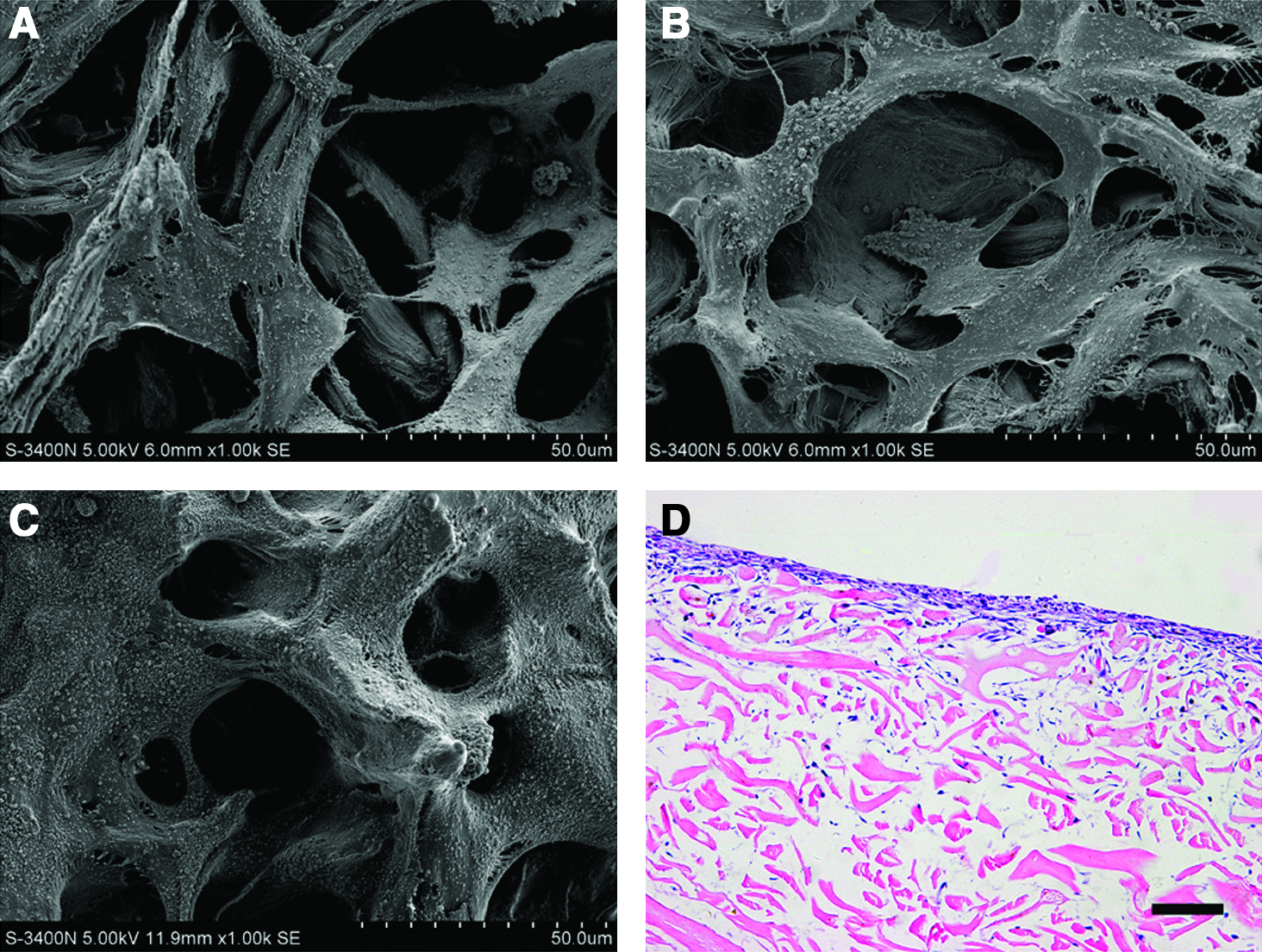
The structure of PADM was evaluated using H&E staining, SEM, and TEM. Analysis of the H&E staining showed that there were no cellular components, cellular debris, or blood vessels in the dermis (Fig. 1A, B). These results were also confirmed by SEM analysis. Further, the collagen fiber bundles, which were arranged in sequential black and white bands (Fig. 1F), became extensively loose after cell extraction (Fig. 1D, E). Masson's trichrome staining was used to detect the preserved ECM after decellularization. The results revealed that PADM not only contained collagen fibrils but also contained elastin fibrils and glycosaminoglycans (Fig. 1C). sodium dodecyl sulfate-polyacrylamide gel electrophoresis also demonstrated that the PADM contained some low-molecular-weight proteins (Fig. 1I) which may be growth factors. Therefore, VEGF, bFGF, and TGF-β1 antibodies are used to identify the growth factors in the PADM using the immunohistochemical analysis. PADM did not stain for VEGF; however, the acellular matrices were almost entirely stained by bFGF antibodies (Fig. 1G), whereas TGF-β1 antibodies also stained positively but was less immunoreactive (Fig. 1H). By western blotting, bFGF but not VEGF or TGF-β1 were positive (Fig. 1J).

The structure and compositions of PADM after decellularization processing. Hematoxylin and eosin staining of PADM
In vivo biocompatibility of PADM
H&E staining revealed that the PADM had been repopulated with rat cells with no evidence of severe acute inflammatory response by the host animal (Fig. 2A–D). After the third week following surgery, some vascular structures were formed at the edge of the PADM scaffolds (Fig. 2C). Moreover, very few capillary networks were sporadically implanted into the middle of PADM at 4 weeks (Fig. 2D).

Histological micrograph of in vivo biocompatibility of PADM at 1
Viability of HUVECs growth on the PADM scaffolds or into the PADM digests
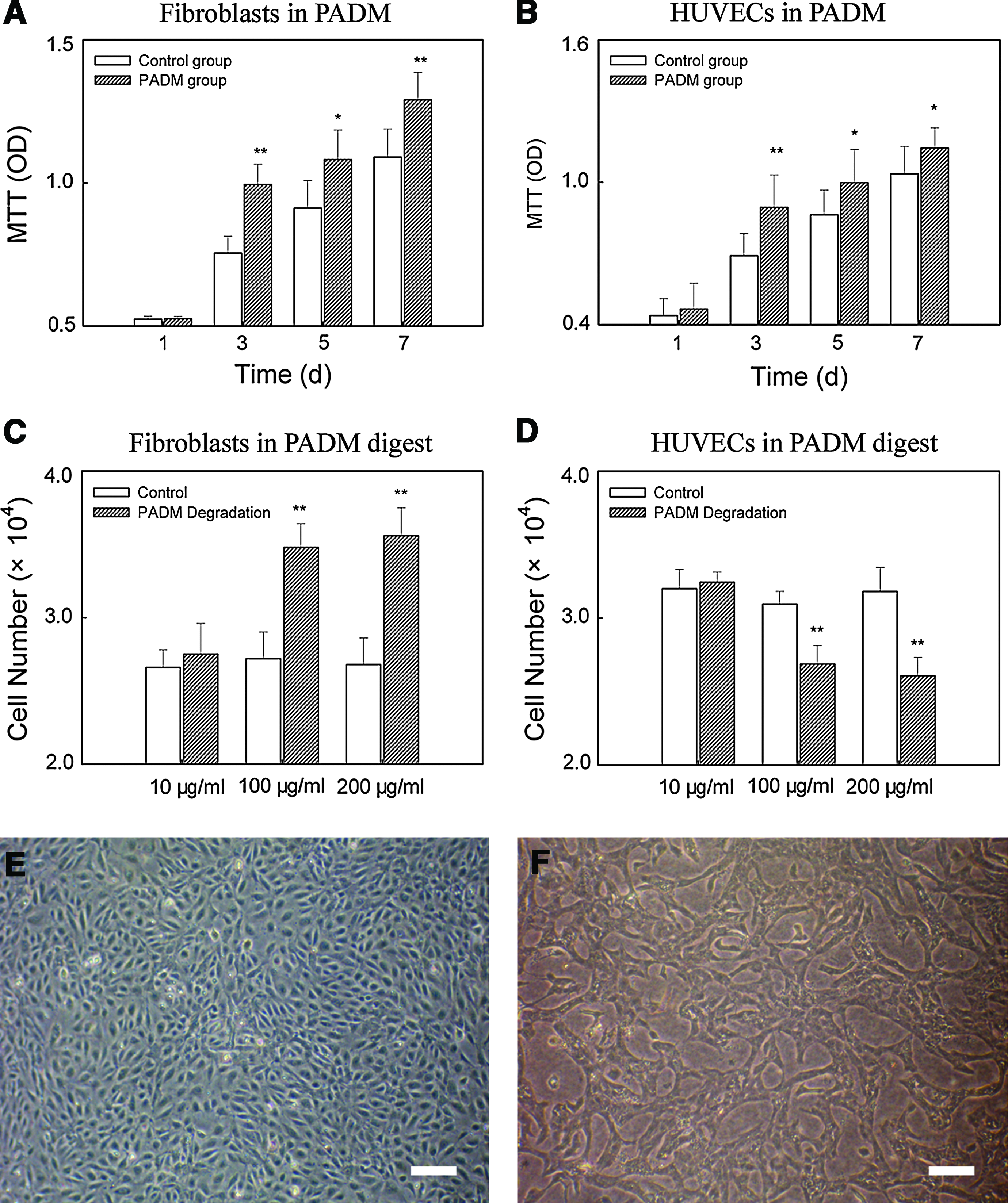
The survival of HUVECs on the PADM was assessed by directly culturing them on the surface of PADM scaffolds or into the PADM digestion product solution and allowing them to grow for various lengths of time. Fibroblasts were also cultured in these two ways as a functional comparison with the HUVECs. The growth of the HUVECs cultured directly on the PADM was significantly better than that on the culture plate only from day 3 to day 7 (p < 0.01). The proliferation rate of the HUVECs on the PADM was about twice that on the plate after a 3-day culture (Fig. 3B). In a pattern similar to that observed for HUVECs, the fibroblasts cultured on the PADM were faster than that on the culture plate (Fig. 3A). Morphologically, no significant morphological changes were observed in both cell types in contact with PADM for all studied time periods. The HUVECs had infiltrated into the PADM after 1 day of culture in vitro (Fig. 4A). After a 3 day culture, there were many HUVECs present within the PADM (Fig. 4B, D), and some tube-like structures had formed in the PADM (Fig. 4C). However, the fibroblasts were chiefly distributed on the upper surface of the PADM (Fig. 5A, B) and formed in layers on the scaffolds after 72 h of culture (Fig. 5C, D), and few cells had infiltrated the interior of the PADM (Fig. 5D).
The viability of cells growth on the PADM scaffolds or into the PADM digests.
The morphology of HUVECs cultured on the PADM for 24
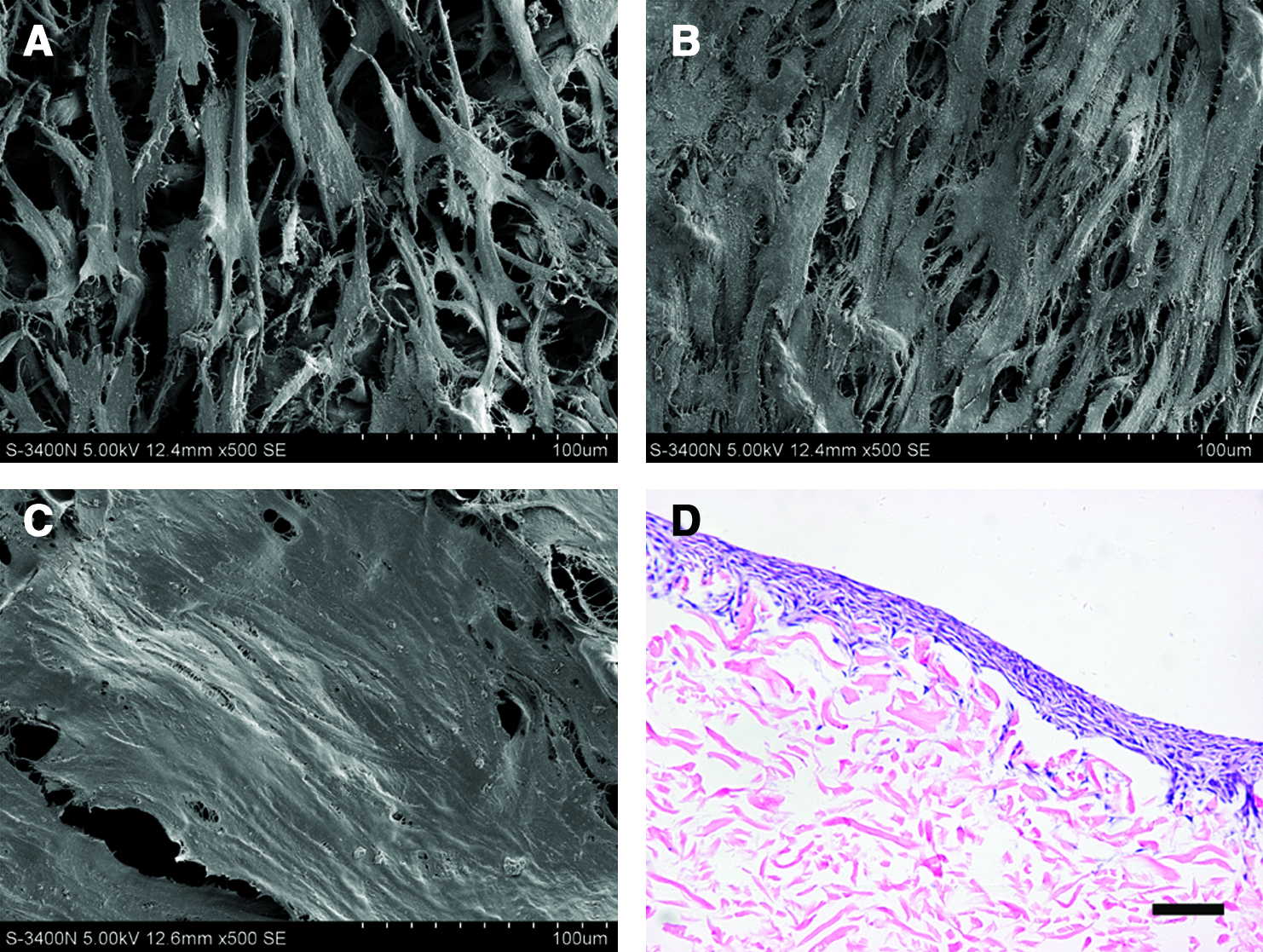
The morphology of human fibroblasts cultured on the PADM for 24
When cultured in the PADM digestion product at the concentrations of 100 and 200 μg/mL, the number of fibroblasts significantly increased. The lower concentration of PADM digest (10 μg/mL) had no effect on cell proliferation (Fig. 3C). Further, the fibroblasts still retained their normal spindle morphology in the PADM digest solution (data not shown). However, the proliferation of HUVECs with the PADM digest decreased compared with that with a pepsin digest control. The number of HUVECs was (1.686 ± 0.127) × 104 at the concentration of 100 μg/mL and (1.607 ± 0.118) × 104 at the concentration of 200 μg/mL, and the percent decrease from control was 24% and 36%, respectively (p ≤ 0.05) (Fig. 3D). However, compared with the initial seeding density (1.0 × 10⁴ cells), the number of HUVECs cultured in PADM digest was enhanced. Interestingly, HUVECs formed similar tube-like structures after 1 day of culture in the 100 and 200 μg/mL PADM digests (Fig. 3F). However, HUVECs exhibited a typical cobblestone appearance, characteristic of endothelial cells when they were cultured in the 10 μg/mL PADM digest and similar to the morphology of endothelial cells in the control digest buffer (Fig. 3E).
Co-culture of HUVECs and fibroblasts on the PADM scaffolds or in the PADM digest in vitro
To evaluate whether HUVECs were able to form capillary-like networks in the PADM scaffolds without the addition of growth factors, HUVECs and fibroblasts were co-cultured onto the surface of the scaffolds or into the PADM digestion product. As shown in Figure 6, many capillary-like networks formed in the PADM after 3 days of culture. These neovessel constructs were chiefly distributed on the upper part of the PADM scaffolds (Fig. 6A). The capillary-like networks were further identified with a PECAM-1 immunohistochemical stain (Fig. 6B). Some capillary-like networks were also formed in the PADM gel when HUVECs and fibroblasts were co-cultured into PADM digest products (Fig. 6C). Moreover, these neovessel constructs were evenly distributed in the PADM gel and stained positive for PECAM-1 (Fig. 6D).
The formation of capillary-like networks in the PADM scaffolds
Subcutaneous transplantation of PADM seeded with HUVECs
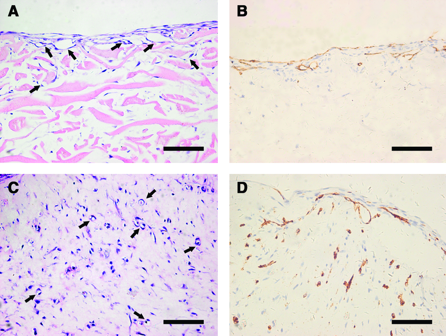
To test the function of the cells on the PADM, PADM seeded with HUVECs or with fibroblasts were subcutaneously transplanted in nude mice. Three weeks after the transplantation, the PADM that were seeded with fibroblasts retained their original structure (Fig. 7A), and few capillary-like networks sporadically formed at the edge of the PADM during this period (Fig. 7B). However, the morphology of the PADM seeded with HUVECs was dramatically modified with the formation of many capillary-like structures and some perfused vascular structures (Fig. 7C). Moreover, all of these structures were reactive to PECAM-1 specific antibodies (Fig. 7D).

Histological micrograph of PADM seeded with human fibroblasts
Cutaneous wound transplantation of PADM seeded with HUVECs and fibroblasts
To investigate whether PADM seeded with HUVECs and fibroblasts were able to form functional microvessels, PADM grafts were implanted into cutaneous wounds on the backs of mice. Histological analysis of grafts harvested 3 weeks after implantation revealed that the grafts were epithelialized and contained numerous blood vessels (Fig. 8A, B). Moreover, some lumena of the neovessels contained erythrocytes (Fig. 8A). The neovessels were further confirmed by adherence of intravenously injected rhodamine-labeled UEA-1 (Fig. 8C). These results demonstrated that the microvessels formed in the PADM could inosculate with the murine vasculature.
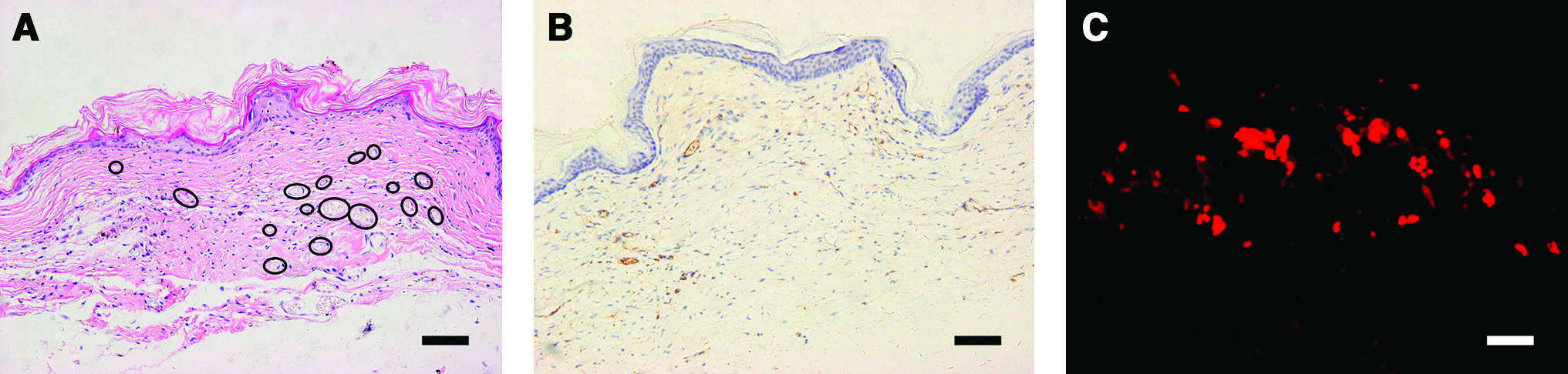
Histological micrograph of PADM scaffolds cocultured with HUVECs and human firoblasts after 3 weeks of cutaneous wound implantation on the backs of mice. Hematoxylin and eosin staining showed that the grafts contained numerous perfused vascular structures
Discussion
Decellularization of tissue has been shown to be an attractive method of obtaining scaffolds for tissue engineering. Removal of cells and cellular components from tissues or organs leaves the natural ECM,37,38 which can provide an analogous in vivo microenvironment for adherence, growth, and differentiation of cells in vitro. Moreover, biological scaffolds composed of ECM have been shown to degrade rapidly after implantation, whereas the ECM remodeling continues for days to weeks after the scaffold degradation is complete. 39 In particular, previous studies have shown that endogenous growth factors such as VEGF, bFGF, and TGF-β remain in naturally derived scaffolds after the decellularization process and terminal sterilization.40–43 These growth factors can promote cellular differentiation and angiogenesis during the remodeling process.
The most effective decellularization protocols include a combination of physical, chemical, and enzymatic approaches. 37 Each of these treatments affect the composition and ultrastructure of the ECM. An optimal decellularization process efficiently removes cells and cellular components while minimizing any adverse effect on the composition, biological activity, and mechanical integrity of the remaining ECM. In our current study, we used a modified decellularization protocol incorporating 0.25% trypsin and 1 M NaOH to prepare PADM scaffolds, and the resulting scaffolds not only retained collagen fibers and elastin fibers but also contained endogenous growth factor bFGF, which is a powerful stimulator of angiogenesis in vivo and a pleiotropic regulator of the proliferation, migration, differentiation, and survival of many cell types, including endothelial cells, in vitro. 44 The results were consistent with previous studies which showed that endogenous growth factors such as bFGF remaining on biological scaffolds are derived from SIS. 43 These growth factors are released during scaffold degradation and exert their biological effects when they are dissociated from their binding proteins and activated. 40 Therefore, the advantage of using PADM scaffolds, rather than exogenously added growth factors, may be attributable to direct angiogenic activity exerted by the scaffolds themselves. The present study showed that PADM scaffolds supported attachment and increased proliferation of HUVECs. HUVECs-PADM constructs transplanted into the subcutaneous tissue of the nude mice showed that many capillary-like networks were formed and some perfused vascular structures were presented in the PADM scaffolds 3 weeks after implantation. This result was in good agreement with the results presented by Schechner et al., who subcutaneously implanted immunodeficient mice with the human ADM seeded with HUVECs that had been transduced with the survival gene Bcl-2. 7 Their studies focused on comparing endothelial cell survival and vascular formation in the human ADM scaffolds with cells transduced with two different genes, but they neglected the effect of the scaffold materials on the survival of the endothelial cells. It is well known that interactions between endothelial cells and their surrounding ECM play a crucial role in modulation of the angiogenic process.15–17 Our study emphasized the effect of the scaffolds themselves on endothelial cell growth, and the results demonstrated that HUVECs without genetic manipulation were able to form vasculature in the ADM scaffold materials. The extent to which the present tube-like structures are due to soluble growth factors cannot be determined. However, the PADM scaffolds supported HUVECs forming tube-like structures in vitro and in vivo without addition of exogenous growth factors.
Biological scaffolds have been shown to degrade rapidly after implantation, 39 but the specific mechanisms of scaffold degradation in vivo remain unknown. The well-characterized enzyme pepsin was selected to digest the PADM to assess the effect of the degradation products of the PADM scaffolds on the endothelial cells. 31 Our studies showed that the in vitro degradation products of PADM scaffolds can also induce HUVECs to form tube-like structures. Moreover, many capillary-like networks were formed when HUVECs and fibroblasts were co-cultured in the PADM degradation products. It is unknown whether the products in the artificial degradation process used in the present study were of the same biochemical composition as those formed during an in vivo degradation process. However, our results, to some extent, demonstrated that the degradation products of the PADM scaffolds were nontoxic to endothelial cells and promoted angiogenesis, unlike the acidic degradation products of the poly(lactic-co-glycolic acid) (PLGA) scaffolds, which may negatively affect the viability of cells near and within the implant in vivo. 45
By implanting PADM grafts into cutaneous wounds in mice, we also showed that the microvessels formed in the PADM could inosculate with the murine vasculature as indicated by detection of erythrocytes in the luminal vascular structures and adherence of intravenously injected labeled UEA-1. That is, the HUVECs could form functional microvessels in the PADM scaffolds. These findings indicate that PADM may provide an appropriate microenvironment for endothelial cells survival, and seeding with endothelial cells may allow ideal vascularization of the scaffold for successful engineering of large, thick, and complex tissues.
However, the PADM scaffold is a three-dimensional sheet that limits its used in other forms of tissue-engineering products. Our previous study showed that PADM can be micronized to particle form, which can be used as microcarriers to expand and deliver human fibroblasts. 46 Further, these microcarriers retained the ultrastructural characteristics of the parent ECM sheet. This study has demonstrated that PADM can support endothelial cells attachment, proliferation, and growth into vascular structures. Therefore, to vascularize large, thick, and complex tissues, endothelial cells can be cultured and form capillary-like networks on PADM microcarriers first. Subsequently, these vascular cell-microcarriers are incorporated into the engineered tissue within an appropriate time to pre-vascularize it. Previous studies also showed that PADM microcarriers will degrade quickly, and the remaining empty spaces will be left for endothelial cells to assemble into blood vessels.39,46 We have great interest in developing this application in the future, as it may be a promising strategy to vascularize various tissue-engineered products such as liver, kidney, and cardiac muscle.
Conclusions
In this study, PADM fabricated with a trypsin-NaOH decellularization method was evaluated as a scaffold to culture HUVECs for the potential application of in vitro vascularization. The PADM supported adhesion and increased proliferation of HUVECs. Further, HUVECs-PADM constructs could form many tube-like structures in vitro and capillary-like networks as well as some perfused vascular structures in vivo. The degradation products of the PADM scaffolds also induced formation of tube-like structures by the HUVECs and supported HUVECs and fibroblasts in forming many capillary-like networks in vitro. In addition, PADM seeded with both HUVECs and fibroblasts were able to form numerous capillary-like networks in vitro. Further, these neovessels could inosculate with the murine vasculature after implantation into cutaneous wounds in mice. These findings indicate that PADM seeded with HUVECs may be a potential system for successful engineering of highly complex tissue.
Footnotes
Acknowledgments
This study was supported in part by the funding support from National High Technology Research and Development Program of China (2006AA02A119) and the Nature Science Foundation of China (60901038).
Disclosure Statement
No competing financial interests exist.