
Abstract
Scaffolds derived from natural polysaccharides are very promising in tissue engineering applications and regenerative medicine, as they resemble glycosaminoglycans in the extracellular matrix (ECM). In this study, we have prepared freeze-dried composite scaffolds of chitosan (CHT) and hyaluronic acid (HA) in different weight ratios containing either no HA (control) or 1%, 5%, or 10% of HA. We hypothesized that HA could enhance structural and biological properties of CHT scaffolds. To test this hypothesis, physicochemical and biological properties of CHT/HA scaffolds were evaluated. Scanning electron microscopy micrographs, mechanical properties, swelling tests, enzymatic degradation, and Fourier transform infrared (FTIR) chemical maps were performed. To test the ability of the CHT/HA scaffolds to support chondrocyte adhesion and proliferation, live–dead and MTT assays were performed. Results showed that CHT/HA composite scaffolds are noncytotoxic and promote cell adhesion. ECM formation was further evaluated with safranin-O and alcian blue staining methods, and glycosaminoglycan and DNA quantifications were performed. The incorporation of HA enhanced cartilage ECM production. CHT/5HA had a better pore network configuration and exhibited enhanced ECM cartilage formation. On the basis of our results, we believe that CHT/HA composite matrixes have potential use in cartilage repair.
Introduction
The main challenge for cartilage tissue engineering is the chondrocytes massive transition into fibroblastic cells during the in vitro culture process. Those dedifferentiated cells form fibrocartilage instead of hyaline cartilage. 5 Additionally, chondrocytes availability is limited because of the size of biopsies that can be collected from the patient's own tissue, without causing the risk of morbidity of the site of explant. Moreover, the development of biodegradable polymers to perform the role of a temporary matrix is an important factor in the success of cell transplantation.6,7
Extracellular matrices (ECMs) provide a microenvironment for cells to maintain homeostasis and to retain the required differentiated state for specific tissues. 8 Among the many different ECM molecules, hyaluronic acid (HA) is the main glycosaminoglycan (GAG) in the mesenchyme during the early stage of chondrogenesis.9,10 In addition, HA is known to influence chondrocytes by triggering a sophisticated signaling pathway leading to enhancement of cellular functions.11,12 However, the concentration of HA must be confined to a relatively low amount, as concentrations may reduce cell adhesion because of its negative charge. 13 This disadvantage can be overcome by combining HA with positively charged polycations such as chitosan (CHT). 14 CHT, a partially deacetylated derivative from chitin, has the ability to interact with negatively charged molecules. 15 Because of its biocompatibility and biodegradability, CHT has been widely applied in tissue engineering strategies.16–19 Additionally, CHT is structurally similar to various GAGs found in articular cartilage. 20 Combining the advantages of both polysaccharides, CHT/HA scaffolds can be suitable candidates for cartilage tissue regeneration. The blend would be stabilized by an ionic interaction between the positively charged CHT and the negatively charged HA.
CHT has been already conjugated with HA to obtain a biomimetic matrix for chondrocytes.16,21–24 Yamane et al. 22 developed CHT-based HA polymer fibers by the wetspinning method. In the hybrid fibers, chondrocyte adhesion, proliferation, and also the synthesis of aggrecan and type II collagen were significantly higher when compared with CHT fibers. Hsu et al. 24 studied CHT–alginate–hyaluronan scaffolds, with or without covalent attachment, with RGD-containing protein. The cell-seeded scaffolds showed neocartilage formation in vitro in the presence or absence of RGD. Tan et al. 21 demonstrated that CHT-HA hydrogels allowed cell survivor, and cells retained the chondrogenic morphology.
The reported studies are focused on the superior biological effects that HA could provide, and its incorporation has not been studied in terms of physicochemical effects. Additionally, to our knowledge, the effect of varying the ratio between CHT and HA, and consequently, its influence in terms of both physicochemical and biological properties, has not been yet reported. On the other hand, because of HA high water uptake and the lack of mechanical strength provided by natural polymers, the existing strategies combining CHT and HA for cartilage tissue engineering are very limited and usually consist in hydrogels.21,25 However, these hydrogels do not have the required mechanical strength to maintain the initial shape of the implanted scaffold. Consequently, they cannot be transplanted into large cartilaginous lesions in advanced degenerative diseases, such as osteoarthritis and rheumatoid arthritis. Moreover, the existing CHT/HA blends are usually used to produce scaffolds by techniques more complex than the freeze-drying method, in which further research is needed to determine the adequate shape, pore size, and mechanical properties of a three-dimensional fabrication for cartilage TE.
The aim of this work was to prepare a new class of hybrid scaffolds composed of CHT as a framework containing low concentrations of HA to mimic cartilage ECM composition. The application of HA as a component of the cartilage scaffold biomaterial could be a reasonable approach for enhancing chondrogenesis. We hypothesized that HA could provide superior effects on the formation and structural and biological properties of CHT scaffolds, by providing a suitable microenvironment where chondrocytes may produce a cartilage-specific matrix for cartilage regeneration. To test this hypothesis, HA was added to CHT solution in a final polymer concentration of 2% (w/v) to prepare three-dimensional scaffolds by freeze-drying method. 19 Using this method, porous scaffolds are easily prepared with controllable pore size, uniform pore distribution, and functional features. Four types of scaffolds were obtained containing either no HA (control, CHT) or 1% (CHT/1HA), 5% (CHT/5HA), and 10% of HA (CHT/10HA). The physicochemical properties and biological behavior of the developed scaffolds were evaluated to study the HA influence in the CHT scaffolds in different parameters, including pore size and geometry, mechanical properties, and cartilage ECM production.
Materials and Methods
Materials
Medium-molecular-weight CHT (Mw: 190,000–310,000, 75%–85% deacetylation degree, viscosity: 200–800 cps) and HA (#81, Lot Dev 00453, Mw: 1.8 million) were purchased from Sigma-Aldrich. Before being used, CHT was purified by recrystallization. CHT was dissolved in 1% (w/v) acetic acid solution and then filtered under vacuum through porous membranes (Whatman ashes filter paper, 20–25 μm) into a Buckner flask. By adding a solution of sodium hydroxide, the pH of the solution was adjusted to 8, resulting in flocculation due to deprotonation and insolubility of the polymer at neutral pH. The polymer solution was then neutralized until the pH was equal to that of distilled water. Samples were frozen at −80°C and lyophilized. Lysozyme from chicken egg white (lyophilized powder, ∼100,000 U/mg, stored at 4°C) and hyaluronidase from bovine tests (Type VIII, 300 U/mg, stored at −20°C) were purchased from Sigma-Aldrich.
Methods
Preparation of CHT and HA scaffolds
CHT and HA were first separately dissolved (1%, w/v) overnight at room temperature in diluted acetic acid (1%, v/v) and subsequently mixed in different ratios (w/v) in a final polymer concentration of 2% (w/v). To obtain a cylindrical shape, the polymeric solutions were placed into plastic tubes and frozen for 1 day at −80°C. The plastic tubes were then cut into small pieces to obtain a scaffold dimension of 5 mm×Ø 6 mm. After freeze-drying, the scaffolds were neutralized with a solution of NaOH (0.1 M) and then freeze-dried again. Four types of scaffolds were obtained, containing 0% (control), 1%, 5%, or 10% of HA (CHT; CHT/1HA; CHT/5HA; and CHT/10HA, respectively).
Physicochemical characterization
Morphology and pore size
The morphology of CHT/HA scaffolds was characterized by scanning electron microscopy (SEM). The scaffolds were gold-coated using a sputter coater (Cressington) for 60 s at a current of 40 mA. Cross-sectional morphologies were viewed using a Philips XL 30 ESEM-FEG operated at 10 kV accelerating voltage. Pore size was measured using a submenu, consisting on a ruler tool, incorporated in the software of the SEM equipment. Once the overall cross-sectional images of the scaffolds were obtained, several pores (>20) were selected. In each selected pore, the ruler tool measured the diameter of the pore by measuring the distance between two points. In each cross-sectional image of the scaffolds, the average pore size was calculated.
Fourier transform infrared spectroscopy imaging measurements
To confirm the presence of chitosan or hyaluronic acid in the different scaffold's formulations, Fourier transform infrared (FTIR) measurements were performed using a Perkin-Elmer Spectrum Spotlight 200 FTIR Microscope System in reflectance mode. As a monochromotic light beam reaches the sample, subsequently, the light reflected is measured for different wavelengths. The reflected light at a determined wavelength is characteristic for each functional group, thus it can be identified, confirming its presence in the sample analyzed. The four scaffold formulations and a sample of pure HA were analyzed without further modifications in the range of 720–1800 cm−1. The selected region for CHT identification was 1650 cm−1, which corresponds to C=O stretching of amide I. 26 The selected region for HA was 1045 cm−1, which corresponds to C-O-C bond stretching vibration of symmetric ester band.27,28 To obtain the chemical maps, a submenu incorporated in the software of the FTIR equipment was used. Two ranges and three colors were selected. For CHT, the range 1630–1670 cm−1 integrates the amide I characteristic peak and it is represented in green. For HA, the range 1025–1065 cm−1 integrates the ester characteristic peak and it is represented in red. The color yellow was selected to represent the presence of the two characteristic peaks simultaneously. Spectra were collected in continuous scan mode for sample areas of 85×85 μm2, with a spectral resolution of 16 cm−1 by averaging 15 scans for each spectrum.
Mechanical properties
Mechanical compression tests of the scaffolds were performed using an INSTRON 5543 (Instron Int. Ltd.) up to 60% of strain, at room temperature. The testing machine was equipped with a 1 kN load cell and the loading rate was 2 mm/min. The compressive modulus was calculated in the initial linear section of the stress–strain curve, when the strain was lower than 10%. The mechanical properties of the scaffolds were tested in both dry and wet states. In the wet state assay, scaffolds were immersed in phosphate-buffered saline (PBS) at pH 7.4 for 24 h in a shaking water bath at 37°C to simulate in vivo conditions and for complete hydration. Compressive mechanical tests were performed in n=6 per scaffold formulation in both assays.
Swelling properties
The water sorption capacity of the scaffolds was determined by swelling of freeze-dried scaffolds (with known weights) in PBS (Gibco) at pH 7.4 for 3 days at 37°C. The swollen scaffolds were removed at predetermined time intervals (15 min, 30 min, 1 h, 2 h, 4 h, 6 h, 8 h, 24 h, 2 days, and 3 days) and immediately weighed with an analytical balance (Scaltec) after the removal of excess of water by keeping the surfaces on a filter paper (Whatman Pergamyn Paper, 100×100 mm). The swelling ratio (SR) was calculated using the following equation (1):
where Ww and Wd are the weights of the scaffolds at the swelling state and the dry state, respectively.
Enzymatic degradation
CHT/HA scaffolds were placed at pH 7.4 in PBS (control) or at pH 7.11 in an enzymatic solution containing 2 mg/mL of lysozyme
29
and 0.33 mg/mL of hyaluronidase
30
in a shaking water bath at 37°C. The medium was replaced every third day. At predetermined time intervals (7 and 14 days), scaffolds were taken from the solutions and washed with distilled water three times to remove salts. The weight was measured after the scaffolds were immersed in 100% ethanol for 2 h and dried for 1 day at room temperature. The percentage weight loss (%WL) of scaffolds was calculated according to the following equation (2):
where Wi is the initial dry weight of scaffold and Wf is the weight of the dry scaffold after incubation in the PBS or enzymatic solution.
Bovine articular chondrocyte culture
Bovine articular chondrocytes were isolated from freshly collected cartilage from a calf knee through enzymatic digestion, under sterile conditions. Briefly, cartilage was minced in small pieces, which were incubated overnight in Dulbecco's modified Eagle's medium (DMEM) with 0.2% collagenase II (Worthington). Afterward, cells were washed with PBS (Gibco) and resuspended in chondrocyte proliferation medium containing DMEM high glucose (Invitrogen), fetal bovine serum (10%; Sigma-Aldrich), nonessential amino acids (0.1 mM; Sigma-Aldrich), penicillin/streptomycin (100 U/100 μg/mL; Invitrogen), proline (0.4 mM; Sigma-Aldrich), and Asap (0.2 mM; Invitrogen). Ultimately, cells were seeded in plastic tissue culture flasks and incubated in a humidified atmosphere with 5% CO2 at 37°C. Adherent chondrocytes were expanded and the medium was changed every third day, until the cells achieved 80% confluence.
Prior to cell seeding, the scaffolds were sterilized with 70% (v/v) ethanol for 2 h, rinsed three times in distilled water, and then immersed in PBS for 2 days. The seeding was performed by injection of a cell suspension, on which the cell concentration was adjusted to 0.5×10⁶ cells in 20 μL of medium (per scaffold). After incubation for 4 h at 37°C in a 5% CO2 atmosphere incubator, chondrocyte proliferation medium or chondrocyte differentiation medium [DMEM, 2 mM glutamax (Gibco), 0.2 mM Asap (Invitrogen), 100 μg/mL penicillin/streptomycin, 0.4 mM proline (Sigma-Aldrich), 100 μg/mL sodium pyruvate (Sigma-Aldrich), and 50 mg/mL ITS+premix (BD biosciences]. Immediately before use, 10 ng/mL TGF-β3 (R&D Systems) and 0.1 μM dexamethasone (Sigma-Aldrich) were added to the seeded scaffolds, according to the type of assay performed.
Biological assays
To analyze cell viability, proliferation, and adhesion of CHT/HA scaffolds, articular bovine chondrocytes were seeded by injection at passage 3 in proliferation medium. Live/dead and MTT assays were performed at 1, 3, 7, 14, and 21 days, according to manufacturer's specifications, and scaffolds were further observed by SEM. To evaluate the chondrogenic phenotype maintenance, articular bovine chondrocytes were seeded by injection at passage 1 in differentiation medium. Safranin-O and alcian blue staining methods, quantitative GAG and DNA assays, and SEM observation were performed. Chondrocyte medium was changed every third day to maintain an adequate supply of cell nutrients.
Live/dead assay
Scaffolds were deposited in a 48-well plate. To perform this assay, chondrocyte proliferation medium was aspirated from the wells and the seeded scaffolds were incubated with ethidium homodimer-1 (6 μM) and calcein-AM (4 mM) (Invitrogen) for 30 min in the dark at 37°C in a 5% CO2 atmosphere incubator. Scaffolds were immediately examined in an inverted fluorescent microscope (Nikon Eclipse E600) using an FITC/Texas Red Filter. The images were captured using a color camera (Nikon FDX-35) and the QCapture software. Calcein-AM is enzymatically converted, producing fluorescent living cells, as live cells have intracellular esterase activity. Ethidium homodimer-1 is only able to enter dead cells, and after binding to nuclei acids, a red fluorescent signal is produced.
MTT assay
Scaffolds were deposited in a 48-well plate and incubated with 1 mL of chondrocytes proliferation medium and 20 μL MTT solution (5 mg/mL; Gibco) per well for 2 h at 37°C in a 5% CO2 atmosphere incubator. MTT is a pale yellow substrate, which is reduced by living cells to formazan, which stains dark purple. This process requires active mitochondria and is thus an accurate measure of the metabolic activity of cells in a culture. At the referred time intervals, MTT solution was added to each well. During this period, viable cells could reduce the MTT formazan. Images were captured using a color camera (Nikon SMZ-10A) and the MATRIX Vison SRGB 32Bit software.
SEM observation
Chondrocytes adhesion on scaffolds was analyzed by SEM (n=2). CHT/HA scaffolds were immersed in 10% (v/v) formalin overnight at 4°C. Specimens were then dehydrated using sequential ethanol series (70%, 80%, 90%, 96%, and 100% (v/v), 1 h in each, and critical point dried using a Balzers CPD 030 machine. The scaffolds were gold-sputtered as previously described for further observation by SEM operated at 10 kV accelerating voltage.
Histology
Safranin-O and alcian blue staining methods were used to analyze cartilage tissue formation. Scaffolds were washed with PBS, fixed overnight in 10% formalin, and then dehydrated using sequential ethanol series [70%, 80%, 90%, 96%, and 100% (v/v), 1 h in each]. Once dehydrated, they were incubated in butanol overnight and then in a solution containing butanol and paraffin (50:50) for 8 h. Ultimately, the scaffolds were embedded in paraffin and 5-μm-thick sections were cut using a microtome. After deparaffinization with xylene and rehydration using a graded ethanol series [from 100% to 70% (v/v)], the samples were stained. For safranin-O staining, sections were counter stained with hematoxylin (Sigma-Aldrich) and fast green (Merk) to visualize cells and cell nuclei, respectively, and safranin-O (Sigma-Aldrich) for visualization of GAGs in red. For alcian blue staining, sections were stained with alcian blue solution (1% in acetic acid) for 30 min to visualize extracellular GAGs in blue. After the washing steps, the samples were counterstained with nuclear fast red for 5 min. Slides were assembled with resinous medium and mounted slides were examined under a light microscope (Nikon Eclipse E600). Representative images were captured using a digital camera (Nikon FDX-35) and QCapture software. Each assay was performed at either 1, 14, 21, and 35 days of culture in duplicate (n=2 per scaffold formulation).
Quantitative GAG and DNA assays
CHT/HA scaffolds were washed with PBS and frozen at −80°C for quantitative analysis of GAG expression and cell number. Subsequently, they were digested with 1 mg/mL proteinase K (Sigma-Aldrich) in Tris/EDTA buffer (pH 7.6) containing 18.5 μg/mL iodoacetamide and 1 μg/mL pepstatin A (Sigma-Aldrich) for 20 h at 56°C. GAG content was spectrophotometrically determined with DMMB (Sigma-Aldrich) staining in PBE buffer [PBS containing 14.2 g/L Na2HPO4 and 3.72 g/L Na2EDTA (pH 6.5)] with a monochromatic microplate reader (TECAN Safire 2) at an absorbance of 520 nm. The standard curve for the GAG analysis was generated using chondroitin sulfate A (Sigma-Aldrich). Quantification of total DNA was determined with the CyQuant DNA kit, according to the manufacturer's description (Molecular Probes), using a fluorescent plate reader (emission: 520 nm; excitation: 480 nm; Perkin-Elmer). The standard curve for DNA analysis was generated with λ DNA provided with the CyQuant DNA kit. The assays were performed at 1, 14, 21, and 35 days of culture.
Statistical analysis
Each experiment was carried out in triplicate unless otherwise specified. All results are presented as mean±standard deviation (SD). The experimental data were analyzed using single-factor analysis of variance technique to assess the statistical significance of results. Statistical significance was set at a p-value of ≤0.05 (*) or ≤0.01 (**).
Results and Discussion
Physicochemical characterization of CHT and HA scaffolds
Scaffolds preparation and microstructure observation
Concentrations of HA-based scaffolds are usually limited to 2% (w/v), or less, because higher concentrations are too viscous for adequate mixing. 31 The morphologies of the scaffolds were investigated based on different ratios between CHT and HA. To characterize the microstructure morphology of CHT and HA scaffolds, cross-sectional SEM images were obtained (Fig. 1). CHT scaffolds showed a closed network pore configuration with sheet-like areas (arrow in Fig. 1C0). In contrast, CHT/1HA and CHT/5HA scaffolds showed a more open network structure. These two formulations had high porosity and uniformly distributed pore structures. In CHT/10HA scaffolds, pore geometry was very irregular with closed or oversized collapsed pores (arrow in Fig. 1C10). This influence on porosity and pore structure when HA was incorporated in scaffolds of polycationic polymers was also found by Ren et al. 13 The differences between the pore architecture are at least partially related to the homogeneity of the polymeric solution, that is, how well are the two polymers mixed and dispersed in the solution. As CHT and HA have an opposite charge, an ionic bond between the carboxyl groups of HA and the amino groups of CHT is expected. 15 As the concentration of HA increased in the CHT/HA formulation, the solution became more opaque. The incorporation of HA up to a concentration of 5% in a CHT solution yielded a well-dispersed colloidal suspension. CHT/5HA suspension was stable, indicating the formation of submicrometer complexes. In contrast, higher concentrations of HA jeopardized efficient mixture of both polymers, resulting in white precipitates. These precipitates tended to aggregate and, consequently, after freeze-drying, resulted in a nonuniform pore geometry (Fig. 1C10). Mao et al. 32 also studied CHT and HA blends and correlated the formation of white precipitates with the high density of carboxyl groups in HA and amino groups in CHT.
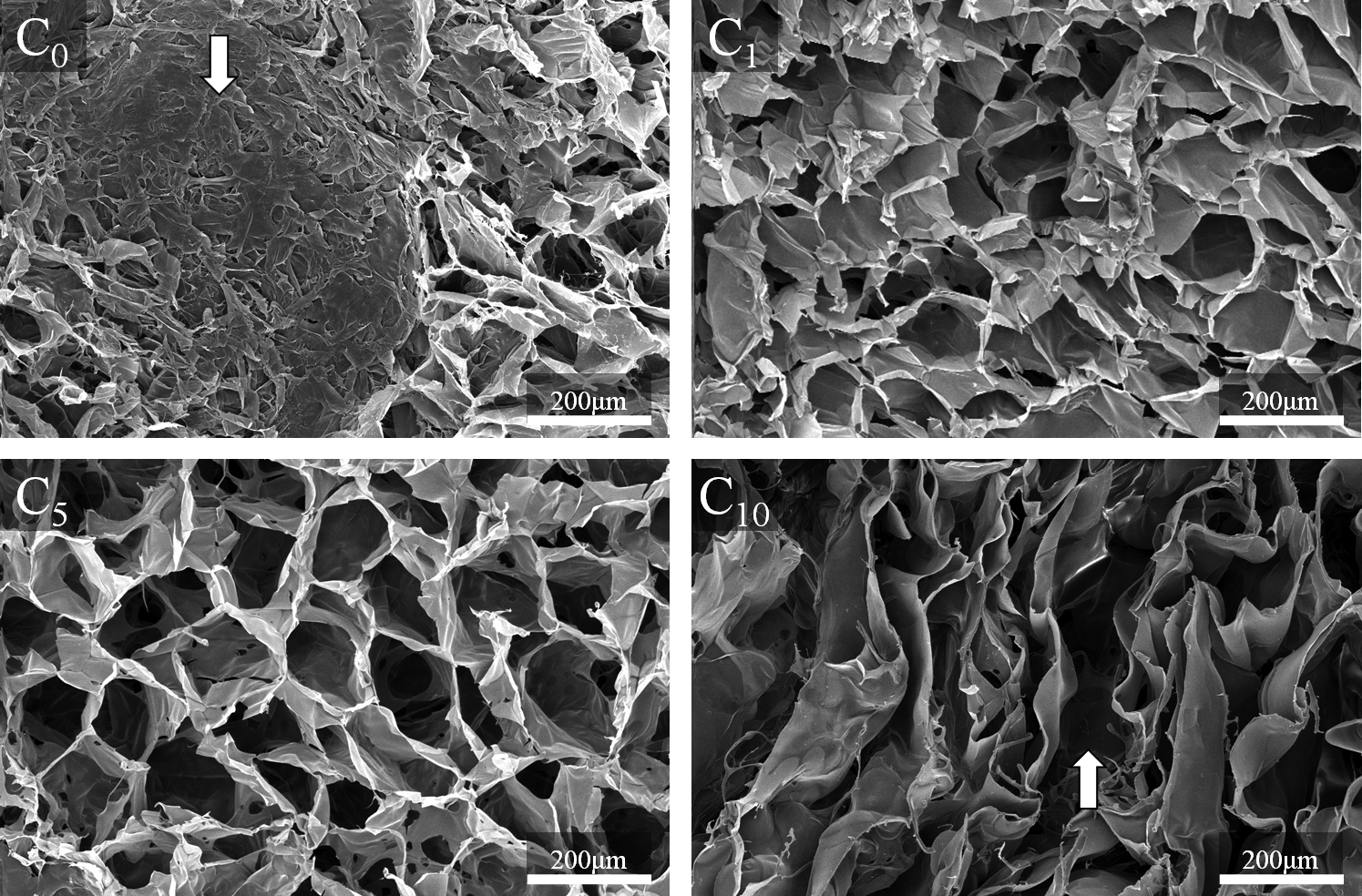
SEM micrographs for cross section (C) of the chitosan (CHT) and hyaluronic acid (HA) scaffolds prepared, using different ratios of CHT and HA (n=3). The subscripts indicate the percentage of HA (0%, 1%, 5%, and 10%). C0 arrow evidences the sheet-like areas of CHT scaffolds. C10 arrow shows the irregular pore geometry of CHT/10HA scaffolds. Original magnification is ×100 and the scale bar represents 200 μm. SEM, scanning electron microscopy.
Pore size increased with concentrations of HA in CHT scaffolds (Table 1). Fan et al. 31 reported the same influence of HA on scaffold pore size. CHT/10HA also exhibited the highest standard deviation in pore sizes because of the nonuniform pore geometry. The open pore network and a range in pore size between ∼77 and 97 μm, as observed in CHT/1HA and CHT/5HA scaffolds, rendered these scaffolds appropriate for cellular infiltration. Particularly, we hypothesize that articular bovine chondrocytes can be incorporated into the aforementioned scaffolds.
Values are reported as mean±SD.
Fourier transform infrared spectroscopy imaging measurements
FTIR measurements (Fig. 2) confirmed the presence of CHT and HA in all CHT/HA scaffolds. Characteristic peaks of both polymers (Fig. 2a) were found, namely a distinct band at 1650 cm−1 (amide I) and at 1580 cm−1 (amide II). 26 The ester group (1045 cm−1), a HA characteristic peak, is the main difference between the two spectra. The referred characteristic peaks were also found in all CHT/HA scaffolds spectra (Fig. 2b). Particularly, CHT/10HA had a notable higher intensity in the ester peak, as it is the formulation with higher amount of HA.

Fourier transform infrared (FTIR) spectra of
The homogeneity of the CHT/HA blends was investigated by FTIR mapping (Fig. 3). As expected, chemical maps of CHT scaffolds (Fig. 3a) and pure HA (Fig. 3b) only showed the selected color for the pure materials (green and red, respectively). As showed in Figure 3d, CHT/5HA scaffolds had an improved polymer mixture, in which the two polymers were well dispersed in the length scale analyzed (tens of microns). On the other hand, in CHT/10HA scaffolds the quality of the blend was compromised. There were more green and red areas, which correspond to CHT and HA alone, respectively, and less yellow areas, which correspond to the homogeneously blended biomaterials.
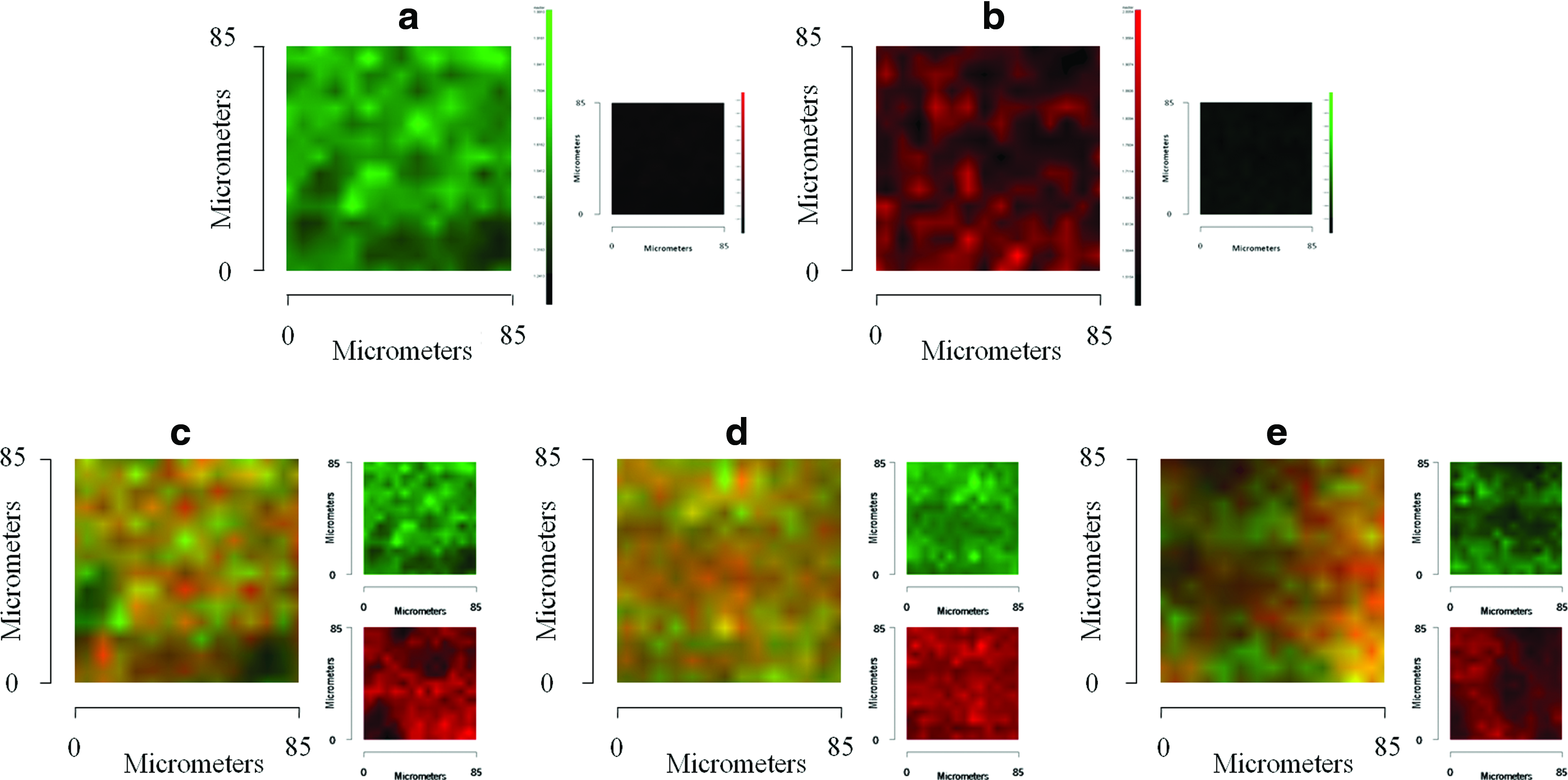
Chemical maps obtained by FTIR microscopy of the cross sections of the scaffolds and pure HA:
Mechanical properties
The mechanical properties of the scaffolds in tissue engineering applications are of great importance because of the necessity of structural stability to withstand stresses incurred during in vitro culture and in vivo implantation. 31
Compressive mechanical tests were performed using acellular scaffolds in dry and wet states. The compressive modulus of the scaffolds is shown in Table 1. All samples exhibited a sponge-like behavior. For both assays, the addition of HA in CHT networks reduced the mechanical strength of the scaffold, while it increased its flexibility. This behavior may essentially be due to the increase in pore size by increasing the HA concentration. It was interesting to note that CHT/1HA and CHT/5HA scaffolds had similar compressive modulus in dry and wet states, probably because of their similar pore network.
In addition to an increase in pore size, O'Brien et al. 33 reported a correlation between decreasing mechanical strength and nonuniform pore shape in HA-based scaffolds. This may explain the reason why CHT/10HA had a much lower compressive modulus.
In the dry state, the compressive modulus of CHT/HA scaffolds were lower but in the same order of magnitude when compared with the modulus of articular cartilage reported by Korhonen et al. 34 The compressive modulus in wet state was much lower compared with the dry state because of HA's ability of high water uptake. As CHT and HA are two natural polymers, this drawback in terms of mechanical strength was expected. However, the principal aim of incorporating HA into CHT scaffolds was to mimic the native cartilage ECM. The compressive modulus may further improve by the deposition of an ECM by chondrocytes in CHT/HA scaffolds.
Swelling properties
Diffusion and exchange of nutrients (e.g., oxygen) and waste throughout the entire scaffold are related to the swelling properties of the scaffolds. Both CHT and HA have an abundant number of hydrophilic groups, such as hydroxyl, amino, and carboxyl groups, which can promote water uptake in the structure. 25 The swelling ability was evaluated by soaking the scaffolds in PBS at 37°C for 3 days (Fig. 4). The ratio between CHT and HA significantly affected the scaffolds' swelling properties. In the first 2 h, all samples rapidly increased their weight. This rapid weight increase continued until 8 h. Then, the swelling ratio of all samples seemed to stabilize, and a slight increase could be observed with time. CHT scaffolds had the lowest values during the entire experiment, whereas CHT/10HA had the highest. CHT/1HA and CHT/5HA had similar swelling. The swelling results indicated that HA incorporation can greatly improve hydrophilicity and wetting of CHT scaffolds. Hence, scaffolds with higher content of HA showed higher values of swelling. This aspect may imply that the absorption and diffusion of solutes through the interior pores in CHT scaffolds are greatly improved by incorporation of low concentrations of HA.
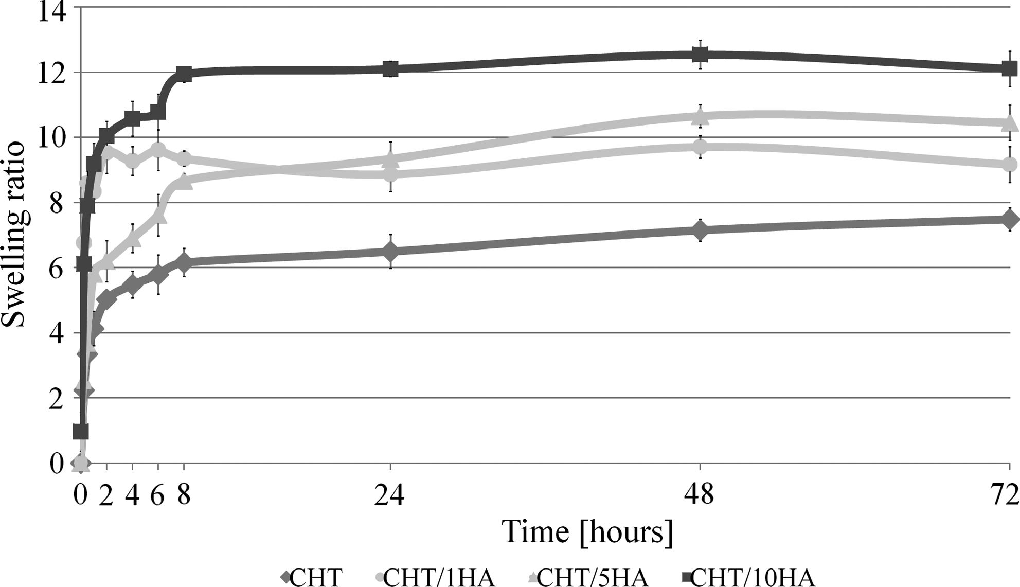
Swelling ratio of CHT and HA scaffolds in phosphate-buffered saline at 37°C for 3 days. Values are reported as mean±standard deviation (SD) (n=3).
In vitro enzymatic degradation
Scaffolds for tissue engineering are usually required to either degrade or be reabsorbed by the body after successful tissue regeneration. CHT/HA scaffolds were incubated in a solution with lysozyme and hyaluronidase or in a solution with PBS (control), both at 37°C for 7 and 14 days, to evaluate the weight decrease in the biopolymers due to enzymatic degradation. The weight loss percentage of scaffolds as a function of incubation time is illustrated in Figure 5.
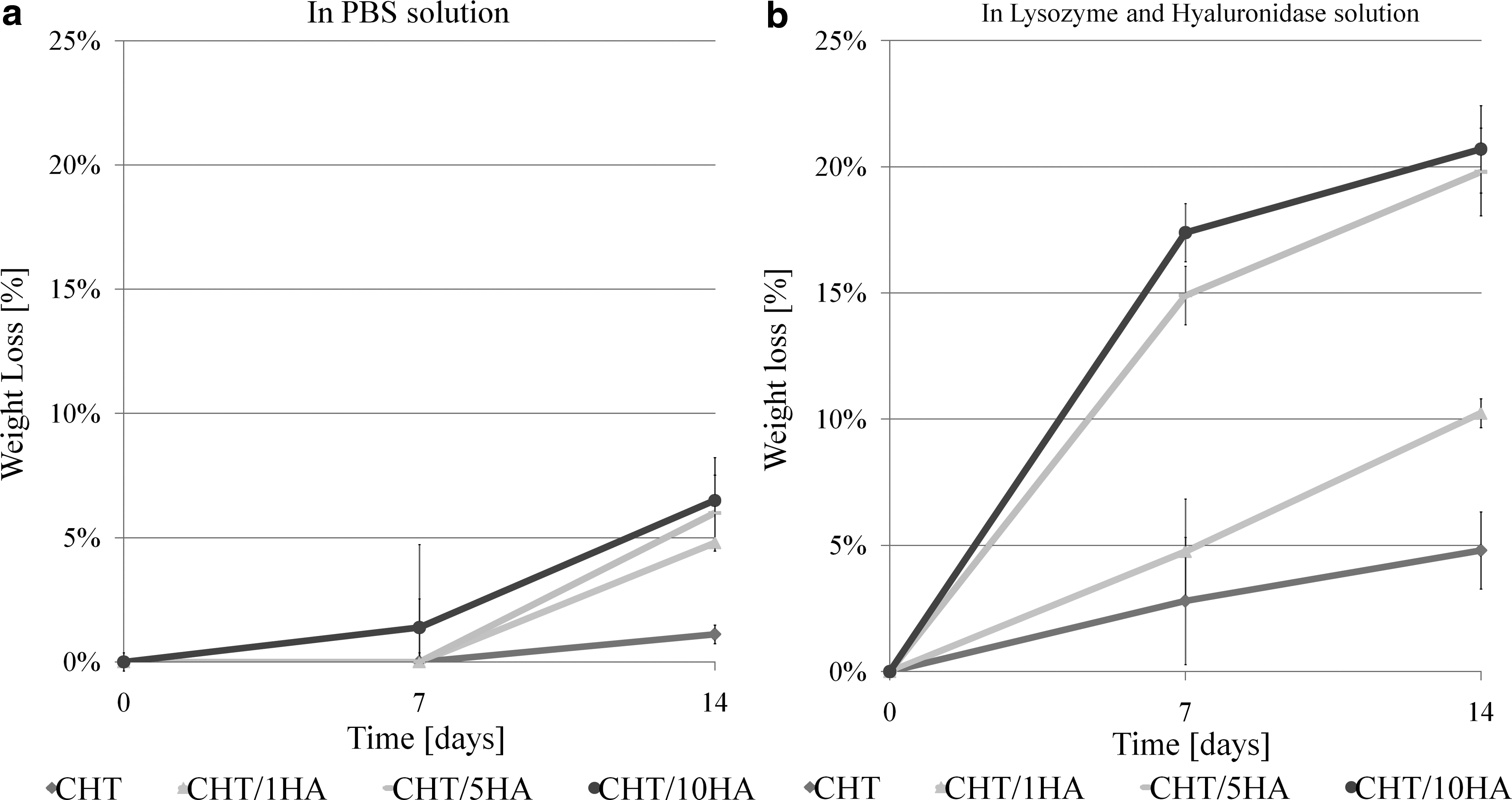
Weight loss percentage of CHT/HA scaffolds as a function of incubation time (7 and 14 days) at 37°C. Scaffolds were embedded in
To distinguish between enzymatic degradation and simple dissolution, we compared the weight loss of samples that had been placed in PBS (Fig. 5a) with those that had been placed in PBS supplemented with lysozyme and hyaluronidase at 7 and 14 days (Fig. 5b). According to Figure 5b, the presence of HA rendered CHT scaffolds more susceptible to enzymatic degradation, which is most likely due to higher porosity and better accessibility of cleavage sites by the enzymes. The hydrophilicity of HA also contributed to a higher degradation of CHT/HA scaffolds as it will enhance the interaction of the biomaterial with the enzymatic solution. After a high initial weight loss at day 7, the degradation rate of the samples generally appeared to slow down at day 14, which is most likely due to the gradual disappearance of hexasaccharide sequences susceptible for enzymatic degradation. The control experiment in PBS (Fig. 5a) showed that practically none of the scaffolds degrade in PBS after 14 days. The degradation of CHT has been already tested in several in vitro studies.35–38 Most of these studies were, however, performed at low pH for optimal lysozyme activity. In our study, the enzymatic degradation experiments were performed at higher pH, which resembles better degradation of the scaffolds in physiological conditions. Hyaluronidase and lysozyme concentrations used in this study (0.33 and 2 mg/mL, respectively) are not representatives of their concentration in vivo. The largest single reservoir of hyaluronidase is the synovial fluid of diarthrodial joints (0.5–4 mg/mL). 39 Lysozyme commonly exists in various human body fluids and tissues, with concentrations ranging from 4 to 13 mg/L in serum 40 and from 450 to 1230 mg/L in tears. 41
In vitro articular bovine chondrocyte culture
Cell viability, proliferation, and adhesion studies on CHT/HA scaffolds
In a live–dead assay, all materials exhibited very good biocompatibility with hardly any detectable cell death (Fig. 6). Further, cells were metabolically active (Fig. 7) and well spread throughout the scaffolds in all formulations. The presence of increasing concentrations of HA did not influence cell loading of CHT scaffolds. This behavior is essentially due to the low capacity for cell attachment of CHT.
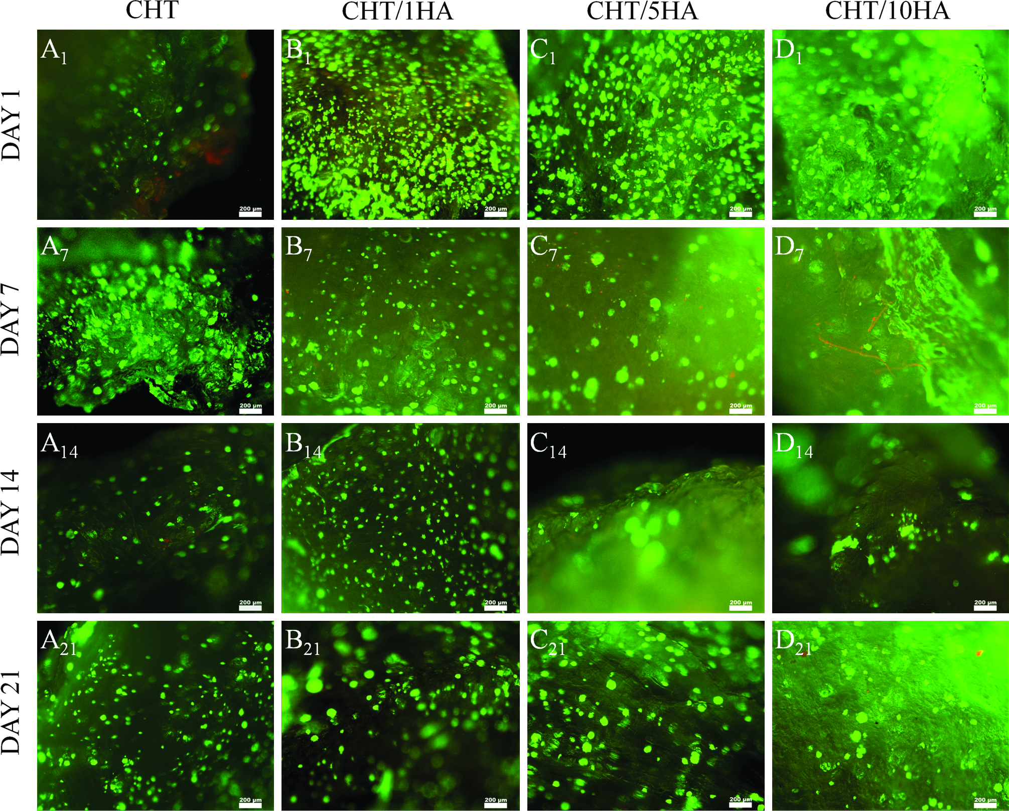
Live–dead assay results of chondrocytes at passage 3 in proliferation medium seeded on CHT/HA scaffolds at days 1, 7, 14, and 21. Live cells were stained green by calcein and dead cells were stained red by ethidium (n=3). Letters
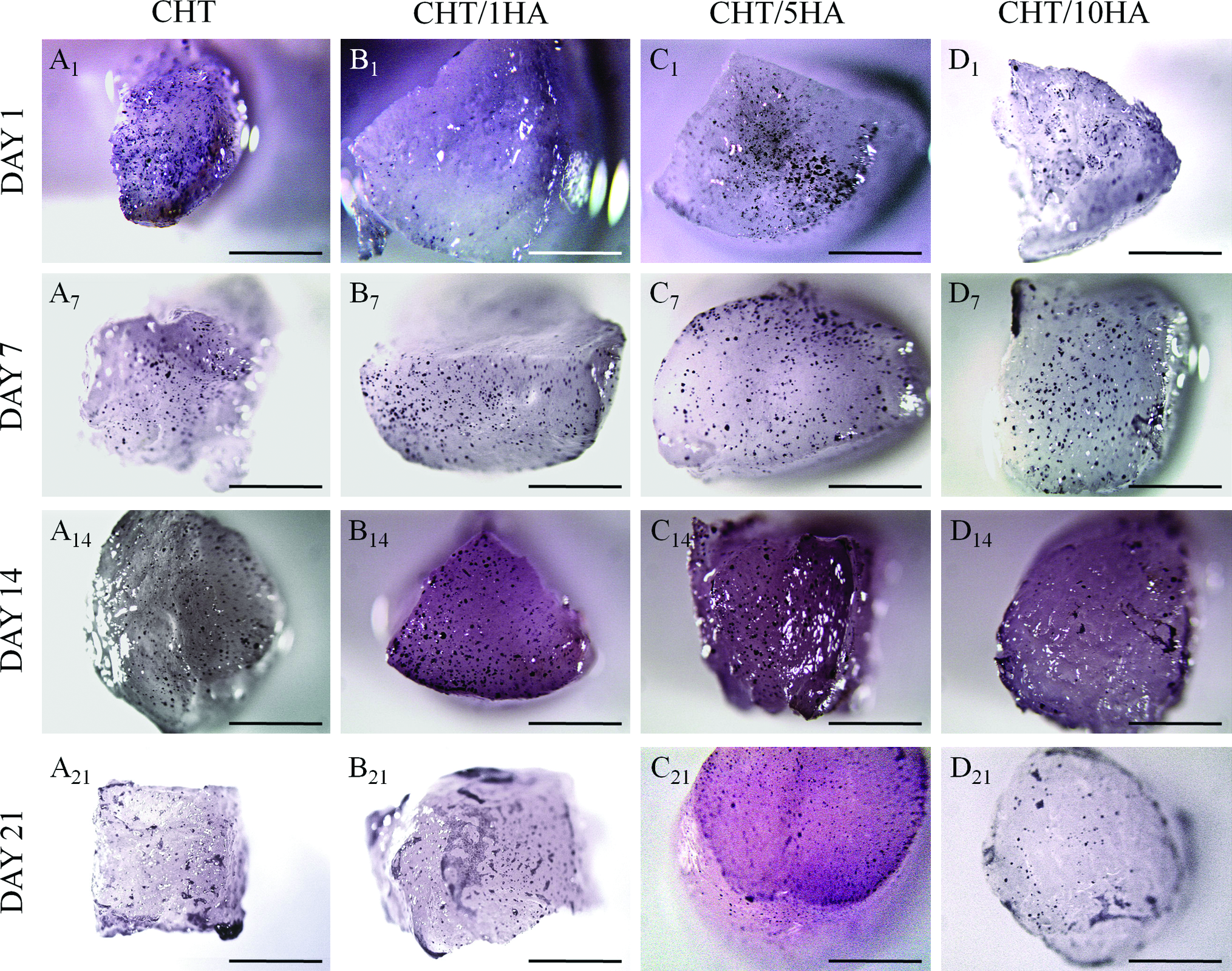
MTT assay results of chondrocytes at passage 3 in proliferation medium seeded on CHT/HA scaffolds at days 1, 7, 14, and 21 (n=3). Metabolic active cells were stained dark purple. Letters
The cell morphology and proliferation on CHT/HA scaffolds was further studied by SEM (Fig. 8). In all scaffolds, chondrocytes were primarily localized in the superficial area at days 1 and 3 and displayed a spherical morphology (cell size diameter: ∼10 μm). At day 7, a limited increase in cell number was observed but cells did not penetrate into the scaffolds. At days 14 and 21, chondrocytes seeded in CHT/5HA scaffolds significantly proliferated into the inner areas of the scaffolds. In all scaffolds, even after 21 days of culture, the cells continued to display a normal spherical morphology, as observed in native cartilage.
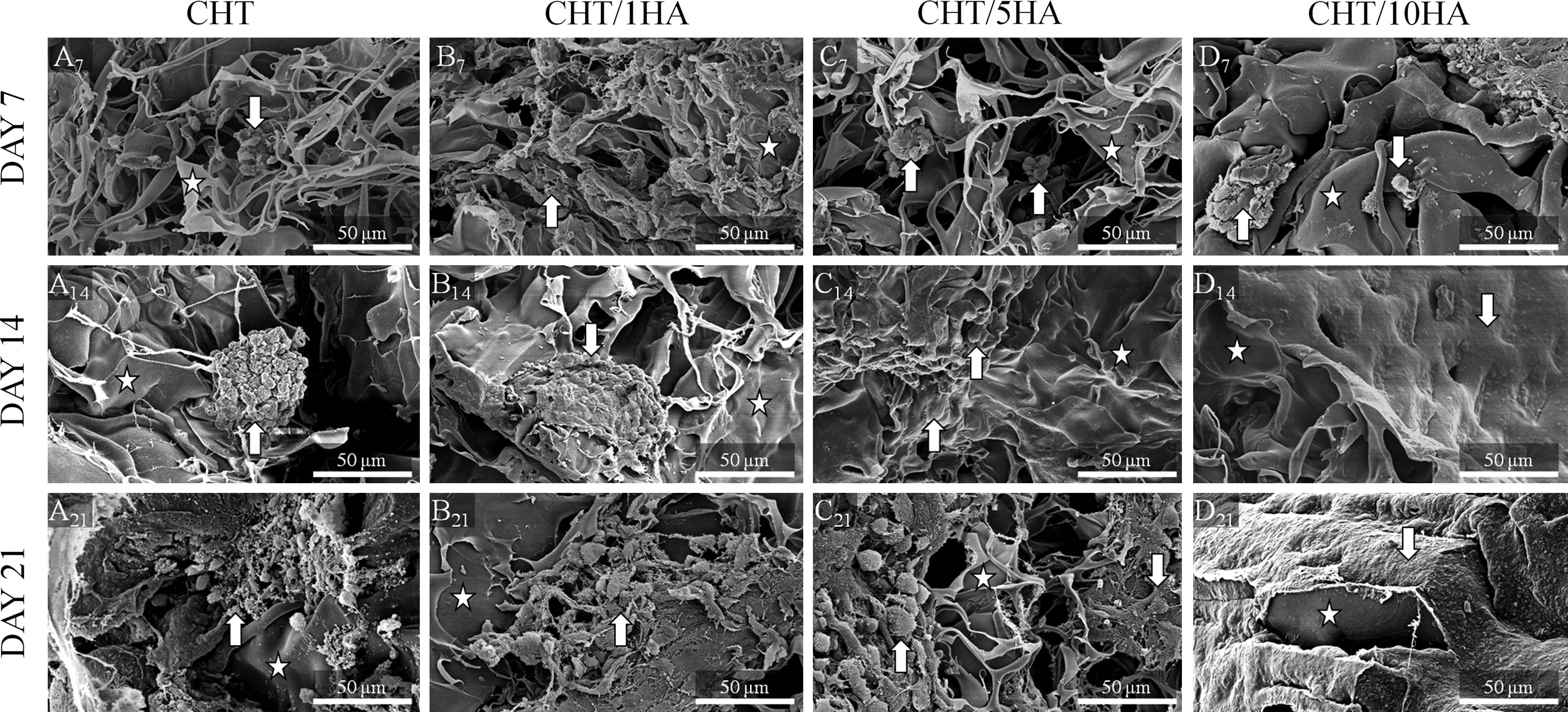
SEM micrographs (10 kV) of CHT/HA scaffolds seeded with articular bovine chondrocytes at passage 3 in proliferation medium (n=2). Letters
Maintenance of chondrogenic phenotype
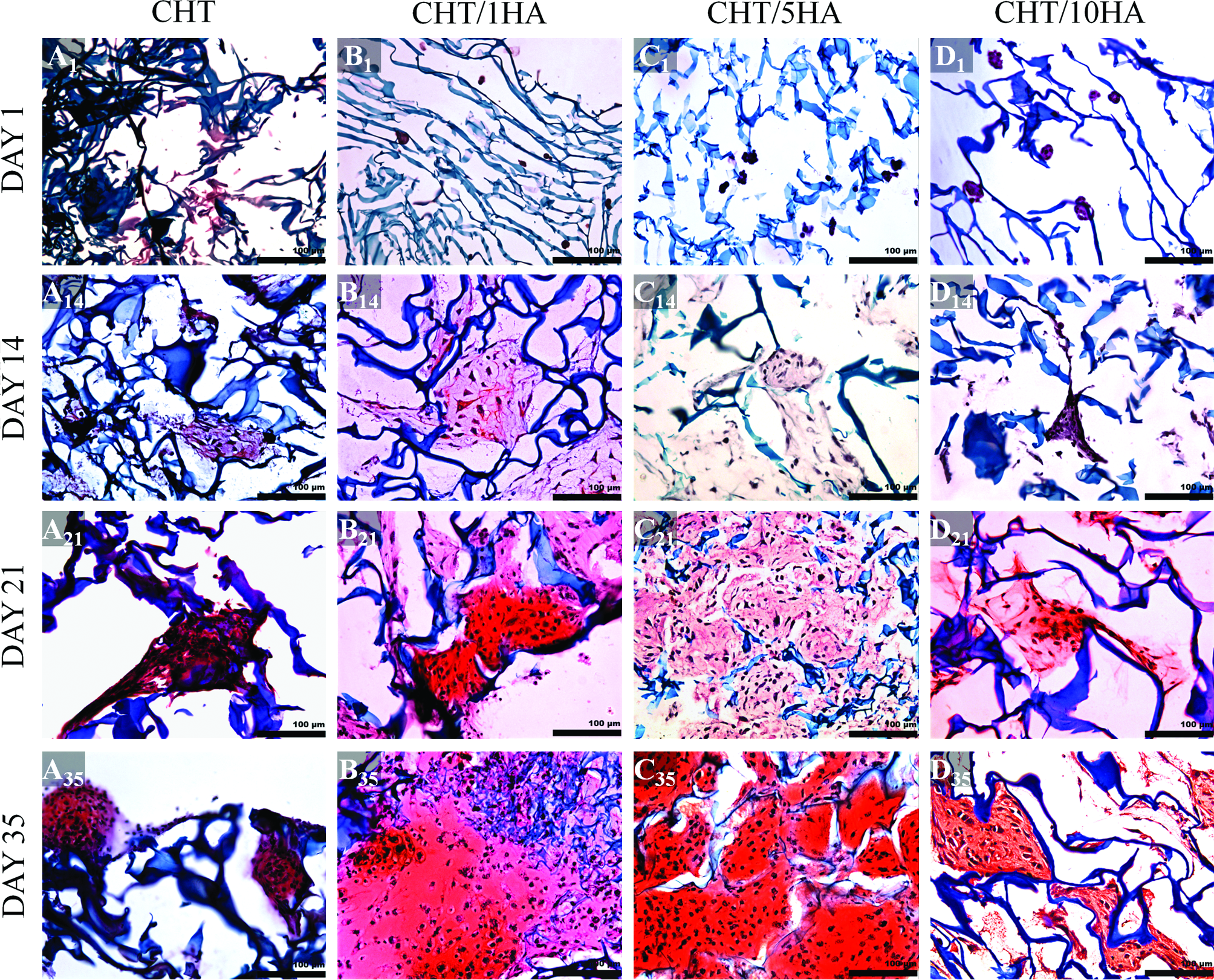
The round shape of chondrocytes is an indicator of phenotype retention and is essential for matrix formation. 42 The newly formed matrix was stained with safranin-O (Fig. 9) and alcian blue (Fig. 10), which showed GAG secretion. As HA is a nonsulfated GAG, it stains blue in alcian blue staining. Low amounts of HA in the scaffold can be easily distinguished from newly deposited ECM by the chondrocytes by comparing alcian blue staining at 1 (Fig. 10A1–A35) with staining at later time intervals. At day 1, chondrocytes did not attach to the walls of CHT scaffolds, whereas in CHT/HA scaffolds, the cells attached to walls, forming small aggregates. At day 14, in CHT/5HA scaffolds (Fig. 9C14 and Fig. 10C14), it was interesting to note that most chondrocytes agglomerated to form very large aggregates adherent to the scaffolds. The aggregates were located on the superficial area. Hardly any penetration of cells in the scaffolds was observed. These events were also reported by Zhao et al. 43 CHT/1HA and CHT/5HA showed enhanced cell–material interactions and ECM production, with enhanced GAG production (Figs. 9 and 10). At day 35, CHT/5HA scaffolds showed better cell dispersion and higher ECM production and GAG deposition when compared with the other formulations. Chondrocytes seeded in CHT scaffolds, even at day 35, remained in small aggregates (Fig. 9A35 and Fig. 10A35).
Histological cross-sections show glycosaminoglycan (GAG) deposition (stained red) in CHT/HA scaffolds by safranin-O staining at 1, 14, 21, and 35 days of chondrocyte culture in differentiation medium (n=2). Letters

Histological cross-sections show GAG deposition (stained blue) in CHT/HA scaffolds by alcian blue staining at 1, 14, 21, and 35 days of chondrocyte culture in differentiation medium (n=2). Letters
Enhanced cartilage tissue formation can be also qualitatively detected by SEM analysis (Fig. 11). At day 1, chondrocytes were in aggregates in all specimens, but at day 14 they were better spread and attached. At days 14 and 21, improved chondrocyte dispersion could be noticed in CHT/1HA and CHT/5HA scaffolds. In all specimens, lacunae formation was seen in the matrix surrounding the chondrocytes. The morphology of these lacunas shows similarities to natural cartilage.

SEM micrographs (10 kV) of CHT/HA scaffolds seeded with articular bovine chondrocytes at passage 1 in differentiation medium (n=2). Letters
Chondrogenic activity evaluation
It is known that when chondrocytes maintain their natural spherical shape they produce more GAGs and higher collagen type II versus collagen type I ratio.44–46 Cell proliferation and ECM formation were quantified by DNA and GAG assays, respectively. In GAG quantification assay (Fig. 12a), the incorporation of HA seemed to initiate an earlier GAG deposition, particularly at day 14 for CHT/1HA and CHT/5HA scaffolds. Results showed that the incorporation of HA seemed to favor GAG deposition, because at day 35 all the scaffolds containing HA had more GAG deposition when compared with CHT scaffolds. For DNA quantification assay (Fig. 12b), CHT/5HA had the highest amount of DNA at day 14. However, at day 35, CHT/1HA scaffolds significantly supported a higher amount of cells when compared with other formulations.
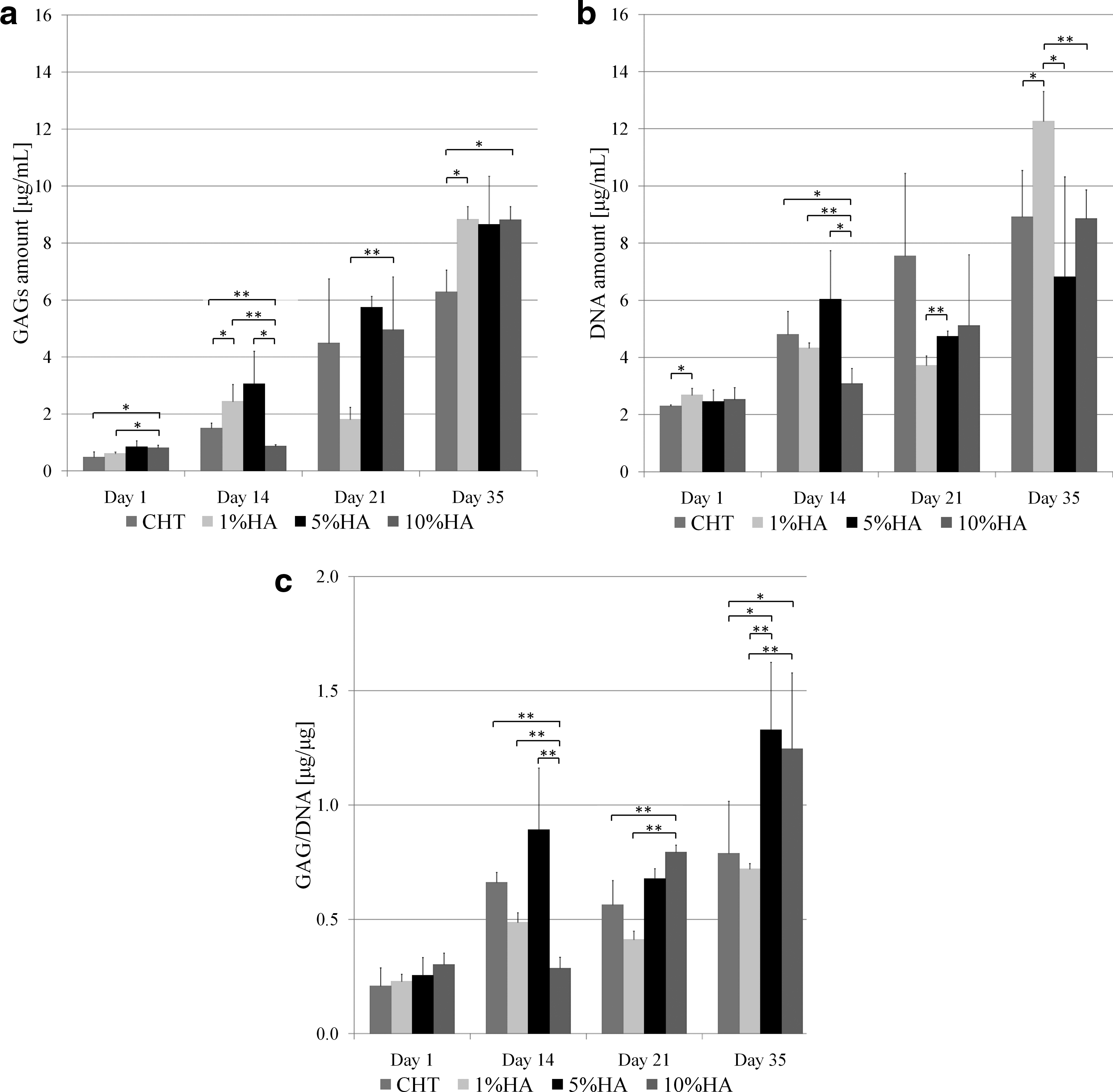
GAG and DNA assays on CHT/HA scaffolds at 1, 14, 21, and 35 days of culture:
A higher value of GAG/DNA ratio indicates a higher degree of differentiation of the cells. 47 For GAG/DNA ratio (Fig. 12c), at day 35, scaffolds with higher amount of HA (CHT/5HA and CHT/10HA) induced a higher ratio. Although CHT/1HA had the highest amount of cells in DNA quantification, CHT/5HA had the highest GAG versus DNA ratio. This may imply that CHT/5HA scaffolds are the formulation that most favors cartilage ECM production, rendering them attractive for cartilage TE.
Physicochemical effects of CHT/HA scaffolds on cartilage regeneration
The key challenge for biomedical researchers is how to design and control material properties to achieve a specific biological response. 48 It has been well established that the physicochemical properties of materials can regulate biological responses, such as differentiation of stem cells, 49 proliferation of fibroblasts, 50 gene delivery, 51 and cell death. 52
CHT/1HA and CHT/5HA scaffolds exhibited a uniform pore network with high porosity (Fig. 1) and a homogeneous polymer dispersion (Fig. 3), rendering them suitable for bovine chondrocyte culture. The pore configuration allowed cellular infiltration into the inner pores of the scaffolds (Fig. 8). On the other hand, CHT and CHT/10HA scaffolds had a very irregular pore network configuration with closed pores. Consequently, in these scaffolds, the adhesion and proliferation of chondrocytes were jeopardized when compared with the other formulations. The scaffolds' high water uptake ability (Fig. 4) allowed the chondrocyte medium to reach the interior pores of the scaffolds. Consequently, diffusion and exchange of nutrients and waste throughout the scaffolds were ensured, allowing cells to proliferate and be metabolically active (Figs. 6 and 7).
Besides appropriate pore network and water uptake ability, CHT/1HA and, especially, CHT/5HA had an improved GAG dispersion and deposition (Figs. 9 and 10). Additionally, the two referred formulations had similar compressive modulus. Hence, physical and chemical factors seem to simultaneously influence the biological outcome. Cartilage regeneration seemed to be favored simultaneously by chemical aspects, that is, intermediate amounts of HA (1% and 5%), and also by physical aspects, that is, the referred scaffold formulations also have similar compressive modulus and a uniform pore network configuration. Therefore, in this case scenario, it is not possible to distinguish case by case the detailed mechanisms of how physical properties affect the biological performance of the CHT/HA scaffolds. Despite this unclear contribution of each parameter, the results obtained in our study are not compromised.
Conclusions
The incorporation of HA up to a concentration of 5% in CHT scaffolds improved the physicochemical properties and biological properties of CHT scaffolds. It enhanced pore network configuration and increased pore size, swelling ratio, and degradation rate when compared with pure CHT scaffolds. Higher concentrations of HA resulted in heterogeneous blends with irregular and collapsed pore networks, particularly CHT/5HA scaffolds favored chondrocyte adhesion, cell proliferation, and cartilage matrix production when compared with pure CHT scaffolds and other CHT/HA blends.
The biological outcome of CHT/HA scaffolds seems to be influenced by physicochemical factors, such as polymer dispersion, pore network configuration, pore size, water uptake ability, and mechanical strength. However, other studies are required to elucidate case by case the dependence of biological response on physical properties and to categorize the relative weight of different physical and chemical factors.
Our results suggest that the freeze-dried scaffolds of CHT and HA could have potential use in the regeneration of cartilaginous lesions caused by various joint diseases, including osteoarthritis and rheumatoid arthritis.
Footnotes
Disclosure Statement
No competing financial interests exist.