
Abstract
A perfusion bioreactor, which was designed based on fluidized bed concepts, was validated for the culture of bone constructs of clinically relevant size. For this study, natural coral has been used as three-dimensional scaffolds. This biomaterial is a microporous, biocompatible, osteoconductive, and absorbable scaffold. This perfusion bioreactor provided a stable environment in terms of osmolarity, pH, and, most importantly, oxidative stress. Bone constructs engineered in this system resulted in significantly higher cell proliferation and homogenous cell distribution than those cultured under static conditions. Particularly relevant to the production of bioengineered bone in a clinical setting, custom-made bone constructs (each one with volume up to 30 cm3) could be produced using a such perfusion bioreactor. Last, but not least, the bone constructs of clinically relevant volume thus produced were shown to be osteogenic when transplanted subcutaneously in sheep.
Introduction
However, these bone constructs are not yet uniform enough or homogenous enough (cell distribution mainly on the exterior surfaces but not within the core of the scaffold) for standard medical application.6–8 Uniform and homogeneous cell distribution in the construct increases the amount of tissue formed, the rate of bone mineralization, 9 and the formation of uniform tissue10,11 within the construct.
Such drawbacks could be overcome by using a bioreactor. This apparatus is a mechanical system in which such parameters as the temperature, pH level, and perfusion flow rate are strictly controlled to nourish biological and biochemical processes. Bioreactors are already used in many operations, including fermentation, water processing, and production of pharmaceuticals. A great advantage to use of the bioreactor, a mechanized and standardized system, in this kind of engineering is the resulting substantial decrease in production costs, and the subsequently wide accessibility to the modern practices developed because of, and coupled to, the utilization of the bioreactor system. Since bioreactors provide cell growth conditions that are reproducible and physiologically relevant, they show significant promise for improving the quality of bioengineered tissues. These systems usually take the form of a closed container housing microporous scaffolds seeded with cells, which can be perfused with a culture medium.
Use of perfusion bioreactors has been advocated for the application of physiologically relevant mechanical forces by fluid flow that stimulates proliferation and differentiation of MSCs.12–16 So far, however, reports of in vivo bone formation in tissue constructs are given in just two experiments carried out in perfusion bioreactors that involved bone constructs of large volume.17,18 Only one of these studies reported how fluid flow affected proliferation of the cells cultured in the bioreactor. 18
When oxygen depletion occurs in the bioreactor, just as in a real biological system, many tissues produce lactate in a process called anaerobic glycolysis. Oxygen metabolism dysfunctions create an excess of very reactive chemical species known as reactive oxygen species (ROS), including free radicals (like ·OH,
Thus, the perfusion bioreactor developed in our laboratory has been designed, based on fluidized bed bioreactor concepts. This type of bioreactor is well known to enhance mass transfer, 22 to address and possibly overcome the inadequacies of existing bioreactors. Our perfusion apparatus was therefore designed (1) to permit maximal nutrient and waste transmission, avoiding unstable washout flow associated with the rotating wall bioreactors and enhancing the perfusion passing into and out of the scaffolds, by limiting the leakage rate of perfusion streaming past the scaffolds, (2) to operate at low perfusion flow rate and low associated shear stresses, thus avoiding the risk of cell detachment of the scaffold, and (3) to engineer more homogenous cellular distribution of the scaffold. Indeed, obtaining bone constructs of clinical grade requires good homogeneity in terms of number of cells and their distribution throughout each scaffold independently of the location of the construct into the bioreactor. Our study aims mainly to investigate the performance of our perfusion bioreactor in designing and engineering implants for large bone defects.
Preamble: Rationale for the Design of the Bioreactor
There are bioreactors designed to evade the problems of limited mass transfer in scaffolds of larger thicknesses. In bone tissue engineering, these apparatuses provide the appropriate mechanotransduction force to foster signal transduction pathways leading to osteogenic differentiation.23,24 Thus, as a major challenge in this type of engineering using a high cell density construct cultured within a bioreactor, a balance has to be found between high supply of medium and sufficiently low fluid shear stresses. Such a trade-off can be achieved in designing, based on the fundamental concepts in fluid dynamics and transport phenomena within porous media, 25 a new perfusion system working as a vertical fluidized bed at low flow regime with constructs deposited at the bottom of the bioreactor.
Mastering the mechanical forces applied
Pressure and shear, among other mechanical loads, drive proliferation and differentiation of osteocompetent cells. The main focus in experimental analyses conducted to date has been on shear, concluded to decisively affect the pattern of how tissue develops. Many experiments on classical one-dimensional osteoblast cell cultures grown under dynamic conditions (during a range of time intervals between some minutes to many hours) have directed their focus toward the response prompted by shear stresses ranging from 1 μPa to 0.05 Pa.26,27 The scientists conducting these experiments report increases of both the rate of proliferation and the rate of mineralization subjected to the aforementioned range of stresses. In spite of the usefulness of these results, not much is known about optimized shear loads in the bioreactor. Long-term studies (lasting between a handful of days and many weeks) have been carried out on cell cultures with shear loads of only 0.01 Pa. 14 At this low loading level, it has been demonstrated that cell proliferation and bone mineralization rates were both enhanced. 28 On the whole, this collection of results shows that continuously applied shear stresses ranging from 1 to 10 mPa incite the osteocompetent cell to grow. 29 It is possible that the integrin cell receptor be activated by such low shear stresses, thereafter contributing to the regulation of target gene expression.27,29 Bioreactors must promote mechanical stimulation without causing detachment of the cells. In vitro studies have demonstrate that cells exposed to shear ranging between 0.6 and 3 Pa30,31 were all detached from their scaffold after 24 h. 28
Solving mass transfer problems
One of the most prominent difficulties in the design of a new bioreactor is addressing the inadequacies in mass transfer, inherent in the fact that engineered constructs must be greater in size than a couple of millimeters to serve as grafts of substitute tissue. To obtain clinically interesting tissue volume, using perfusion bioreactors could be beneficial to the cell culture. In this case, the flow induces increased nutrient flux, oxygen supply, and waste removal by supplementing diffusion transport processes with convection transport processes, which are typically of greater intensity. Indeed, the flow rate within the implant in bioreactors (and, as a result, the associated convective transport) is a function of the applied pressure gradient and the implant's hydraulic permeability depending upon the microscopic architecture of this porous medium.
Choice of the bioreactor type
At present, many bioreactors, including spinner flask, perfusion, and rotating wall vessel, have been considered and researched to test their suitability in bone tissue engineering. Each has its inadequacies. The spinner flask model provided enhanced fluid flow,32,33 but also harbored a turbulent environment potentially injurious to a seeded cell.34,35 This model solves the external diffusion problems, but not those experienced inside the scaffold itself, where solvent and nutrients have difficulty in passing through the pores. As a result, cell growth and development of the matrix only occurs on the outside of the scaffold. 35 Osteogenic differentiation and proliferation of MSCs in porous scaffolds are augmented by the perfusion bioreactor.14,36 However, in this type of bioreactor, the cells at the periphery of the scaffold wash away with the more elevated perfusion flows located there, 37 resulting in a nonhomogeneous cell distribution, a problem that can be overcome with an appropriately chosen bioreactor geometry. The rotating wall vessel bioreactor,38–40 characterized by three-dimensional (3D) high rates of mass transfer and small shear loads, similarly experiences nonhomogeneous cell growth and formation of the extracellular matrix.34,41,42 Additionally, studies have shown that scaffolds repeatedly impact the bioreactor wall, inflicting many perplexing, unquantifiable physical disturbances on the cultured cell. It has been demonstrated that these recurrent impacts greatly constrain the cell density attained and the mineralized matrix created in the rotating wall vessel bioreactor.35,42
Materials and Methods
Bioreactor and bioreactor system
A custom-made perfusion bioreactor was designed, assembled, and employed in the experiments that comprise the present study. This laboratory setup (schematic shown in Fig. 1A) consisted of a vertical cylindrical (3.3 cm in diameter; 21 cm in height) polycarbonate chamber. The perfusion bioreactor was connected to a reservoir containing the culture medium in a closed loop arrangement. The connections between the bioreactor and the reservoir and between the bioreactor and a peristaltic roller pump (Masterflex) were made of platinum-cured silicone tubing (Masterflex tubing; Cole Parmer) to minimize leaching of chemicals from the polymer and protein adsorption on the surface of the tubing. With the exception of the pump, the whole perfusion bioreactor was placed in an incubator and operated under sterile conditions at 37°C for the duration of the experiments. Another advantageous characteristic of the type of tubing chosen was its high gas permeability. As a result of this permeability, oxygenation and preservation of physiological pH in the circulating medium was assured during operation of the perfusion bioreactor. Due to concerns regarding the mechanical resistance properties inherent in the silicone, neoprene tubing (Pharmed; Cole Parmer), a stiffer material, though lower in gas permeability, was employed in a small (25 cm in a total length of 200 cm) segment inside the pump.
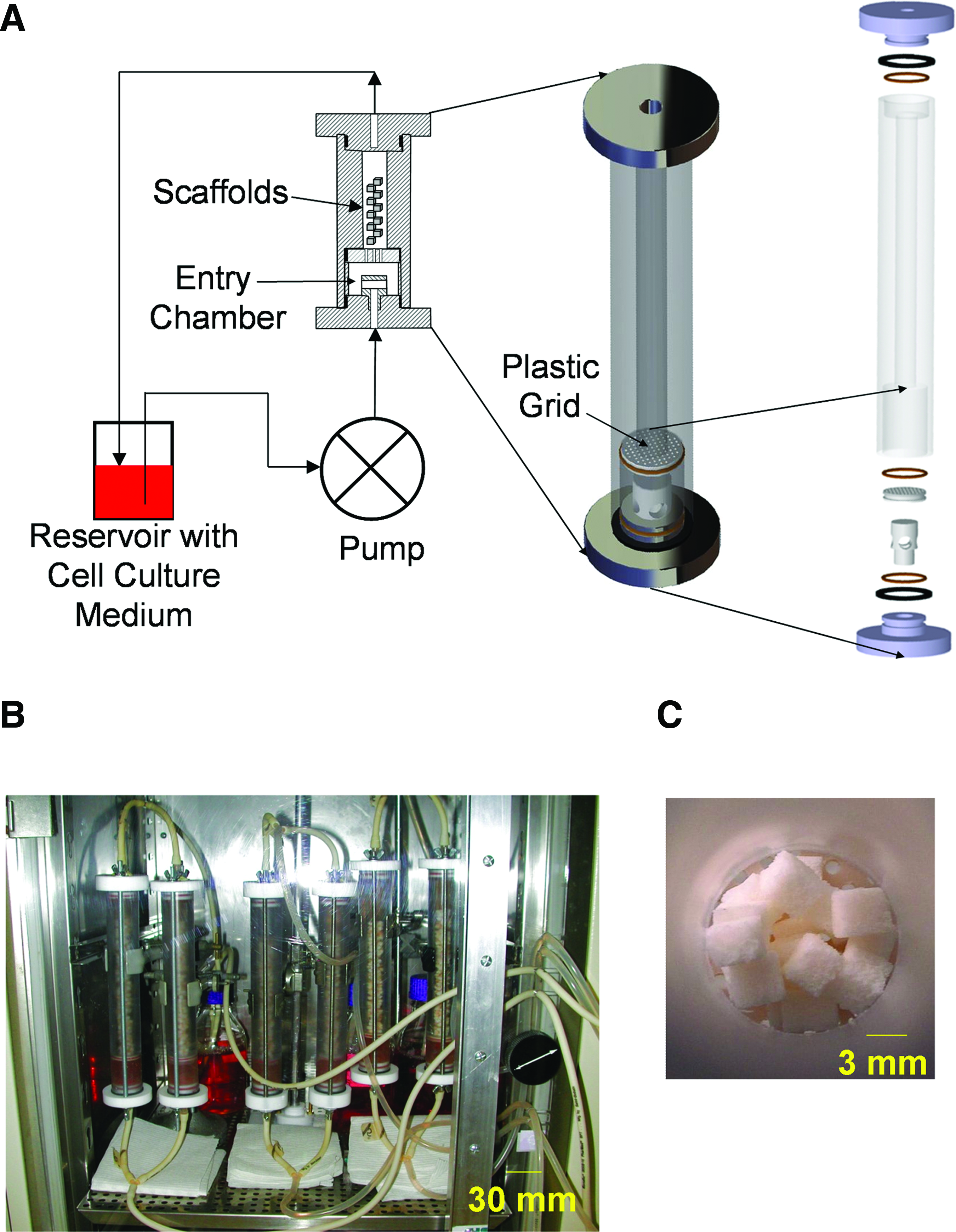
The perfusion bioreactor used in the present study.
A peristaltic roller pump was employed for the circulation of cell culture medium from the reservoir through the perfusion bioreactor in the direction schematically shown in Figure 1A. The mean velocity of the fluid inside the bioreactor, corresponding to the domain of flow rate delivered by the peristaltic pump, was ranged from 1 to 100 mL/min. To maintain reproducible flow conditions and prevent fluid flow disturbances, the medium from the reservoir entered an entry chamber in the lower portion of the perfusion bioreactor (Fig. 1A). A plastic grid (with pores of 1 mm in diameter) separated the entry from the perfusion bioreactor chamber (where the cell-containing scaffolds were placed during experiments). Although the perfusion bioreactor could accommodate many more scaffolds, only up to 160 scaffolds (each 3 × 3 × 3 mm3) at a time were used in the experiments of the present study. Multiple such perfusion bioreactors were operated during experiments (Fig. 1B). The circulating medium was changed every 3 days under sterile conditions; for that purpose, the tubing and perfusion bioreactor were emptied of medium for a very short (1 min) time; a fresh cell culture medium was added in the reservoir and the perfusion system was reinstalled.
Coral scaffolds
Coral, a scaffold shown in prior experiments to exhibit biocompatibility, osteoconductivity, and absorbability, has served as a bone replacement material for over 10 years. 43
The 3D (3 × 3 × 3 mm) scaffold varieties employed in our experiment comprised natural coral of the genera Porites and Acropora, respectively. 44 That derived from Porites was characterized by an architecture of interlinked pores (average diameter 250 μm) and a higher porosity (49% ± 2%); that derived from Acropora was characterized by an architecture of pores (average diameter 500 μm) in communication and a lower porosity (12% ± 4%). 44 Preceding the cellular seeding process, the scaffolds were autoclaved for sterility (at 120°C for 20 min).
Upon placement in the perfusion bioreactor chamber, which contained the flowing cell culture medium, the constructs (cell-containing scaffolds) did not exclusively align in either the vertical or horizontal direction; instead, they settled in a random fashion with no predominant orientation. This arrangement created passages in-between and around the individual constructs through which the cell-culture medium flowed (Fig. 1C).
In our case, both the coral substrate chosen and the randomized localization and orientation of the constructs associated with their initial sedimentation process in the bioreactor lead to the complex design of the scaffold structure. As a rough estimation, it result is therefore that the overall substrate contained in the perfusion bioreactor is a porous medium presenting a large spectrum of pore dimensions, from 100 μm to 1 mm and an overall porosity >50%. Accounting for the value of the perfusion mean velocity as previously given (10–103 μm/s), plus the architectural characteristics of the substrate as mentioned here and the rheological properties of the perfusion fluid, an approached evaluation of the applied shear stress would be between 10−2 and 10 mPa. These values are commonly advanced in case of the mechanotransduction effects of cells embedded within 3D substrates without risk of cell detachment.
Transfection and initial culture of GFP-C3H10T1/2 cells in tissue culture flasks
The C3H10T1/2 cell line (purchased from ATCC) was derived from 14- to 17-day-old C3H mouse embryos. 45 The cell type found in this line is mesenchymal. Enhanced green fluorescent protein (eGFP)-C3H10T1/2 cells, derived from a single cell clone (that had been stably transduced with rMLV-LTR-eGFP lentivirus), 46 were cultured in the basal essential medium (BEM; Sigma) supplemented with 10% fetal bovine serum (FBS), penicillin (100 U/mL), and streptomycin (100 μg/mL) (all from PAA Laboratories) at pH 7.4 and under standard cell culture conditions (37°C, 5% CO2). Passaging of the GFP-C3H10T1/2 cells, following standardized, accepted protocols, 46 was carried out at 80% confluence.
Seeding and culturing of GFP-C3H10T1/2 cells in 3D scaffolds
Coral scaffolds (described in the Coral scaffolds section) were maintained in the cell culture medium (so that they would be prewetted, and so that proteins from the fetal bovine material in the medium would be adsorbed on the scaffold material), under standard incubator conditions overnight. At day 0, GFP-C3H10T1/2 cells (introduced in Transfection and initial culture of GFP-C3H10T1/2 cells in tissue culture flasks section) were enzymatically released from tissue-culture polystyrene flask surfaces (using a 0.5% trypsin EDTA solution), suspended at a density of 1 × 10⁶ cells/mL (of BEM, supplemented with 10% FBS and 20 mM HEPES [Sigma] at pH 7.4) transferred in a 15-mL polypropylene tube containing the coral scaffolds (105 cells per scaffold), and thus maintained under static standard cell culture conditions for 24 h. Viable GFP-C3H10T1/2 cell distribution on the coral scaffolds was qualitatively assessed using 3-(4,5-dimethylthiazol-2-yl)-2,5-diphenyltetrazolium bromide (MTT) staining. To accomplish this, the individual constructs were rinsed twice with PBS and maintained in MTT solution (1.5 mg/mL) at 37°C for 4 h. Each construct was then photographed using a Micro-nikkor 60 mm objective linked to a DXC-930 P Sony camera and a Leica Q500 IW computer.
In vitro investigation of the perfusion bioreactor performance
Assessment of fluid flow influence in the perfusion bioreactor
GFP-C3H10T1/2 cells were seeded and cultured on coral scaffolds; the process is explained above in Seeding and culture of GFP-C3H10T1/2 cells in 3D scaffolds.
Twenty-four hours postseeding, the constructs were transferred into the perfusion bioreactor in BEM (containing 10% FBS and 20 mM HEPES) and cultured under media flow conditions at various flow rates (specifically, 5, 10, 40, and 80 mL/min). At day 3, each construct was frozen at −80°C, thawed, and then grinded using a crushing device (RETSCH, MM 301 model) in lysis buffer (1 mM MgCl2, 0.1 M Na2CO3, 0.1 M NaHCO3, and 0.1% Triton × 100; pH 10.2) for 5 min. The intensity of the fluorescence signal (BIO-TEK; FL600 FA Microplate Fluorescence Reader) varied linearly with the initial quantity of cells loaded in coral scaffolds (Supplementary Fig. S1A; Supplementary Data are available online at
Cell proliferation under optimal flow rate conditions
GFP-C3H10T1/2 cells loaded onto coral scaffolds were cultured under 10 mL/min media flow rate in the perfusion bioreactor. On the 3rd, 6th, 9th, 12th, 15th, 18th, and 21st days of culture, the respective cell count was established by measuring eGFP fluorescence intensity as described in the Assessment of fluid flow influence in the perfusion bioreactor section.
Influence of location of the constructs in the perfusion bioreactor on cell number per scaffold
To investigate the influence of construct location under media flow conditions in the perfusion bioreactor on cellular proliferation, we laid 140 cell-loaded scaffolds in each perfusion bioreactor and cultured them under 10 mL/min of medium flow rate. At day 21, all constructs were collected and carefully labeled by successive numbering (as a function of the vertical location of the construct, starting from the top of the perfusion bioreactor and continuing downward); the location of each construct inside the perfusion bioreactor during the experiments was thus identified. This location was expressed as the distance (in centimeters) of each bone construct from the plastic grid (Fig. 1A). We established the number of cells per construct as per the Assessment of fluid flow influence in the perfusion bioreactor section. Cell number reported was the average of the content of 10 successive constructs.
Assessment of lactate dehydrogenase, pH, osmolarity, lactate, and ammonia
On the 3rd, 6th, 9th, 12th, 15th, 18th, and 21st days, an aliquot (6 mL) of culture medium was collected from the reservoir, centrifuged, aliquotted, and kept in the freezer (−20°C) for about 3 weeks, until assessment. At the end of this period, lactate dehydrogenase (LDH) activity, lactate concentration, ammonia concentration, osmolarity, and pH were determined using a clinical analyzer (Architect C8000; Abbott Diagnostic) according to manufacturer's instructions.
Assessment of the redox status (total antioxidant status and thiobarbituric acid-reactive substances)
On the 3rd, 6th, 9th, 12th, 15th, 18th, and 21st days, an aliquot (6 mL) of culture medium was collected from the reservoir, centrifuged, aliquotted, and kept in the freezer (−20°C) to await analysis. We established total antioxidant status, using a kit (Randox Laboratories Ltd.) and following manufacturer's instructions. The presence of malondialdehyde, which is an indicator of lipid peroxidation, was detected in the culture medium using the SOBIODA test kit (SOBIODA Laboratories Ltd.), which measures thiobarbituric acid-reactive substances; this assay was performed following the manufacturer's instructions.
Histology
On the 3rd, 6th, 9th, 12th, 15th, 18th, and 21st days of culture in the perfusion bioreactor, constructs (n = 2) were randomly selected, processed for undecalcified histology, and stained with Stevenel Blue according to established techniques.43,47
Statistical analysis
The number of experiments carried out for the above cases is noted in the respective figure legends.
Numerical data were analyzed statistically using the Student's t-test and StatView software. p-Values of <0.05 were considered significant.
In vivo investigation of the osteogenic potential of bone constructs prepared in the perfusion bioreactor
Bone constructs preparation
Bone marrow was aspirated from the iliac crest of ewes using established techniques mixed in α-minimal essential medium α-MEM; containing 10% FBS) and was transferred to tissue-culture plates 43. Sheep MSCs adhered to the tissue-culture polystyrene surfaces and after 1 week were enzymatically released using a 0.5% trypsin-EDTA solution and, subsequently, cultured in vitro in α-MEM (containing 10% FBS) under standard cell culture conditions until confluence. Sheep MSCs (passage 1) suspended in α-MEM (containing 10% FBS, 100 mM ascorbate, 10 mM β-glycerophosphate, and 10−8 M dexamethasone) were seeded (105 cells per scaffold) onto 3D (each 3 ×; 3 × 3 mm3) Acropora coral scaffolds, transferred, and cultured with α-MEM containing 10% FBS, 100 mM ascorbate, 10 mM β-glycerophosphate, 10−8 M dexamethasone, and 20 mM HEPES (as described in the In vitro investigation of the perfusion bioreactor performance section) under media flow conditions in the perfusion bioreactor in vitro. The medium circulated at a 10 mL/min flow rate and was changed every 3 days. Control constructs were maintained in vitro in static culture conditions as described in the section In vitro investigation of the perfusion bioreactor performance.
In vivo assessment of the osteogenic potential of the constructs
Two-year-old female Pre-Alpes sheep, purchased from a certified seller (INRA), were raised following procedures established and published by the European Committee for Care and Use of Laboratory Animals (Directive du Conseil 24.11.1986. 86/609/CEE). Experimental tests involving animals were executed in accordance with the requirements and moral standards for laboratory treatment of animals established by European legislation. The animal experiments received specific approval by the official Committee of Animal Research Surveillance at the Denis Diderot University, Paris.
Constructs were implanted using an established 48 model that mimics the Masquelet procedure. 49 Briefly, cylinders (height = 25 mm; diameter = 15 mm) of polymethylmethacrylate (Sulfix 6®) were implanted bilaterally into paraspinal subcutaneous sites. After 6 weeks, the implanted polymethylmethacrylate cylinders were removed by opening the induced membrane; each resulting cavity was filled with a mean of 61 ± 7 Acropora coralline scaffolds, either unloaded (n = 4) or loaded with MSC cultured in the perfusion bioreactor for 1 week (n = 1) and 3 weeks (n = 3). The specific location of the various implants on the dorsum of each animal was randomly assigned. Animal subjected to the surgical procedure experienced a successful recovery, remaining healthy for the entirety of the experimental period. All subcutaneous implants remained in situ for the duration of the experiment. After an 8-week interlude postimplantation, the pouches containing the constructs were excised, fixed in 10% formalin neutral buffer, processed for undecalcified histology, and stained with Stevenel Blue according to established techniques. 47
Results
In vitro experiments
Cell proliferation
First, to determine the influence of perfusion flow rate, C3H10T1/2 cells seeded on coral scaffolds were cultured under dynamic conditions in the bioreactor system for 3 days.
The effect of the flow rate of circulating medium (specifically 5, 10, 40, and 80 mL/min) on C3H10T1/2 cell proliferation was assessed and compared to results obtained from cell cultures under static conditions (control) for 3 days; after 3 days, the cell number was highest for constructs exposed to circulating medium at the 10 mL/min flow rate (compare a cell number of 0. 9 ± 0.3 million cells per construct exposed to a flow rate of 10 mL/min and a cell number of 0.3 ± 0.08 million cells per construct under static conditions; p < 0.05). Cell proliferation at 5 and 40 mL/min was 0.7 ± 0.9 and 0.6 ± 0.4 million cells per construct, respectively. It should be noted that no more cells were observed when 80 mL/min medium flow rate was used. Exposure of cells to high medium flow rates resulted in high magnitude of the wall shear stress within the bioreactor that is detrimental for cell proliferation and survival. Based on these results, a culture media flow rate of 10 mL/min was chosen as optimal and used for the present study. At this flow rate the bioreactor provided a biomechanical environment in which mammalian cells survived and proliferated.
The apparatus underwent further testing to verify that it was operationally reliable in raising culture during longer durations, as long as a 21-day period. During the various tests carried out, the apparatus and experimental environment did not fail mechanically or become contaminated. Whereas cells maintained under static culture conditions (control) reached a plateau as early as 6 days, proliferation of cells exposed to 10 mL/min medium flow continued through day 21 (Fig. 2A). R 2 values show that the relationship between time and the number of GFP-C3H10T1/2 cells during a 21-day cell culture period within the bioreactor was linear (R 2 = 0.9773). This coefficient potentially aids in the prediction of bioreactor cell count during a cell culture period. The cell number was highest in individual constructs exposed to a circulating medium flow rate of 10 mL/min on day 21 of culture (2.6 ± 0.3 million cells per construct compared to 0.6 ± 0.1 million cells per construct under static conditions; p < 0.001). The number of cells per construct was fourfold higher in constructs cultured under such dynamic conditions than in constructs cultured under control static conditions. This result demonstrated that mechanical stimulation (i.e., shear and normal stresses applied to the cells by the fluid flow) was involved in the enhancement of cellular proliferation.
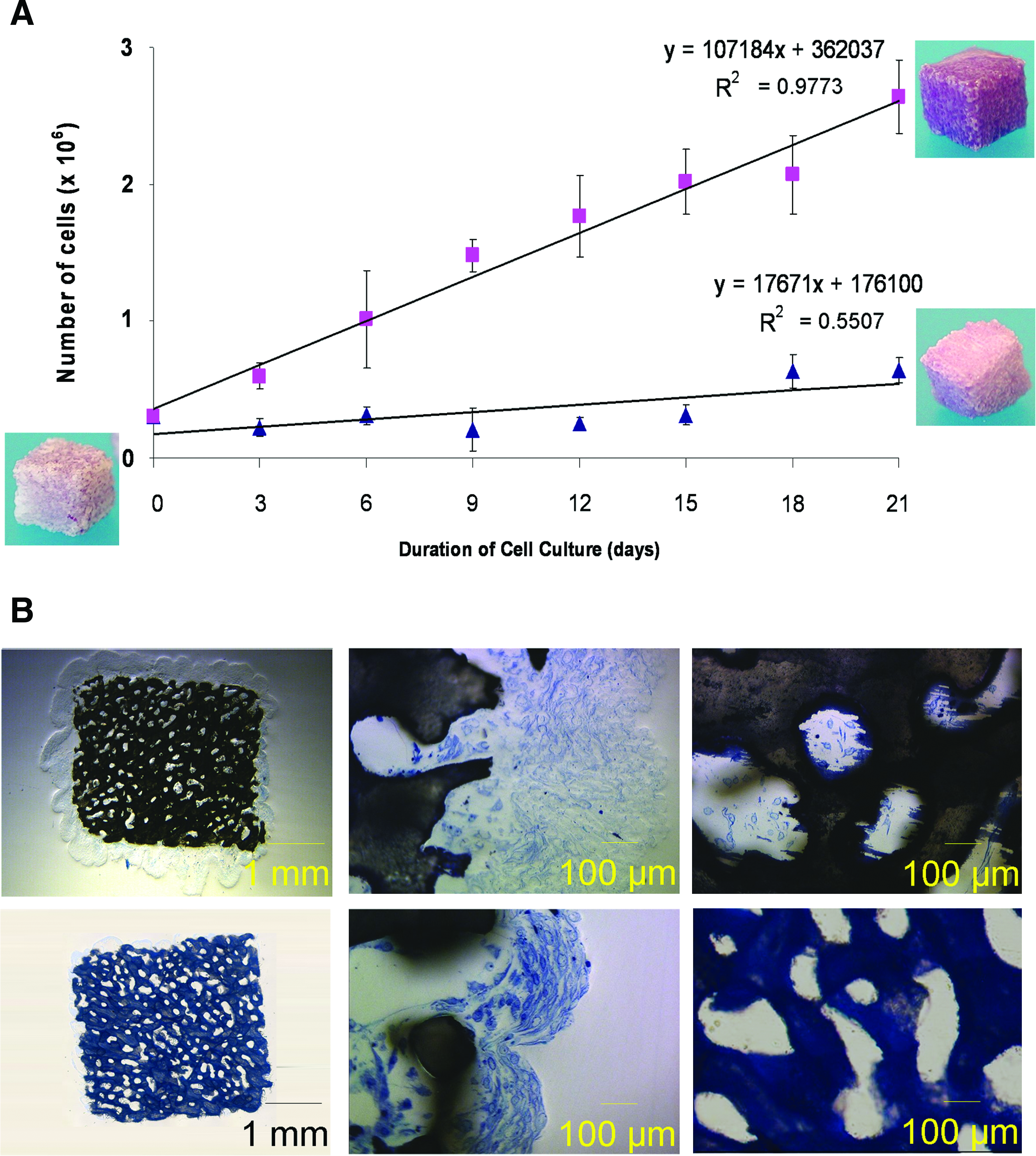
Effect of medium flow on GFP-C3H10T1/2 cell proliferation.
MTT staining (an index of viable cells) was homogeneous on all outside surfaces of the constructs cultured in the perfusion bioreactor under these conditions for 21 days (Fig. 2A).
Staining intensity is proportional to the number of cells; it increases with an increase in cell count. Constructs cultured under dynamic conditions were darker than those cultured under static conditions. Histological examination of the constructs exposed to 10 mL/min medium flow over the 21-day period revealed a uniformly thick (600 μm) multilayer of cells on the external surfaces of the scaffolds (Fig. 2B, upper row, left, and center), but only individual cells within the scaffolds (Fig. 2B, upper row, right). Cells at the core of the scaffolds (i.e., 1.5 mm from the outside surface) exhibited morphological signs of necrosis (Fig. 2B, upper row, right). In contrast, under static culture conditions, the cells formed colonies on the external surfaces of the scaffolds (Fig. 2B, lower row, left, and center), but, most notably, no cells were found in the interior of these scaffolds (Fig. 2B, lower row, right). The scaffolds cultured in the perfusion bioreactor displayed increased cell density, measured by both light microscopy and eGFP and indicative of the favorable consequences in using this type of bioreactor.
Another important characteristic of the perfusion bioreactor tested in this study was its capacity to obtain similar cell proliferation behavior in the individual constructs, independently of their specific location and alignment along the height of the containing chamber. In addition, when constructs were placed into the bioreactor, they did not align exclusively in either the vertical or horizontal direction; instead, they settled in a random fashion with no predominant orientation. This arrangement created passages in between and around the individual constructs through which the cell culture medium flowed (Fig. 1C). On day 21, the number of cells present within individual constructs cultured under a medium flow rate of 10 mL/min was homogenous, ranging from 2.1 to 2.5 million cells (Fig. 3). Results indicated that the culture medium flow (i.e., viscous shear stress) distribution was fairly homogenous within the bioreactor.
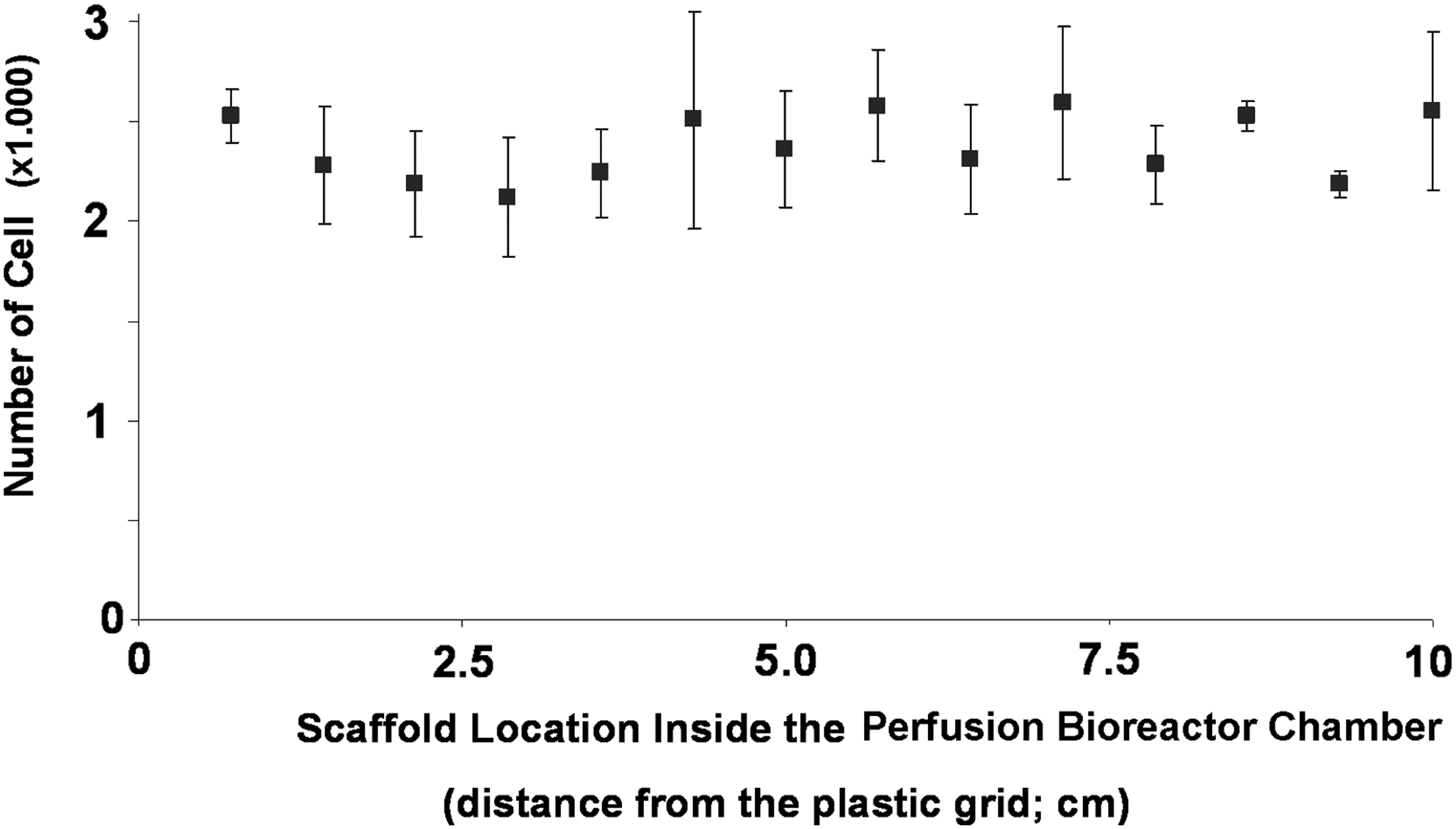
Effect of location of individual constructs in the perfusion bioreactor on GFP-C3H10T1/2 cell number per coral (Porites) scaffold after 3 weeks of culture. The medium flow rate was 10 mL/min (n = 6).
Assessment of biochemical parameters
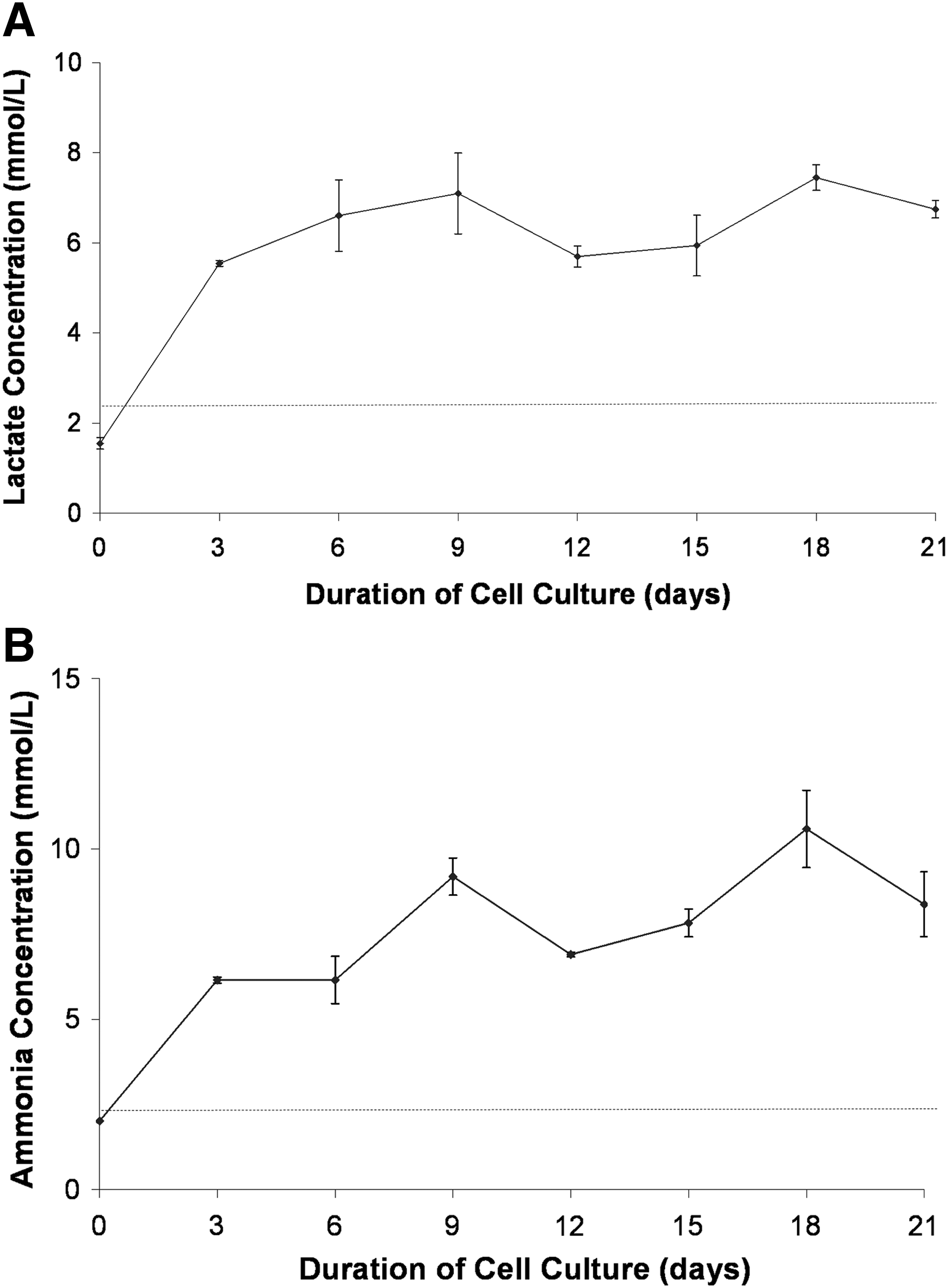
Measurements of pH and osmolarity in the medium, as well as of LDH, lactate, and ammonia released from cells, proved that the 10 mL/min circulating medium flow condition had established an environment appropriate for cell culture. The pH of the cell-culture medium in the reservoir, measured after 3-day intervals throughout the duration (up to 21 days) of the experiments, remained steady (7.1–7.4) throughout the study.
In addition, the osmolarity remained steady at 300 mosm/kg during the 21 days of the study, and the LDH level (elevated LDH activities in the culture medium indicate cell injury) was similar to that of fresh medium (11 ± 5 vs. 19 ± 4 IU/L, respectively). During that time period, the concentration of lactate ranged from 5 to 7 mM (Fig. 4A), whereas the concentration of ammonia ranged from 5 to 10 mM (Fig. 4B). These two metabolites, that is, lactate and ammonia, could inhibit cell growth. Within our experimental framework, high lactate concentrations (5–7 mM) in the bioreactor during experiments did not inhibited but increased cell proliferation (Supplementary Fig. S2A). Results indicated that high lactate concentrations did not influence the pH and osmolarity of the perfusion cell culture medium during the experiments. In addition, the high ammonia concentration (5–10 mM) did not affect cell growth in the perfusion bioreactor (Supplementary Fig. S2B). These results demonstrated that GFP-C3H10T1/2 cell proliferation capacity was not influenced neither by high lactic acid nor by ammonia concentrations in our bioreactor cell culture medium.
During the 21 days of the present study, the level of thiobarbituric acid-reactive substances (a marker of lipid peroxidation, which is potentially induced by an increased production of ROS into the perfusion culture medium) remained low and similar to that measured in control cell culture medium (0.4 vs. 0.2 μM, respectively; Fig. 5A). During the same time period, the overall total antioxidant status (which encompasses antioxidant activity in all biological components that comprise the cell culture medium) was similar to the antioxidant status in a fresh medium (0.24 ± 0.06 mM vs. 0.16 ± 0.08 mM, respectively; Fig. 5B). These results indicated that application of shear stress has no significant effect (when compared to a fresh culture medium) in ROS production, suggesting that shear forces had no toxic effect on cells hosting by the bioreactor.

Assessment of the redox status in the perfusion bioreactor during GFP-C3H10T1/2 cell culture under 10 mL/min medium flow rate for 21 days (n = 6).
In vivo osteogenicity
In contrast to constructs containing sheep MSCs cultured for 1 week in the perfusion bioreactor under 10 mL/min media flow rate, which were collected as separate units, the constructs cultured for 3 weeks in the perfusion bioreactor under 10 mL/min media flow rate were collected as a group of interconnected units linked with an extracellular matrix (presumably of collagenous nature) produced by the cultured cells (Fig. 6A).
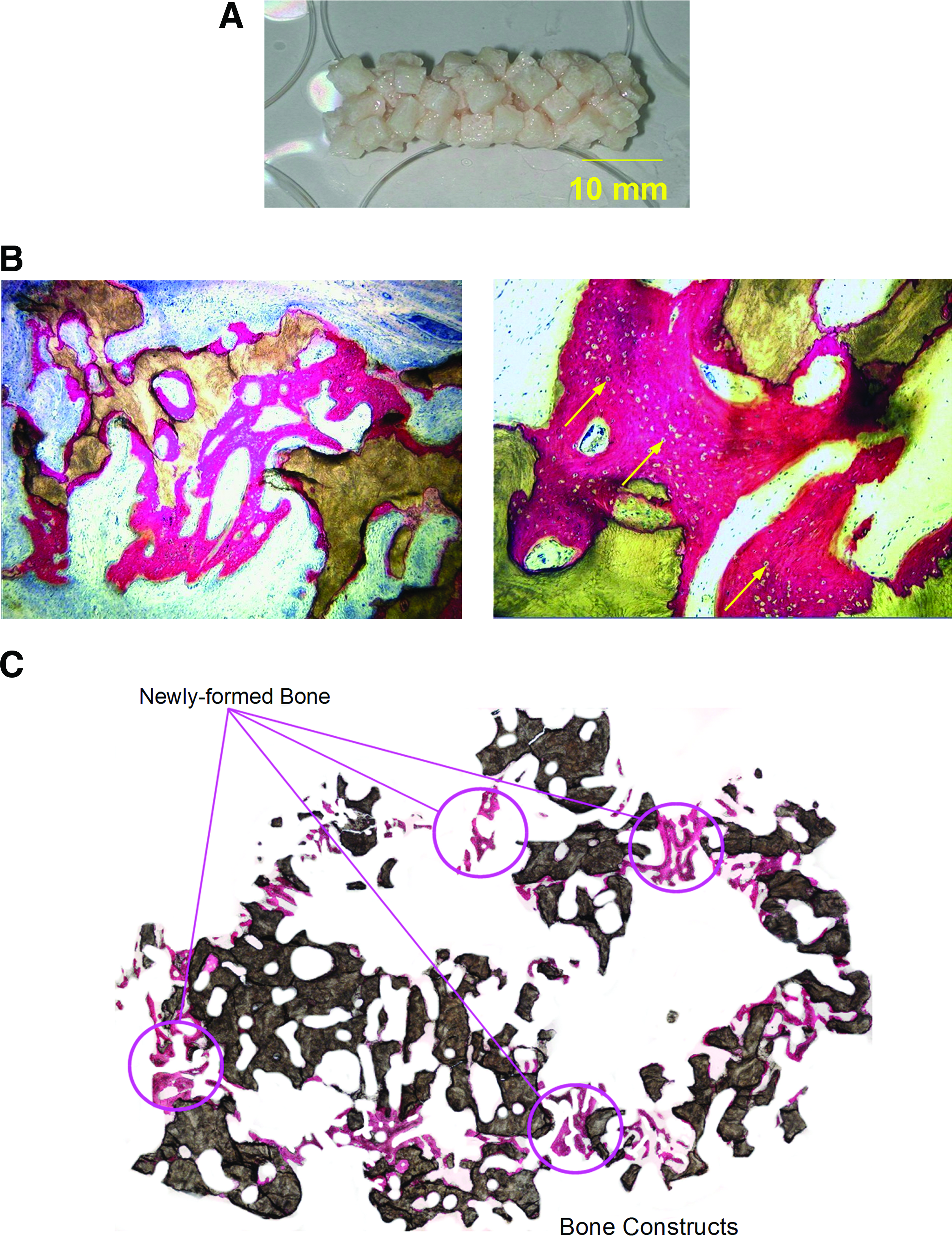
Histological assessment of the osteogenicity of cell-containing constructs (Acropora) cultured in the perfusion bioreactor for 3 weeks and then implanted subcutaneously in sheep for 8 weeks.
After 8 weeks of subcutaneous implantation in sheep, histological examination of the excised constructs that had previously been cultivated under dynamic conditions for 1 or 3 weeks (Fig. 6B and C, respectively) revealed freshly formed bone deposited on the surfaces of the individual constructs tested.
The bone matrix in all retrieved samples had areas of mineralized bone and osteoid, with osteocytes located inside the matrix and a film of osteoblasts coating the newly deposited bone. Blood vessels were often associated with, and were in close proximity to, the new bone. There were variations from explant to explant, but, in most cases, the new bone deposits were mainly observed on the exterior construct surfaces (Fig. 6C). No correlation was perceived between the duration of culture and the amount of newly formed bone. This result permitted the hypothesis that there was no link between initial construct cell count and new bone formation.
Discussion
This bioreactor provides significant advantages for expanding cells in vitro: a regulated biochemical environment, a facility in reproducing procedural protocols adapted for the experiments, and a reduced risk of contamination by viruses or bacteria. Our experimental results have shown in vitro that cells proliferated and were distributed homogenously to a substantially greater extent in bone constructs engineered in our perfusion bioreactor (which provided a suitable physiological environment) than those cultured in static conditions. Bioreactors are crucially needed to engineer bone 2 in a consistent and cost-effective manner.
Where complex and automated robotic systems have been envisioned for engineering tissues in the context of allogenic implants, the present study has aimed at presenting a simple and user-friendly system of preparing bone constructs directly in hospitals, eliminating the alternative dependence on industrialized central fabrication sites.
The present study established that such a perfusion bioreactor (which was designed based on fluidized bed concepts) provided an environment in which mammalian cells survived and proliferated. The scaffolds cultured in these conditions (in comparison to those cultured in static ones) displayed increased cell density, measured by both eGFP and light microscopy and indicative of the favorable consequences in using this type of bioreactor. In the bioreactor, shear forces generated by the fluid flow, which have been indicated as a prominently stimulating factor in cell proliferation,24,50 are effective both within the volume of the scaffold and on its outside surfaces during the first days of culture, an advantage over the spinner flask type bioreactor introduced earlier. The cells collectively developing in the bioreactor are exposed to stresses whose mean magnitude can be adjusted by regulating the global amount of perfusion flow rate. Naturally, however, the porosity and thus micro-layout of the different scaffolds determine the local behavior and its resulting impact on the discrete cell. These qualities of the perfusion bioreactor that result from agitation of the medium exceed those of the spinner flask type in that they include internally, as well as externally, eliciting cell proliferation via nutrient delivery and mechanical stimulation. Throughout our experiments, cells thrived and multiplied on coral-based scaffolds in the culturing environment of our 3D perfusion system. The circulating medium flow rate positively affected GFP-C3H10T1/2 cell proliferation; as noted earlier in the text, culturing GFP-C3H10T1/2 cells in the perfusion bioreactor at a 10 mL/min medium flow rate led to a cell count that was 4-fold higher than the results obtained from cells maintained under static culture conditions for 21 days. It should be noted that exposure of cells to high medium flow rates (specifically, 80 mL/min) resulted in massive cell detachment. Most importantly, our study established that culturing cell-loaded scaffolds in the perfusion bioreactor resulted in constructs with similar cell density, independent of the construct position inside the chamber. Progressively, though, the formation of a uniformly thick multilayer of cells on the external surfaces of the scaffolds prevented additional mass transfer into and out of these same scaffolds. Continued viable cell growth at the core of the scaffolds, 1.5 mm from their outside surfaces, depended upon the capacity of the bioreactor to enhance nutrient transfer via mass convection. 23 Convection increases the nutrient stock and improves the removal of metabolic components from inside of the scaffolds, thus preserving local cell viability.
Although our main purpose was not to investigate the long-term mechanotransduction cell response of the perfusion bioreactor, we note the importance of the abundant production of extracellular matrix (presumably of collagenous nature) observed in engineered sheep constructs after 21 days of culture in the bioreactor.
The cell uses glutamine and glucose in the production of energy. The toxic byproducts of this energy production, ammonia and lactate, may impede the development of the cell, 51 in part by disrupting the pH of the electrochemical gradient, which is essential in transport across the cell membrane. 52 Disproportionate lactate accretion increases the basicity of medium, thereby impeding development of the cell. Because ammonia (NH3) does not carry a charge, it diffuses easily across the cell and organelle membranes, increasing the basicity inside the cell. 52 These two byproducts have an inhibitive impact on the development of cells, in some cases51–53 over a broad range of concentrations ranging between 10 and 40 mM for lactate and from 0.5 to 12 mM for ammonia, and seemingly dependent upon the particular cell line. In light of this, lactate and ammonia concentrations seem to be vital in the optimization of the MSC expansion procedure carried out in a bioreactor. Within our experimental framework with the C3H10T1/2 cell line, it was shown that lactate concentrations in the bioreactor during experiments (7 mM) did not inhibit cell proliferation, but rather increased cell proliferation. Further, results indicated that such lactate concentrations did not influence the pH and osmolarity of the perfusion cell culture medium during the experiments; these two parameters remained constant over time. In the same manner, the high ammonia concentration (10 mM) did not affect cell growth in the perfusion bioreactor. From these results, it is clear that neither high lactic acid concentration nor high ammonia concentration affected the proliferation of cells in our bioreactor cell culture medium.
As discussed earlier, oxygen metabolism dysfunctions in mammalian cells create an excess of ROS, including H2O2, ·OH, and
Lastly, bone constructs comprised of coralline scaffolding and sheep MSCs having been raised for 1 or 3 weeks in the perfusion bioreactor, excised after an 8-week subcutaneous implantation in sheep, showed plentiful and freshly formed bone development upon examination. No correlation was perceived between the duration of culture and the amount of newly formed bone. This result is interesting in that it permits the hypothesis that there is no link between initial construct cell count and new bone formation. However, just one animal was used in this experiment, nullifying the possibility of interpreting the findings quantitatively. Ultimately, restoration of bone mass in wounds that are classified as critical-size defects from bioreactor-grown bone constructs will require that the proof of principle be a much larger animal model sample size.
Conclusion
A perfusion bioreactor, which is easy to manufacture and operate, and whose low cost makes possible a disposable unit, was designed and validated for the preparation of cell-containing constructs of clinically relevant size. This perfusion bioreactor provided a stable environment in terms of osmolarity and pH, with low levels of lactate and ammoniac and ROS level remaining similar to that determined in a fresh culture medium. Bone constructs engineered in this system resulted in significantly higher cellular proliferation than those cultured under static conditions (p < 0.001). Most importantly, the constructs thus prepared were of clinically relevant volume and promoted new bone formation when implanted subcutaneously in sheep.
Footnotes
Acknowledgments
The authors thank Biocoral, Inc., for donating the coral implants and Prof. R. Bizios for her comments on the article. The authors also acknowledge the financial support from Fonds d'Amorçage Biothérapie BTH06003, the ANR Therabone, and Contrat d'interface AP-HP/INSERM. Apart from the donation of the implants, no financial support was received from Biocoral, Inc.
Disclosure Statement
No competing financial interests exist.
References
Supplementary Material
Please find the following supplemental material available below.
For Open Access articles published under a Creative Commons License, all supplemental material carries the same license as the article it is associated with.
For non-Open Access articles published, all supplemental material carries a non-exclusive license, and permission requests for re-use of supplemental material or any part of supplemental material shall be sent directly to the copyright owner as specified in the copyright notice associated with the article.