
Abstract
The concept of contact guidance utilizes the phenomenon of anchorage dependence of cells on the topography of seeded surfaces. It has been shown in previous studies that cells were guided to align along the topographical alignment of the seeding substrate and produced enhanced amounts of oriented extracellular matrix (ECM). In this study, we aimed to apply this concept to a three-dimensional full silk fibroin (SF) hybrid scaffold system, which comprised of knitted SF and aligned SF electrospun fibers (SFEFs), for ligament tissue engineering applications. Specifically, knitted SF, which contributed to the mechanical robustness of the system, was integrated with highly aligned SFEF mesh, which acted as the initial ECM to provide environmental cues for positive cellular response. Mesenchymal stem cells seeded on the aligned hybrid scaffolds were shown to be proliferative and aligned along the integrated aligned SFEF, forming oriented spindle-shaped morphology and produced an aligned ECM network. Expression and production of ligament-related proteins were also increased as compared to hybrid SF scaffolds with randomly arranged SFEFs, indicating differentiative cues for ligament fibroblasts present in the aligned hybrid SF scaffolds. Consequently, the tensile properties of cultured aligned constructs were significantly improved and superior to the counterpart with randomly arranged SFEF. These results thus show that the aligned hybrid scaffold system is promising for enhancing cell proliferation, differentiation, and function for ligament tissue engineering applications.
Introduction
Despite the high occurrence rate, the ACL does not heal spontaneously when torn and surgical reconstruction has been the standard treatment modality in the field of sports medicine. 7 Even though this may be the case, ACL heals poorly in response to repair by simply suturing the injured tissue back together and very often grafts are required for ACL reconstruction. 8 Although surgical reconstruction of ACL, using autografts, allografts, or synthetic grafts, has been practiced for the restoration of knee joint function, several disadvantages and risks persist, such as ligament laxity, donor-site morbidity, pathogen transfer, mechanical mismatch, poor tissue integration, and foreign body inflammation.9–11 Consequently, there is an increased need to research for alternative treatment solutions12,13 and tissue engineering has evoked much interest as it offers the potential of regenerating functional tissues of autologous origin.6,14–18
One of the key challenges in tissue engineering of ligament resides in the design of a scaffold that is effective both as a load-bearing construct and a structural template for neo-ligament development. As a load-bearing construct, the scaffold for ligament regeneration must possess adequate mechanical strength postimplantation and degrades at a rate corresponding to that of new tissue deposition. Paradoxically, the scaffold should have sufficient void volume for cell infiltration and extracellular matrix (ECM) deposition to promote gradual load transfer from scaffold to the neo-tissue.14,19 Silk fibroin (SF) of Bombyx mori origin has been shown to be particularly promising for this application after removal of the hyper-allergenic sericin component from raw silk14,16,17,20–22 and has a compatible degradation rate that involves a gradual loss of tensile strength over 1 year in vivo due to proteolytic actions.14,23 More importantly, SF has outstanding and customizable mechanical properties, making it suitable for use in constructs with high porosity without compromising the overall mechanical robustness of the construct.14,19,20,22,23 As a structural template, SF has been shown to bear equivalence to collagen in supporting cell attachment, inducing appropriate morphology and growth since it is a natural protein as well.23–25 To further mimic the ECM structure, SF has been successfully electrospun to form nonwoven meshes of sub-micron diameters and is found to enhance cell adhesion and spreading of type I collagen due to its high surface-to-volume ratio.21,26,27
From a biomimetic perspective, ligament anatomy suggests that apposite fibrous structural template is an important consideration for ligament scaffold design. Although it may be unclear as to what dimensional order should structural cues be present for collagen-hierarchy reconstitution such that neo-ligament function can be optimized, recent studies have preliminarily demonstrated the positive effects of fiber alignment at the nanometer to sub-micron level on cell morphology and ECM production.19,28,29 The correlation of cellular spreading and orientation with electrospun fiber topography is demonstrated by Lee et al. 29 They have found that human ligament fibroblasts cultured on electrospun aligned polyurethane fibers (657±183 nm) were spindle-shaped, oriented in the fiber direction, and secreted significantly more collagen than on randomly oriented fibers. Under cyclic strain in the direction of alignment, 150% more collagen was produced. The alignment and elongation of cells along electrospun fibers sensitize them for effective mechano-transduction by tensioning cytoskeletal filaments. 30 The orientation of fibroblasts along a ligament has also been shown to improve its tensile strength. 28 Nevertheless, there has been limited work in translating and utilizing this fundamental knowledge for functional ligament constructs that are viable in both the mechanical and cellular aspects to allow early implantation without the need for long-term ex vivo culture. Furthermore, assessments for the differentiative potential of bone marrow-derived mesenchymal stem cells (MSCs) down the ligament fibroblast cell lineage as induced by aligned three-dimensional (3D) scaffolds have also been limited and inconclusive.
In this study, a full SF hybrid scaffold system composed of a knitted SF fibrous mesh integrated with SF electrospun fibers (SFEFs) with an aligned arrangement was investigated as scaffold for functional ligament tissue engineering. The aligned hybrid SF scaffold would be fully characterized before evaluating its feasibility in vitro for application in ligament tissue engineering. It was anticipated that the novel aligned hybrid SF scaffold would combine the excellent biomechanical properties of the knitted SF mesh with the inducing ability of aligned SFEFs (AL-SFEFs) for fibroblastic differentiation of the seeded MSCs to regenerate functional tissue engineered ligaments.
Materials and Methods
Fabrication of hybrid SF scaffolds
Raw B. mori silk fibers (Nang Lai silk) were obtained from the Silk Innovation Center at Mahasarakham University, Thailand. Raw silk knits of dimensions about 40×20 mm (12 needles and 27 strokes) were each fabricated from three yarns of raw silk (80 fibroins/yarn) using a knitting machine (Silver Reed SK270 Suzhou). After which, the raw silk knits were degummed using the degumming protocol optimized for SF structural and mechanical preservation as described previously. 20 Briefly, the process involved mechanical agitation of raw silk knits in degumming solution consisting of aqueous Na2CO3 and sodium dodecyl sulphate, 0.25% (w/v) each (Sigma-Aldrich), at 100°C for 30 min. After removal from degumming solution, the degummed silk or SF knits were rinsed in warmed distilled water and left to air-dry for at least 24 h before further processes were conducted.
Electrospinnable SF solution was obtained from a series of steps involving an initial dissolution of dried SF in saturated lithiumthiocyanate (250%, w/v; Sigma-Aldrich) to a final SF concentration of 20% (w/v). Upon full dissolution, any undissolved SF or particles were removed by centrifugation at 3000 rpm for 10 min. The supernatant obtained was dialyzed for 3 days against distilled water using Snakeskin™ pleated dialysis tubing (10,000 MWCO; Thermo Fisher Scientific). The dialyzed aqueous SF solution was then lyophilized to obtain regenerated SF sponge. The SF sponge was then dissolved in hexafluoro-2-propanol (Fluka, Sigma-Aldrich) at 9.5% (w/v) to obtain the electrospinnable SF solution.
Two types of SFEFs were fabricated: AL-SFEFs and randomly arranged fibers (RD-SFEFs). AL-SFEFs were produced using a customized rotational electrospin setup involving a combination of positive electric field plates and a rotating collector device. Using such a setup, the path of the polymer jet was restricted such that highly AL-SFEFs could be obtained on the grounded rotating frame comprising of two parallel steel rods. To minimize large angular deviations (ADs) caused by electrical repulsion and isotropic deposition, AL-SFEFs were collected at regular short intervals (5 min) onto a glass slide and amassed to the desired amount as standardized by final mass (∼12 mg). RD-SFEFs were collected from a grounded metallic collector and amassed to a comparable mass of ∼12 mg. The two groups of SFEF meshes were collected on 44×22 mm glass slides.
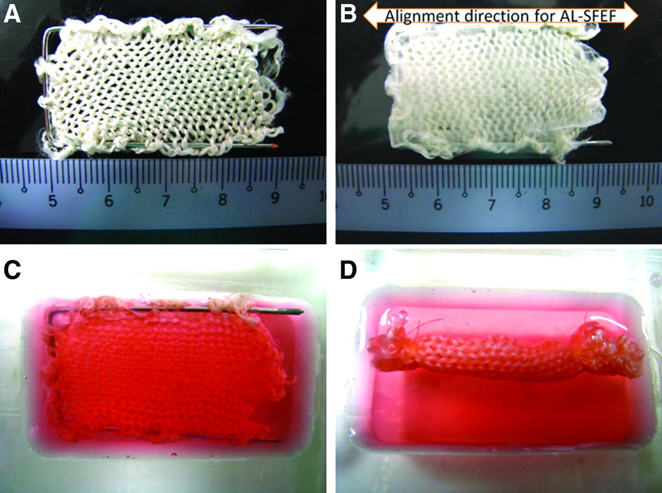
Subsequently, the meshes were integrated with the degummed SF knits (Fig. 1A) to produce the complete hybrid SF scaffolds. To produce aligned hybrid scaffolds (AL), two pieces of AL-SFEF meshes were obtained by gently peeling off from their respective glass slides and laid flatly on both sides of a degummed SF knit, taking note that the SFEFs' alignment direction was in line with the longitudinal axis of the knit (Fig. 1B). Methanol treatment was performed next to allow crystallization and insolubilization of SFEF meshes by transforming amorphous silk I into regular β-sheet secondary structures.23,26,31 As SFEFs have been reported to form a denser structure by methanol treatment, 26 it was exploited to allow the contracting SFEF meshes bind the knitted SF. A two-step process was used to securely bind SFEF meshes onto the knitted SF. Briefly, the first step involved applying methanol selectively to the borders where the two layers of SFEF meshes overlapped to generate localized wetting and contraction, which formed a tight seal between contacting SFEFs at the edges. Such boundary contraction further tensioned the mesh over the knit, forming a well-integrated structure. The hybrid scaffolds were dried before proceeding to the second step, whereby they were immersed in methanol for 30 min under vacuum to promote further integration of the meshes into the SF knit. Hybrid scaffolds with RD-SFEFs (RD) were produced similarly using RD-SFEF meshes. Before cell seeding, the hybrid scaffolds were sterilized by means of formaldehyde (37%) (Mallinckrodt Baker) gassing for 24 h. All other sterile equipment was sterilized by autoclaving.
Scaffold characterization
The general morphology of the hybrid scaffolds was observed and determined via phase-contrast microscopy (IX71 Inverted Research Microscope; Olympus). Scanning electron microscopy (SEM) was conducted to observe the surface morphology of the knitted SF postdegumming and the hybrid scaffolds. Samples were gold-sputtered before observation in a SEM (JEOL Ltd, JSM-5600 LV). Plain knitted SF were observed after the degumming process for presence of sericin remnants to ascertain removal of sericin, whereas the hybrid scaffolds were observed for the integration of the SFEFs with the knit and to determine the diameter and directional distribution of the SFEFs using image analysis software (ImageJ 1.38×; Wayne Rasband, NIH). Fiber diameters (300 sampling points) and the AD (500 sampling points) were determined independently for both the RD and AL scaffolds. For AD analysis, fiber orientations were referenced from a defined vertical direction (0°) and ranged from −90° to 90°. The AD value was then calculated from the angular measurements of fiber orientation using circular statistics as described by Fisher. 32 MATLAB (ver. 6.5 Mathworks) was used to implement the circular statistics algorithms. 33
Fourier-transformed infrared spectroscopy, using the attenuated total reflection method (FTIR-ATR), was performed to determine the secondary structure of SF at different processing stages and verify any conformational changes in the SFEFs as compared to the original degummed SF. Three samples—degummed SF, methanol-treated SFEF mesh, and hybrid SF scaffold—were tested to obtain FTIR-ATR spectra in the spectral region of 1000–2000 cm−1 (Thermo Nicolet, Avatar 360 FTIR spectrometer).
Mechanical tests were conducted on the different scaffold types (RD and AL) with the knitted SF acting as control using a universal testing machine (Instron 3345 Tester; Instron) at standard environmental conditions (20°C, 60% relative humidity). For each group of scaffold, cylindrical specimens (5 mm outer diameter, 20 mm gauge length, n=5) were rolled from laminar form of the hybrid scaffolds along the widths. The specimens were kept moist with phosphate-buffered saline (PBS) solution and loaded to failure by increasing the tensile load continuously at a crosshead speed of 10 mm/min without any pretension or preconditioning. Tensile load and elongation were recorded as represented by the load–displacement curves, where the failure load, linear stiffness, and extension at failure load were determined from. The extent of the toe region relative to displacement was also determined to indicate the degree that different scaffold types could be stretched before linear extension. To eliminate multi-factor contributions such as failure load and stiffness, toe region was determined based on the extent of displacement made with incremental stiffness changes before linear extension instead of the absolute stiffness values involved. In other words, only the extent of the toe region, in terms of the extension, was evaluated and not the rate of stiffness change.
Isolation and culture of MSCs
MSCs were generated from bone marrow aspirates of New Zealand White (NZW) rabbits based on a protocol approved by the Institutional Animal Care and Use Committee, National University of Singapore, using the techniques as reported. 34 Briefly, bone marrow was aspirated from the iliac crest of anesthetized (12 weeks old, 2.5–3.0 kg) NZW rabbits and mononuclear cells were separated by means of their short-term selective adherence to tissue culture polystyrene (TCP) and cultured in complete culture medium containing Dulbecco's modified Eagle's medium with low glucose (Gibco, Invitrogen) supplemented with 10% fetal bovine serum (HyClone Logan), L-glutamine (580 mg/L), and penicillin–streptomycin (100 U/mL). Cultures were incubated at 37°C with humidified 5% CO2. After 72 h, nonadherent cells were removed by changing medium. Upon attaining 70%–80% confluence, adherent cells were detached using 0.05% trypsin and serially subcultured. A homogenous MSCs' population was obtained after 2 weeks of culture and MSCs (P3) were harvested for further use.
Cell seeding efficiency, viability, and proliferation
The MSCs (P3) were resuspended in complete culture medium containing Dulbecco's modified Eagle's medium with high glucose (Gibco, Invitrogen) supplemented with 10% fetal bovine serum (HyClone Logan), L-glutamine (580 mg/L), and penicillin–streptomycin (100 U/mL). About 1.5×10⁶ cells in 1 mL were then seeded by simply pipetting onto one laminar side of each sterile hybrid scaffold, and topped up with 5 mL complete culture medium after 1 h of initial seeding. The same amount of cells were placed into T175 TCP flasks and cultured concurrently as negative control group. The two experimental groups of hybrid scaffolds (RD and AL) were cultured in a laminar manner (Fig. 1C) for 3 days before being rolled up with the cell-seeded surface in the inner core. The rolled-up scaffolds were then continued to be cultured separately in customized six-well polycarbonate dishes, in 8 mL complete medium for each well (Fig. 1D), for another 11 days (total experimental period of 14 days), with medium being changed twice a week.
Cell seeding efficiency was determined at 36 h after initial seeding, whereby the culture medium was collected from the wells or TCP, filled into separate centrifuge tubes, and concentrated and unattached cells were counted. The cell seeding efficiency was expressed as a percentage of the number of cells attached to the scaffold to the total number of cells seeded.
Each group of cultured scaffolds and TCP was assayed for viability and cell proliferation using Alamar Blue™ assay following vendor's instructions (BioSource International) at different time points after culturing for 3, 7, and 14 days. Briefly, the seeded scaffolds (n=5) were incubated in complete culture medium supplemented with 10% (v/v) Alamar Blue™ fluorescent dye for 3 h. Two hundred microliters of medium was then extracted from each sample and measured at 570/600 nm in a microplate reader (Sunnyvale). Cell-free culture medium supplemented with 10% Alamar Blue™ was used as a negative control for this assay.
Cell morphology
Cell morphology was assessed for the hybrid scaffold groups to specifically observe the cellular orientation, distribution, and its interaction with the scaffold architecture. To achieve this, at each time point, both groups of the cultured samples were fixed in 4% paraformaldehyde for at least 15 min and permeabilized with 0.1% Triton-X100 in PBS for 1 min. The F-actin filaments were stained with Texas Red®-X phalloidin (Molecular Probes, Invitrogen Corporation) diluted 1:100 in PBS for 15 min and nuclei stained with 4′,6-diamidino-2-phenylindole, dihydrochloride (Molecular Probes, Invitrogen Corporation) with working concentration of 300 nM in PBS for 5 min. Samples were thoroughly washed three times with PBS before inspection with laser scanning confocal microscopy (Zeiss LSM 510 Meta, Germany). To further observe the cell morphology and cellular interaction with the scaffolds, samples were carefully unrolled and the seeded surface examined by SEM (JEOL Ltd, JSM-5600 LV).
Collagen quantification
The collagen production and deposition by the MSCs in the cultured hybrid scaffolds and TCP were quantified using a picrosirius red-based colorimetric assay (SirCol™ collagen dye binding assay kit, Biocolor Ltd.) following the vendor's protocol. Briefly, at the various time points (3, 7, and 14 days), the cultured hybrid scaffolds (RD and AL) were finely cut and digested with 500 μL of pepsin solution (0.25 mg/mL). For the TCP control, cell culture was removed from the seeded surface via mechanical cell scraping and suspended in PBS. After centrifugation of the cell suspension, 500 μL of pepsin solution (0.25 mg/mL) was added to the cell pellet likewise. Suspensions from the three groups were then shaken at room temperature for 2 h. One milliliter of dye reagent was added to 200 μL of digested solution and mixed for another 30 min at room temperature. The pellet of dyed collagen was then precipitated by centrifugation at >10,000 g for 10 min and then dissolved in 1 mL of releasing reagent. The absorbance of redissolved dye was measured in 96-well plates at absorbance wavelength of 540 nm, from which the collagen amount was derived by extrapolation from standard curve.
Histological assessment
MSC-seeded RD and AL scaffolds (n=3) were harvested for hematoxylin and eosin staining after 7 and 14 days of culture. The specimens were fixed in 10% neutral buffered formalin, paraffin blocked, and sectioned longitudinally (along the lengthwise axis of the rolled-up scaffold). As it was of interest to examine the core of the hybrid scaffolds for cell morphology and continued viability over the 14 days experimentation, longitudinal sections were taken from the core region and stained for histological evaluation. The slides were dehydrated before being mounted on glass cover slips.
Real-time quantitative reverse transcriptase–polymerase chain reaction analysis
To assess fibroblastic differentiation of the seeded MSCs, gene expression for ligament-related ECM proteins such as collagen type I, collagen type III, tenascin-C, and tenomodulin was analyzed and evaluated. Gene expression of bone-related proteins such as osteonectin and osteopontin was evaluated to ascertain the level of osteogenic differentiation and was compared with the ligament-related markers. After 7 and 14 days of culture, total RNA was extracted from the cultured hybrid scaffolds (RD and AL) and TCP controls (n=3) using the RNeasy Mini Kit® (Qiagen) according to the vendor's protocol. RNA concentration was determined by using nanodrop (NanoDrop Technologies) and 200 ng RNA was used to synthesize cDNA with Iscript cDNA synthesis kit (Biorad Laboratories). Quantitative reverse transcriptase–polymerase chain reaction (QRT-PCR) was performed using QuantiTect SYBR-Green PCR kit (Qiagen) to quantify the transcription level of ligament-related genes, including collagen I, collagen III, tenascin-C, and tenomodulin, and bone-related genes including, osteonectin and osteopontin, using glyceraldehyde 3-phosphate dehydrogenase as the reference gene. The primer sequences used, as summarized in Table 1, were obtained from published literature18,35–37 and were synthesized by Aitbiotech Pte Ltd. cDNA (1 μL) from each sample was mixed with 10.0 mL of QuantiTect SYBR Green PCR master mix, 0.25 mL of each primer, and 8.50 mL of RNase-free water. Quantitative real-time PCRs were carried out and monitored using a Stratagene Mx3000P system. Reaction was done at 95°C for 15 min, followed by amplification for 40 cycles, which included a denaturation step at 95°C for 15 s and an extension step at 60°C for 1 min. The amplification was performed in duplicates, and transcription levels of the target genes were normalized to glyceraldehyde 3-phosphate dehydrogenase before analysis using the 2ΔCt formula with reference to undifferentiated MSCs (P3).
Collagen I, Collagen III, and GAPDH sequences obtained from ref. 18
Tenascin-C sequences obtained from ref. 35
Tenomodulin sequences obtained from ref. 36
Osteonectin and osteopontin sequences obtained from ref. 37
GAPDH, glyceraldehyde 3-phosphate dehydrogenase.
Western blot analysis
After 7 and 14 days of culture, cultured hybrid scaffold groups were digested with pepsin (200 mg/mL in 0.5 N acetic acid; Sigma-Aldrich) for 72 h at 4°C for total protein extraction. Upon pepsin inactivation using 10 N NaOH, the protein extract was concentrated using a Microcon 30 centrifugal filter (30,000Mw cutoff; Millipore Co). The concentrated protein extracts of each sample were then individually mixed with laemmli buffer and 50 mM dithiothreitol solution, put to 3%–8% sodium dodecyl sulfate–polyacrylamide gel electrophoresis, and blotted onto nitrocellulose membranes. Subsequently, Western blot was carried out using the Western blot kit following vendor's protocol (Zymed Laboratories, Invitrogen). Briefly, the membranes were blocked with blocking buffer for 1 h and incubated at 4°C overnight with diluted (1:500) primary monoclonal antibodies. The specific primary antibodies used were mouse anti-type I collagen monoclonal antibody (Sigma-Aldrich), the mouse anti-type III collagen monoclonal antibody (Millipore), and the mouse anti-tenascin-C monoclonal antibody (Millipore). The membranes were then washed with washing buffer five times before incubating with secondary antibodies diluted to 1:200 in blocking buffer for 30 min. After washing with washing buffer again, the membranes were incubated with enhanced chemiluminescence working solution for 5 min. Band signals were detected and relative band intensities were obtained and compared among the groups.
Biomechanical test on cultured hybrid scaffolds
Tensile tests were performed for both groups of hybrid SF scaffolds at different time points (7 and 14 days) following the aforementioned protocol. Unseeded hybrid scaffolds (blank RD and blank AL) were tested as control group. The load (N) and extension (mm) data were collected over five samples for each group and time point, and the failure load, elastic region stiffness, extension at maximum load, and extent of toe region were determined after plotting the load displacement curves.
Statistical analysis
Single-factor analysis of variance technique and post-hoc Tukey tests were used to assess the statistical significance of multiple comparisons. For pair-wise comparisons, two-tailed, unpaired Student's t tests were used. GraphPad Prism ver. 5 (GraphPad Software) was used to implement the statistical analysis. All data were expressed as means±standard deviation, and p<0.05 was considered statistically significant.
Results
Scaffold characterization
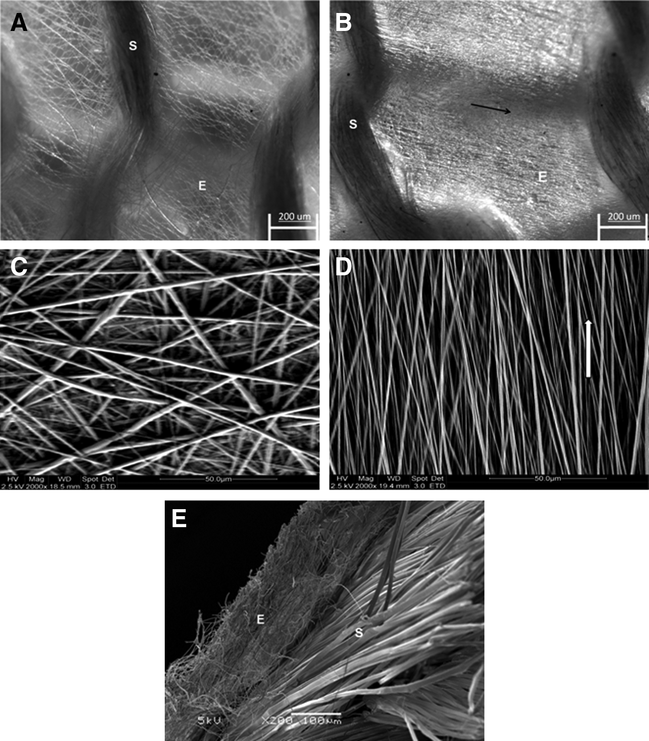
Knitted SFs, as observed using the SEM, had smooth SF fibers and were clear of sericin after the degumming process. Gross observations showed that the degummed knitted SFs had macro-pore sizes of about 1.0 mm diameter (Fig. 1A), which was covered uniformly by the SFEF mesh on both sides of the knit to facilitate cell seeding (Fig. 1B). Both groups of the hybrid SF scaffold were revealed to be highly porous with interconnected pores uniformly distributed throughout the scaffold (Fig. 2A–D), with pore sizes ranging from 1 to 60 μm for the equiaxed pores of RD scaffolds, and 1 to 20 μm (minor axis) and 10 to 100 μm (major axis) for the elliptical pores of AL scaffolds. The SFEF meshes were also observed to be well integrated with knitted SFs after the two step binding process, which involved utilizing the contractile forces from methanol treated SFEF (Fig. 2E). The dimensional difference of the SFEFs and the knitted SF fibers, both of which composed the hybrid SF scaffold, was also exhibited in this figure. Specifically, the SFEFs had diameters of 1211±441 nm (RD-SFEFs) and 796±111 nm (AL-SFEFs), whereas the degummed SF fibers had diameters of 11.7±1.69 μm. The smaller AL-SFEF fiber diameter was due to the rotation of the grounded rotating frame, which collected and exerted a pulling force on the electrospun jet, and consequently reduced the dimensions of the AL-SFEF fibers.
Scaffold morphology of hybrid SF scaffolds:
SFEFs of the AL hybrid scaffolds were observed to exhibit single directional alignment, whereas there was no observable alignment in the RD types (Fig. 2A–D). Histograms plotted for the angular distributions of SFEFs (Fig. 3) indicated significant alignment of the AL group (AD=4.8°) as compared to RD group (AD=51.8°).

Histograms representing angular distributions of SFEFs:
Conformational analysis of SF at various stages of the scaffold fabrication process was performed using FTIR-ATR. Specific to the secondary protein structure of SF, strong absorption bands at 1655 cm−1 (amide I), 1540 cm−1 (amide II), and 1230 cm−1 (amide III) were attributed to the random coil conformation, whereas absorption bands of 1628 cm−1 (amide I), 1533 cm−1 (amide II), and 1260 cm−1 (amide III) were assigned to the β-sheet conformation.38–40 From the FTIR-ATR spectra obtained (Fig. 4), it was observed that SF upon degumming exhibited peaks at 1655 cm−1, which corresponded to the amide I region of the random coil structure (Fig. 4a). Peaks of the amide I region for random coil was not apparent in the methanol-treated SFEF mesh (Fig. 4b). Upon overall methanol treatment to the hybrid scaffold, there was an overall conformational transformation by reduction in random coils to greater proportion in β-sheets, as observed from the FTIR-ATR spectra (Fig. 4c). This was indicated by the absence of peaks corresponding to random coil structures, especially at the amide I regions. The spectra indicated that the dissolution and electrospinning process did not significantly alter the SF protein conformation as it was preserved as β-sheets, which is the native conformational state of SF fibers.
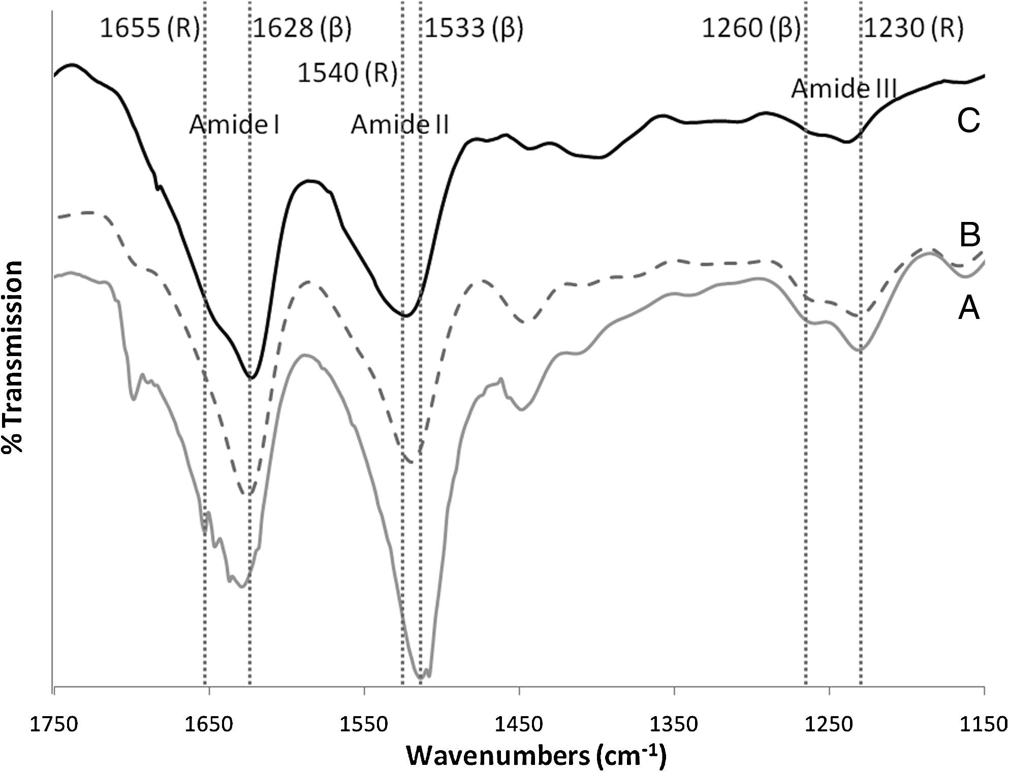
Fourier-transformed infrared spectroscopy, using the attenuated total reflection method of (A) degummed SF, (B) methanol treated SFEF mesh and (C) hybrid SF scaffold.
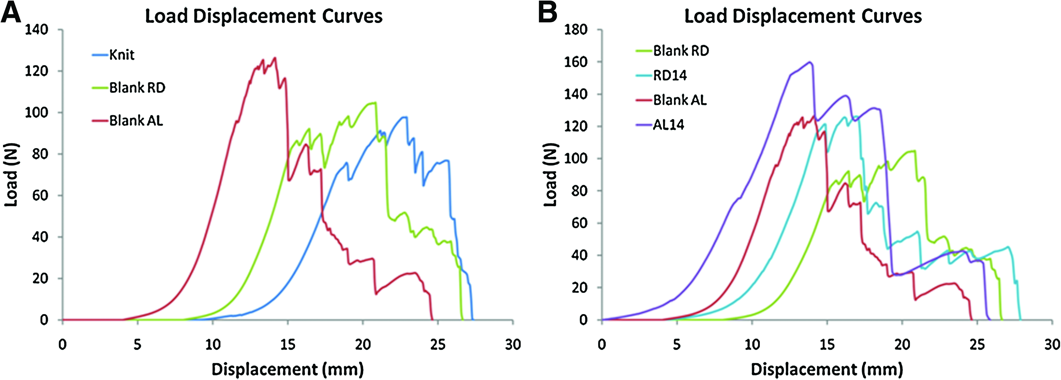
Rolled-up blank RD and AL hybrid scaffolds were tested for their tensile properties against rolled-up knitted SF (Table 2). All specimens were tested to failure and the rupture sites were noted to consistently initiate at the mid zone of the gauge length. The load–deformation curves recorded comprised the toe region, linear region, microfailure region, and failure region, which were similar to the curve of native ACLs (Fig. 5A). The microfailures were generally attributed to the failure of knitted SF microfibers in tandem with extension of the construct. The tensile properties of blank AL were significantly better (p<0.05) than both the knitted SFs and blank RD in terms of the failure load (129.21±7.43 N) and stiffness (22.12±1.22 N/mm). No significant difference was noted for the extension at maximum load across the three groups (p>0.05). The extents of toe regions were 2.44±0.94 mm, 3.30±1.04 mm, and 2.70±1.11 mm for knit, blank AL, and blank RD, respectively, with no significant differences identified among the three groups (p>0.05).
Representative load-displacement curves for different
p<0.05 when compared with knitted SF (for blank scaffolds) or compared with RD scaffolds at each time point (for cultured scaffolds). SF, silk fibroin; RD, randomly arranged; AL, aligned.
MSC adhesion, viability, and proliferation
Cell attachment rate for all three groups (RD, AL, and TCP) after 36 h from seeding was around 93% of the total amount of seeded MSCs per scaffold. Alamar Blue™ assay revealed that, as compared to the RD and TCP groups, cell viability was significantly higher in the AL hybrid scaffolds after 7 days of culture (24% more on day 7 and 25% more on day 14 when compared to RD; p<0.01; Fig. 6). There was also consistent proliferation in the AL group, whereas the proliferations in the RD and TCP groups were not statistically significant (p>0.05) within the 14 days of culture period.
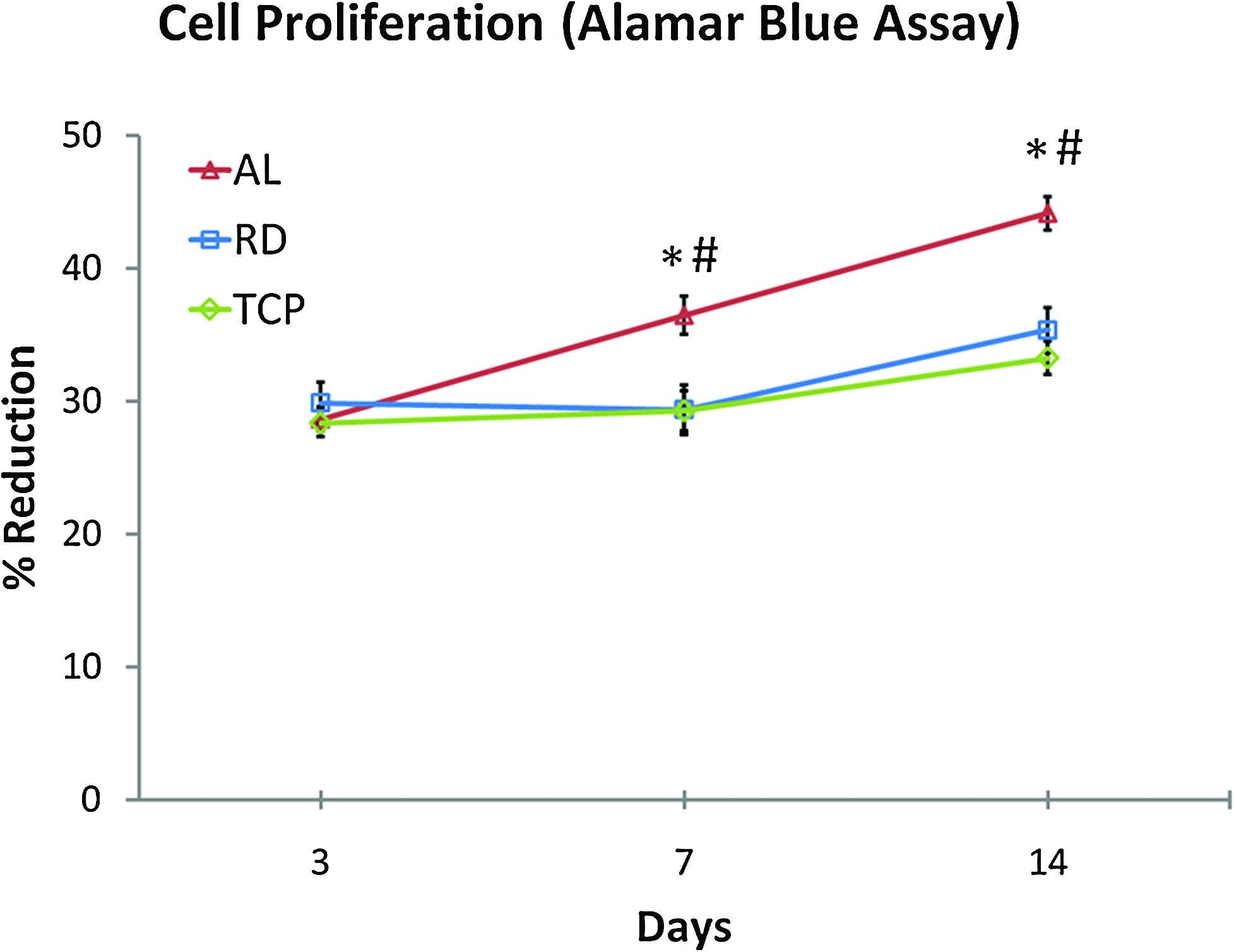
Alamar Blue™ assay illustrating consistent and significantly more viable cells in the AL group compared to other groups, respectively, from day 7 onward (#p<0.01, Student's t-test, n=5), with significant cell proliferation (*p<0.05, analysis of variance and post-hoc Tukey tests, n=5) through the 14-day culture. Color images available online at
Cell morphology
After 3 days of culture, the MSCs seeded onto AL had already developed spindle-shaped morphologies and were oriented in the direction of SFEF alignment (Fig. 7B). This was apparent when compared to the MSCs' equiaxed cell morphology with minimal ellipticity when cultured on RD (Fig. 7A). From the confocal micrographs taken on days 7 and 14, cells cultured on AL continued to align along the length of the scaffolds (Fig. 7D, F) after the hybrid scaffolds were rolled up to form ligament analogs on day 3, whereas no apparent directionality was observed in cells cultured on RD (Fig. 7C, E). Qualitatively, increase in cell density was observed in both groups over the 14-day culture period (Fig. 7C–F), with near confluence observed at the cell-seeded layers by day 14.
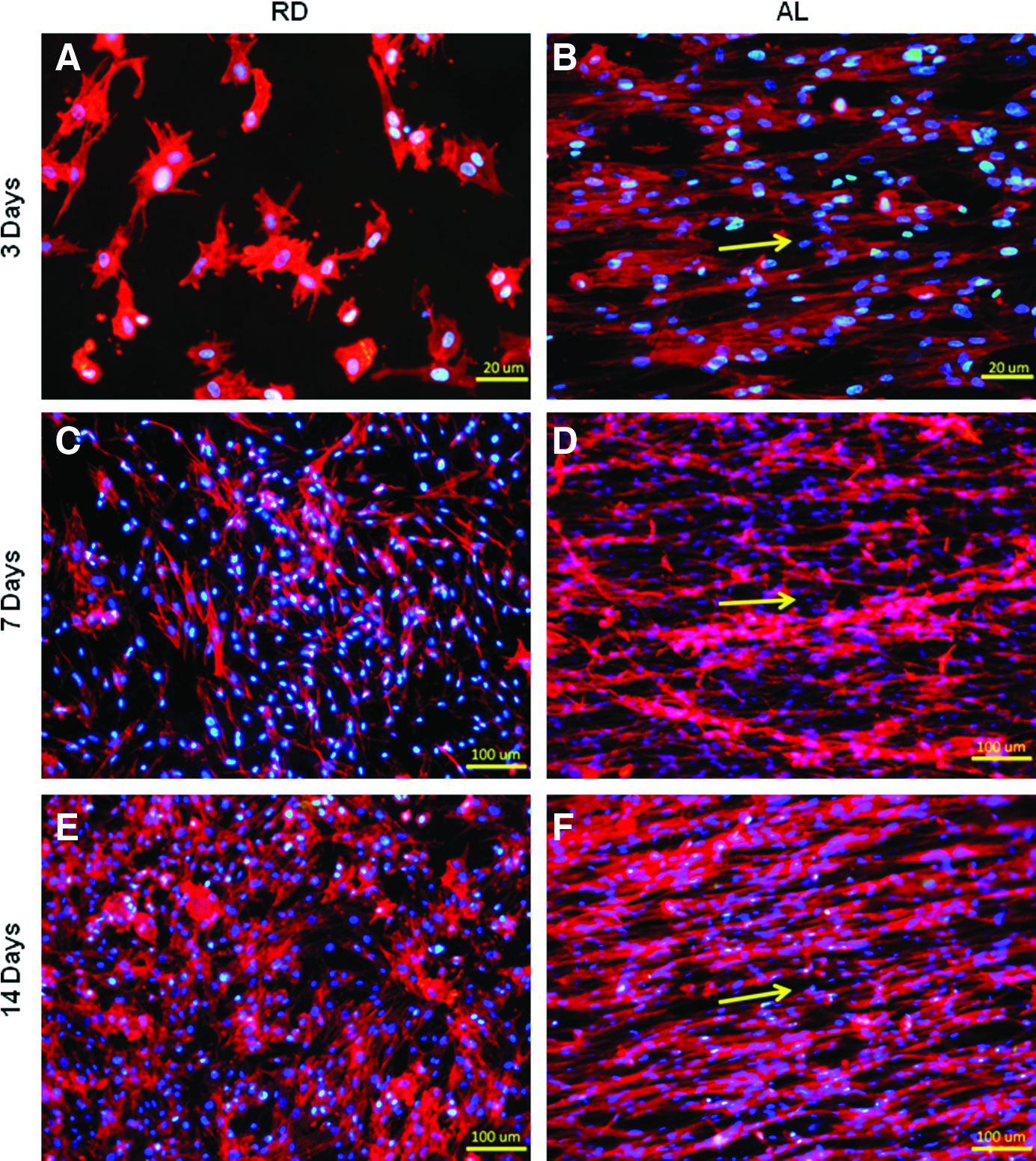
Confocal micrograph illustrating actin fibers (red) and nuclei (blue) of fluorescent-stained MSCs seeded on
SEM images taken for seeded surfaces, after carefully unrolling the hybrid scaffold constructs, revealed that MSCs were attached to and elongated along the direction of localized SFEF orientation (Fig. 8A, B). As compared to the AL scaffolds, MSCs attached to RD formed projections along the SFEFs they were attached to, thereby unable to generate elongated morphologies as the surrounding SFEFs themselves were randomly arranged. As a result of cell alignment at an early stage of culture (3 days post seeding), cells on AL began to form ECM network across cell colonies, whereas no obvious ECM network was formed in RD (Fig. 8C, D). More extensive ECM networks were seen at day 14, with more uniform and continuous ECM network observed for AL as compared to RD (Fig. 8E, F). In addition, cells on AL began to form 3D tissue-like oriented bundles along the direction of SFEF alignment by day 14, which was not apparent in the RD group (Fig. 8G, H). Such 3D structures indicated that cross-layer ECM networks were likely to be forged, and consequently, cellular extensions were made to bridge with the adjacent AL-SFEF layer of the rolled-up AL hybrid scaffold.
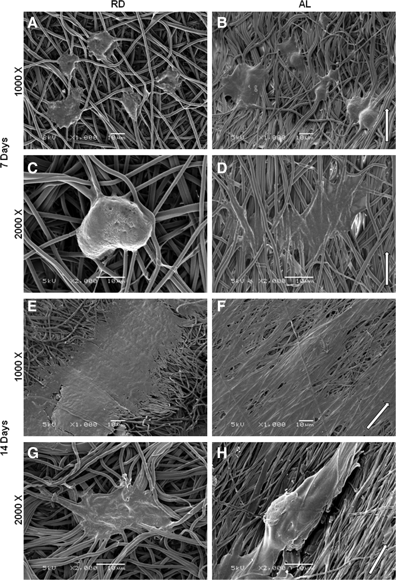
SEM images of MSC-seeded
Collagen synthesis
Insoluble collagen assay was performed to determine the amount of deposited collagen on the hybrid scaffolds and TCP as an indication of the extent of ECM formation. This quantification assay revealed that there was significant increase (p<0.05) in collagen production and deposition in the hybrid scaffolds over the culture duration (Fig. 9). MSCs cultured on TCP, however, did not have significant increase in collagen deposited (p>0.05) until 14 days after seeding. Between the groups, significant difference was found between AL and both RD and TCP at day 14, with AL having 33.5% more collagen deposited than RD and 66.3% more than TCP (p<0.01).
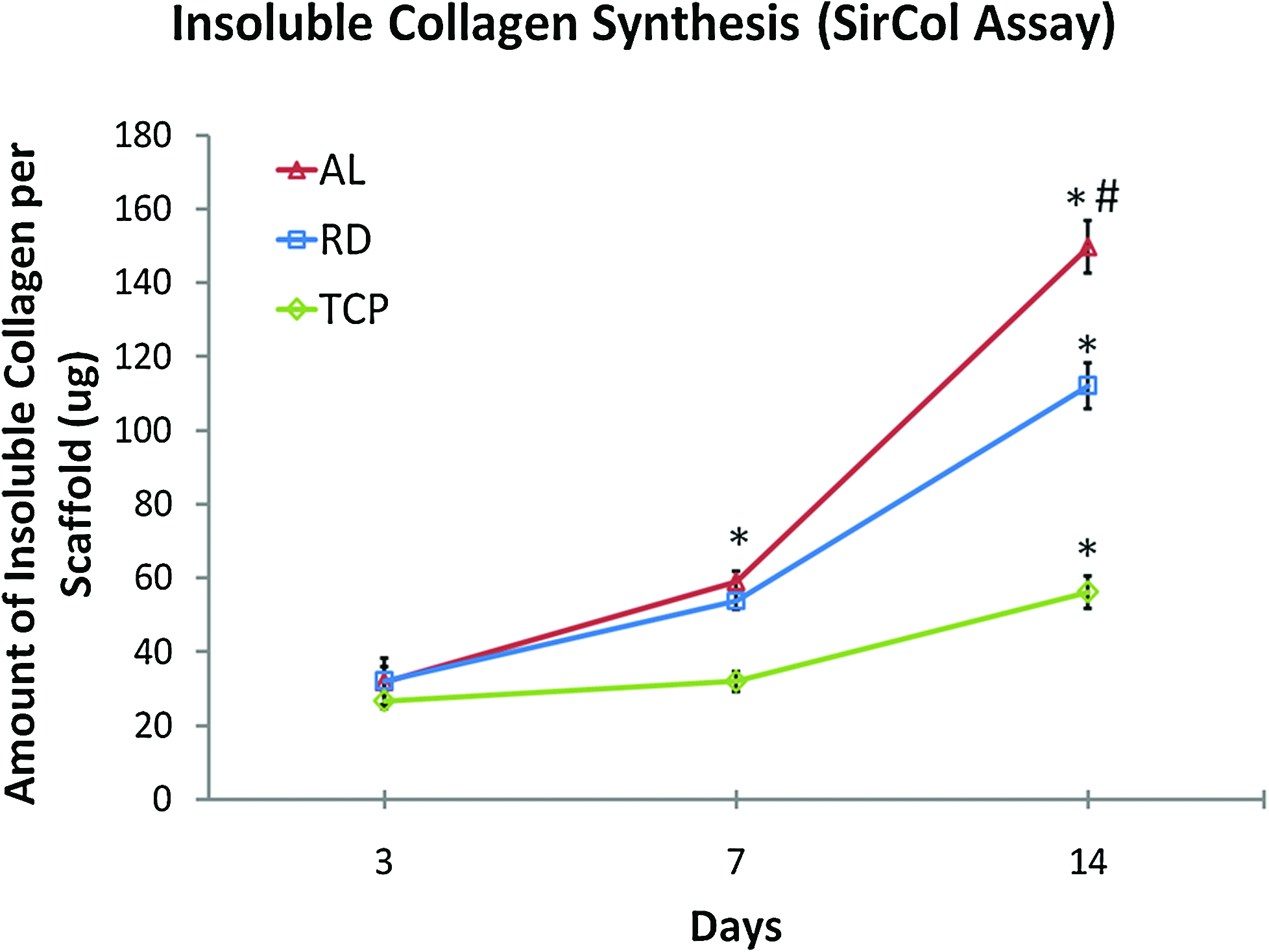
SirCol™ assay for amount of collagen deposited per scaffold/culture sample. Significant increase in collagen deposition was observed in the RD and AL groups at both day 7 and day 14 from the respective previous time points (*p<0.05, analysis of variance and post-hoc Tukey tests, n=3). Significantly more collagen was deposited in the AL group as compared to other groups, respectively, at day 14 (#p<0.01, Student's t-test, n=3). Color images available online at
Histological analysis
Hematoxylin and eosin staining was performed on longitudinal sections of the inner core of RD and AL after 7 and 14 days of culture to assess cell proliferation and ECM production in the two groups qualitatively (Fig. 10). Consistent with previous observations, aligned spindle-shaped cells with elongated processes were also observed in the AL core sections (Fig. 10B, D), whereas spherical or equiaxed cells were observed in the RD core sections (Fig. 10A, C). Histological images also revealed observable increase in cell density and distribution of the core of AL, whereas, interestingly, no observable improvement in cellular distribution was found at the core of RD. This indicated that the cells remained viable and proliferative in the core of AL constructs but for RD, and it was limited to just an increase in cell colony sizes, with minimal interconnected colonies formed. Despite the difference in cellular distribution between the two types of hybrid scaffolds, both were shown to be able to support cellular viability at the core due to the interconnected porous structures of the hybrid scaffolds.
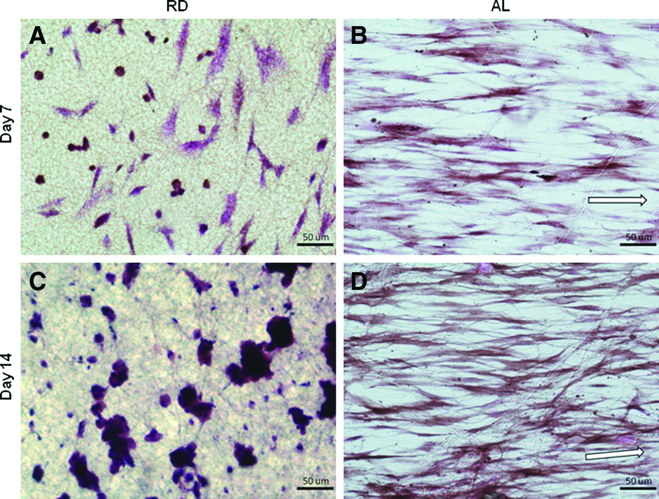
Histological evaluation of MSC-seeded
Gene expression of ligament-related ECM proteins using real-time QRT-PCR
The expression of ligament-related genes in TCP, RD, and AL were evaluated via real-time QRT-PCR. It was revealed that expression levels for collagen I were significantly upregulated in the AL group compared to the RD and TCP groups from day 7 onward (Fig. 11A), whereas collagen III and tenascin-C expression levels were significantly upregulated in the AL group compared to the other two groups on day 14 only (Fig. 11B, C; p<0.05). However, there was no significant difference in tenomodulin expression between the groups through the 14-day culture period (Fig. 11D, p>0.05). All target genes were significantly upregulated in the AL group from day 7 to day 14 (collagen I: 92.7% higher, collagen III: 146.8% higher, tenascin-C: 19.6% higher and tenomodulin: 32.8% higher; p<0.05), which was not the case for the other two groups.
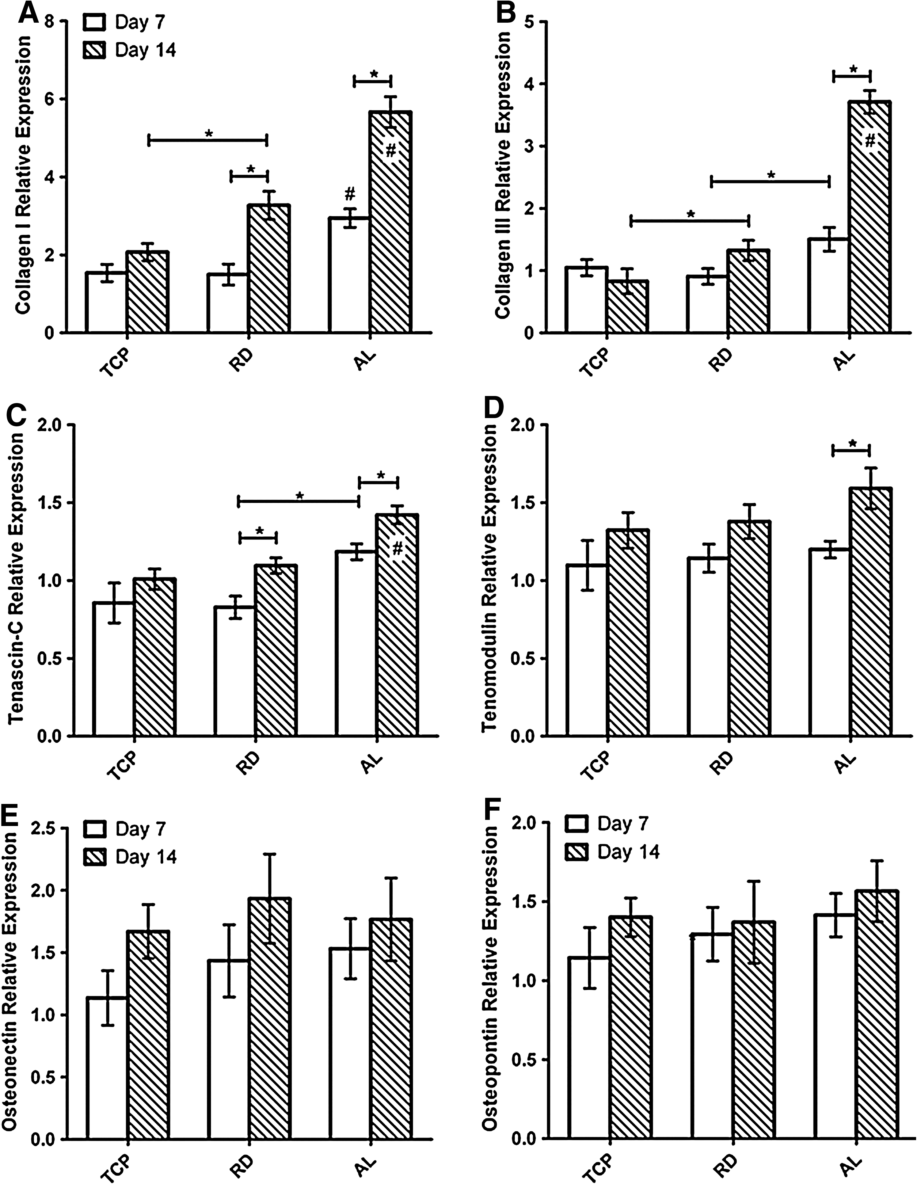
Gene expression for MSCs cultured in TCP, RD and AL groups for 7 and 14 days.
On the other hand, gene expression for osteonectin and osteopontin was not significantly upregulated in all the groups (Fig. 11E, F; p>0.05). Although osteonectin expression was slightly upregulated as compared to osteopontin during this experimental period, the upregulation was not significant. Relative to the ligament-related gene expression, the low expression of osteogenic markers indicated that ligament fibroblast remained the dominant lineage in the hybrid SF constructs.
Collectively, these results indicated that AL hybrid scaffold could stimulate upregulation of ligament-related gene expression at a faster rate than the other two groups and enhance differentiation of MSCs to ligament fibroblasts.
Western blot analysis
Protein expression for collagen I, collagen III, and tenascin-C was detected for RD and AL groups after 7 and 14 days of culture. Analysis was performed on densitometric data of the optical intensity of each lane expressed in the western blot membranes, which was normalized to RD cultured for 7 days. The results demonstrated that the matrix of the cultured ligament analogs composed of mainly type I and III collagen and tenascin-C, with collagen I being predominant as its expression was consistently higher in both RD and AL when compared to collagen III and tenascin-C (Fig. 12). Significantly more collagen I was expressed in AL than RD from day 7 onward (day 7: 169.7% more, day 14: 99.4% more), whereas significance was found for collagen III and tenascin-C on day 14 (collagen III: 121.8% more and tenascin-C: 123.6% more; p<0.05). For RD, significant increase from day 7 to day 14 was found for collagen I only (246.9% more), but for AL, it was found for all three proteins tested (collagen I: 156.5% more; collagen III: 134.8% more; tenascin-C: 73.2% more; p<0.05). Similar trends were observed in the RT-PCR results as shown previously.
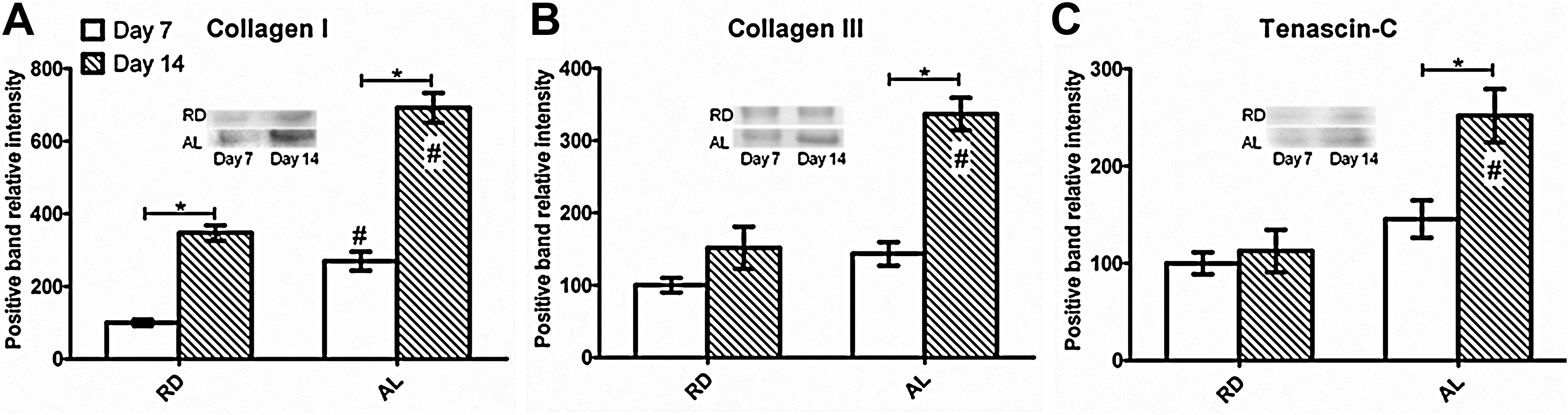
Western blot analysis of ligament-related ECM proteins, including
Tensile properties of cultured hybrid scaffolds
Cultured rolled-up hybrid scaffolds (RD and AL at day 7 and 14) were tested for their tensile properties against rolled-up blank RD and AL hybrid scaffolds (Table 2). The specimens were tested to failure under wet conditions and rupture was noted to initiate from the center region of the entire gauge length, though exact rupture site was inconsistent across samples. Load–displacement curves plotted revealed the toe region, linear region, microfailure region, and failure region, similar to that of native ACLs (Fig. 5B). Similar to the blank scaffolds, microfailures were observed in the cultured hybrid scaffolds, which were generally attributed to SF knitted microfiber failure in tandem as the construct was extended. For both RD and AL, there was significant increase in maximum load (RD: 18.5% increase and AL: 22.6% increase) and stiffness (RD: 11.7% increase and AL: 18.5% increase) from the respective blank hybrid scaffolds only after 14 days of culture (p<0.05). Through the 14 days culture period, AL had significantly higher maximum load and stiffness than its RD counterpart of the same culture duration (day 7 and day 14; p<0.05), with AL being 25.9% stronger and 22.2% stiffer than RD after 14 days of culture. Both the RD and AL groups had significantly larger extents of toe region after the 14 days culture than the respective blank hybrid scaffolds, with RD having 6.48±1.45 mm and AL having 5.05±1.23 mm by day 14 (p<0.05). This indicated that after culture for 14 days, both RD and AL could be stretched over significantly larger displacements before linear extension than their cell-free counterparts. However, no significant difference was found in the toe region extents between RD and AL after 14 days of culture (p>0.05). No significant differences were noted for the extension at maximum load across the groups and at different time points (p>0.05).
Discussion
From literature review, we are confident that this study is the first to report on the development and characterization of a mechanically functional full silk scaffold with customizable aligned topographical cues for the regeneration of ligament. Enhanced cell proliferation, cell viability, and ECM production were observed in the AL hybrid scaffold when compared with both the RD type and the two-dimensional (2D) culture on TCP. MSCs were observed to attach in an aligned fashion in the direction of SFEF alignment from as early as 3 days postseeding and continued to expand in cell density over the 14 days culture period. More importantly, there was no loss of directionality in cellular elongation as aligned spindle-shaped cells were observed consistently after 7 and 14 days of culture whereby the hybrid scaffolds were rolled up into the cylindrical ligament analogs. The construct stimulated upregulation of gene expression for ligament-related ECM proteins, which consequently induced increased deposition of collagen and aligned ligament-related ECM components. The increased aligned ECM deposition subsequently improved the overall mechanical properties of AL constructs. These results indicated that the hybrid SF scaffolds with AL-SFEFs are suitable for functional tissue engineering of the ligament.
The mechanical advantage of the hybrid SF scaffold is largely attributed to the knitted SF mesh. SF has been shown to have superior mechanical properties and suitable for use in tissue engineering of load-bearing structures,14,19,20,22,23 particularly when SF is obtained from an optimal degumming process that focuses on retention of SF structure. 20 The protein structural arrangement of SF is critical in determining the mechanical properties of the natural polymer. It depends on whether SF exists as Silk I, which consists of α-helix and random coil structures, or Silk II, which consists of anti-parallel β-sheets, whereby the polypeptide main chains are aligned and adjacent chains connected by hydrogen bonds.41–44 Of the two, Silk II is considered the more mechanically viable form due to its high crystalline state. The SF fibers used to fabricate the knitted silk mesh of hybrid SF scaffolds were generally of the Silk II conformational state. However, even though the degumming process had been controlled and optimized to reduce conformational transition of SF, a small level of conformational shift from Silk II to Silk I occurred in the degummed SF knits as shown from the FTIR-ATR analysis performed. It is believed that through the degumming process, certain degree of SF degradation had taken place via penetration of the degumming agents into microvoids and amorphous noncrystalline region of SF's intermolecular structure.45,46 With this disruption in the SF secondary structure, leading to an increase in the amorphous random coil regions, the tensile behavior and physical properties of SF might be altered. This is consistent with Jiang et al.'s results, whereby it was found that during degumming, the original molecular order of SF was disrupted, leading to reduction in crystallinity. 47 The conformational change can be reversed upon immersion of methanol as observed in the FTIR-ATR performed for methanol treated hybrid SF scaffold (Fig. 4). From the FTIR-ATR results obtained, it was also verified that the structural conformation of SFEFs was preserved as Silk II after methanol treatment.
The knitted architecture was chosen from a variety of textile architectures. Twisted and braided scaffolds, comprising near-parallel microfiber bundles, are close analogs of a functional ligament in terms of morphology and mechanical characteristics.14,23,48 However, limited inter-fiber space in such configurations impeded cell infiltration especially at the early tissue regeneration phases.15,49,50 In the study conducted by Cooper et al.,
15
cells remained concentrated at the periphery of braided poly-
The AL hybrid scaffold not only has the micron-scale SF knit, which caters to the mechanical aspect of the construct, but also composes of the sub-micron AL-SFEF mesh, which acts as a seeding substrate that provides topographical cues to stimulate cellular alignment and consequently aligned ECM deposition. The aligned surface topology is the typical native environment of ACL fibroblasts. In fact, preferential alignment forms the basis of anisotropicity, which is fundamental to musculoskeletal tissues. 51 This is apparent when normal ligaments are compared with repaired ligaments. The reparative process of injured ligaments typically involves three phases: the acute inflammatory or reactive response phase, the regenerative or repair phase, and the tissue remodeling phase. 4 Upon acute inflammatory response, healing matrix consisting of randomly aligned collagen and amorphous ground substance begin to form. The repair and regeneration phase then follows, whereby the healed matrix becomes gradually more organized with time. Tissue remodeling thus follows lasting a year or longer. Although the whole healing process can take longer than a year with discrete reparative phases, the collagen fibrils laid down by the fibroblasts in fact remain relatively disorganized and surrounded by amorphous ground substance. As a result of that, the properties of repaired ligaments are inferior to the normal ligaments. In actual fact, the lack of collagen alignment can shift the dynamics of continuous ligament remodeling toward degradation and reduction in the load-bearing capacity of newly formed tissue. 14 In view of this, the hybrid SF scaffold with AL-SFEFs for early cellular alignment and consequently alignment of the ECM and collagen fibrils may be relevant.
Using electrospinning, aligned fibers have been produced to act as topographical cues or guidance for cells. Xu et al. 52 compared the cell–cell adhesion and proliferation of human coronary artery smooth muscle cells on TCP, polymer film, and aligned polymer fiber. They observed that smooth muscle cells migrated along the axis of the aligned fibers and expressed spindle-like contractile phenotype. On the other hand, Baker and Mauck 53 employed poly(ɛ-caprolactone) fibrous scaffolds for the tissue engineering of the meniscus. They seeded MSCs on aligned and random fibers, with results demonstrating that the aligned fibrous scaffolds can serve as a micro-pattern for directed tissue growth and produce constructs with improved mechanical properties compared to random scaffolds. Yim and Leong 54 had also presented a comprehensive review of cellular interaction with nanoscale topography. Their findings also indicated that cells respond to topography of synthetic substrates of the nanometer and sub-micron range in terms of adhesion, proliferation, migration, and gene expression. Other than these studies that utilized synthetic polymers, contact guidance had also been implemented via naturally derived bioscaffolds. Almarza et al. 55 had demonstrated the use of elongated small intestinal submucosa to create aligned small intestinal submucosa fibers and applied it as bioscaffold for tendon/ligament tissue engineering. Positive responses were seen in the cultured bone marrow-derived cells seeded in such bioscaffolds as well. Nevertheless, issues pertaining the lack of the source, difficulty in controlling the quality or degree of fiber alignment, and infection due to cross or intra-species transplantation persist.
The novelty of this study thus involves the development of a consistent and reproducible aligned hybrid SF scaffold that not only supports accelerated formation of aligned collagen fibers, but is also mechanically functional, making it suitable for ligament tissue engineering applications. The advantages of the AL hybrid SF scaffold were exemplified in the cellular viability and proliferation results, whereby cells seeded on AL were significantly more viable after 7 days of culture and had consistent proliferation throughout the 14 days culture, which was not apparent in the other 3D RD system or the 2D TCP system. As compared to 2D culture systems, the 3D culture systems could provide environmental cues closer to the native tissue to stimulate cell-surface receptors and adhesion sites that regulate cell cycle and gene expression for key ligament ECM components.14,56–58 The AL hybrid SF scaffold further enhances these responses by bearing surface chemistry and topographical similarities with the native ECM. The aligned architecture also facilitates medium intake into the core of the scaffold construct through active capillary action rendered by the AL-SFEFs. Consequently, higher cell densities with uniform cellular distribution were observed in the inner core of AL than RD.
With the ability to stimulate elevated proliferation, high cell density of aligned spindle-shaped cells could be formed within the 14 days culture, contributing to the formation of extensive networks of aligned ECM as observed. Further to that, the formation of early stage collagen fiber bundles was apparent in the 3D culture of AL as bridging structures were observed in the SEM images of carefully unrolled AL scaffolds. These bridging structures were possibly adhesion points with adjacent aligned ECM layers in the attempt to form 3D collagen fiber bundles. This observation was complemented with the gene expression results, which indicated significant upregulation of ligament-related genes in the AL group after 14 days of culture. These ligament-related genes were indicative of the differentiative phase that the seeded MSCs underwent. Similar method for evaluating MSCs differentiation to the ligament fibroblast was performed by several other groups as well.17,18,34,35 Although tenomodulin was not significantly upregulated in the AL group, there was significant upregulation at day 14 from day 7, indicating imminent increase in expression of tenomodulin, which is associated with increase in collagen fibril diameter during ligament development. 59 Coupled with the increase in ligament-related ECM protein detected in the AL group, it was clear that AL stimulated accelerated ligament tissue formation and maturation, with close structural similarity to the native tissue.
Other than the advantageous aligned topographical cues, MSCs were stimulated to differentiate toward the ligament fibroblast lineage, instead of other lineages such as bone, possibly due to the presence of unbalanced stress states within the rolled up scaffold. Such stress states arose as lengthwise tension and circumferential relaxed states existed when the laminar scaffold was being rolled up. This internal stress state might provide some mechanical cues to trigger ligament fibroblast differentiation. However, further studies should be done on different scaffold conformations to ascertain this effect.
The formation of aligned ECM structure was then translated to the superior mechanical properties of the AL constructs as compared to the RD constructs. Specifically, AL was observed to be significantly stronger and stiffer after being cultured with MSCs for 7 and 14 days as compared to the RD scaffold type. Upon culture for 14 days, the AL and RD hybrid scaffolds exhibited significantly extended toe regions of nonlinear increase in load–displacement than the respective blank hybrid scaffolds. It could thus be deduced that the ECM deposited in both the AL and RD contributed to the tensile loading, with the discontinuous and nonmature ECM structures being loaded before the hybrid scaffold. Interestingly, such loading pattern is also very similar to the native ACL, whereby wave or “crimp” patterns of the fibrils exist in the matrix to provide loading of fibrils in tandem via recruitment to “buffer” for slight elongations without incurring overall fibrous damage60,61 and to provide shock absorbance for sudden lengthwise loading. 62 Tensile tests performed on native ACL also revealed similar nonlinear load–displacement curves, as a consequence of gradual increase of tissue stiffness. 63
Although the regenerated tissue contributed to the increase in tensile properties of the cultured hybrid scaffolds, majority of the loading capacity came from the hybrid SF scaffold itself. The hybrid SF scaffold was designed to accommodate proteolytic degradation in vivo with the knit being customized to support more than twice the maximum ACL force in rabbits. The maximum rabbit ACL force experienced was estimated at 40 N, which was about 138.6% of rabbit body weight (2.5–3.0 kg). 64 Although proteolytic degradation of SF was minimal for in vitro cultures, drastic decrease in tensile strength had been reported to range from 55% at 6 weeks to 73% at 30 days after subcutaneous implantation of silk scaffolds.23,65 A 50% decrease in SF scaffold failure load was also observed after 24 weeks in an in vivo study conducted by our group for rabbit ACL replacement. 17 Therefore, the knitted SF structure in this study could withstand the maximum ACL load even if a 50% decrease in failure load due to proteolytic degradation were to be effected within 24 weeks of implantation. This was notwithstanding the fact that the loading capacity was increased due to the SFEFs incorporated and the ECM that would be deposited. The tensile stiffness of knitted SF, on the other hand, was less than half of the native ACL tissue. 17 Stiffness was increased after SFEFs were incorporated to the SF knits, which was especially so for the AL hybrid scaffold type as its stiffness was about 50% of the native tissue. Having lower scaffold stiffness as such can prevent stress shielding and allow mechanical forces that are subjected to the scaffolds to be effectively conducted to the attached cells. This strategy fall in line with the principles of functional tissue engineering 66 and will be especially important when the cell-seeded scaffolds are to be mechanically conditioned either in vitro or in vivo to further enhance the proliferative and differentiative potential of MSCs on the hybrid SF scaffolds. This stiffness property can complement the effective mechano-transduction property of AL-type hybrid scaffold to promote functional regeneration of the ligament tissue.
While the production of ligament-related proteins (collagen I, collagen III, and tenascin-C) was clearly improved in the seeded AL constructs, studies relating to the distribution pattern of these proteins within the scaffold will be necessary. It will also be interesting to investigate collagen fiber formation and distribution in the transverse cross section of the constructs to identify the distribution pattern of mechanically contributing collagen fibers and ascertain collagen bundles formation. It is speculated that an essential benefit of culturing seeded AL scaffolds is the ability for AL-SFEFs to effectively transfer axial strain to the attached aligned MSCs. From the works conducted by Lee et al., 29 enhanced ECM production as a result of effective mechano-transduction due to aligned electrospun polyurethane fibers were observed for human ligament fibroblasts. As such, it will be of interest to investigate further the effect of differentiative cues, in the mechanical form, on fibroblastic differentiation of MSCs in our 3D aligned SF construct and be studied over longer durations. Apart from dynamic loading provided by a mechanical bioreactor in vitro, in vivo studies will also be necessary in the future to fully assess the functionality of the regenerated ACL with specific emphasis on the cellular and protein distribution within the scaffold construct and the biomechanical properties generated. By conducting the in vivo studies, long-term ECM buildup within the scaffold system can also be studied to ascertain that the construct remains viable even with ECM buildup. As part of the future work to further enhance vascularization, growth factors such as vascular endothelial growth factor can be bound to the SF scaffold system via co-axial electrospinning techniques.
Conclusion
In this study, a method of fabricating an aligned hybrid SF scaffold for ligament tissue engineering was presented. The hybrid SF scaffold consisting of knitted SF mesh with AL-SFEFs was shown to support MSC proliferation and provided favorable topographical and surface chemistry for cellular and ECM alignment. MSCs were subsequently stimulated to produce elevated amounts of ligament-related proteins, indicative of ligament fibroblast differentiation. The resultant construct was stronger, stiffer, and displayed similar biomechanical characteristics as the native ACL, making the aligned hybrid SF scaffold suitable for functional repair and regeneration of the ligament tissue.
Footnotes
Acknowledgments
This study was supported by the Biomedical Research Council, Singapore, and Faculty of Engineering, National University of Singapore. The authors thank Mr. Alfred Gan Tau Liang for his assistance in scaffold fabrication and characterization.
Disclosure Statement
No competing financial interests exist.