
Abstract
Purpose:
To develop a centrifugal cell seeding method for rapid and efficient reconstruction of ocular surface with limbal stem cell deficiency (LSCD) in rabbits.
Methods:
The orthogonal design method was used to optimize centrifugation parameters for cell seeding. Methylthiazol tetrazolium proliferation assay, colony-forming efficiency, and flow cytometry were used to study cell viability. Histology, electron microscopy, and immunocytochemistry were evaluated for centrifugation-constructed cornea epithelial sheets (CCCESs). The rabbit eyes with LSCD were treated with or without CCCES for in vivo evaluation.
Results:
The 80.04% attached cells with 98.04% viability were achieved using optimal cell seeding density at 9 × 105 cm−2 with centrifugation at 1800 rpm for 4 min. The 0.4% glycerin was added in the medium to increase the surface tension and osmotic pressure to optimal condition for obtaining higher cell density. The three-layer epithelial sheets were rapid constructed, which displayed the characteristics of normal corneal epithelium. In vivo transplantation, labeled cells of CCCES were detected at 30 days. CCCES reconstructed the LSCD corneal epithelia without conjunctivalization and neovascularation, evidenced by positive K3 and negative K4, Muc5AC.
Conclusion:
The scaffold-free corneal epithelial sheets were rapidly constructed using optimal centrifugation procedure, which was demonstrated to reconstruct ocular surface with LSCD.
Introduction
Therefore, a rapid and effective method for cell sheet construction is required to enhance the performance of the engineering tissue. In this work, cell sheets constructed by applying centrifugal driving force were studied. Orthogonal design experiments were used to produce optimization centrifugation parameters. Glycerin that can increase cell surface tension was used as an additive to the medium to gain high seeding efficiency and high-density cell sheet. The seeded epithelia cells were monitored for their growth. The effects of centrifuge-constructed cornea epithelial sheets were investigated in vivo experiment.
Materials and Methods
Cell culture
Human corneal epithelial cell line (SDHCEC1) was subculture as we previously reported. 5
Amniotic membrane preparation
Proper informed consent in accordance with the principles of the Declaration of Helsinki on Biomedical Research was obtained from all amniotic membrane (AM) donors. For the cornea epithelia cultures, the AM was prepared as we previous described, 6 cut into pieces ∼2.3 cm × 2.3 cm and put on six-well Transwell culture inserts (0.4 μm; BD Biosciences).
Procedure to construct multilayer human corneal epithelia sheets by centrifugal driving force
Optimization centrifugation parameters
Orthogonal design [L9(34); Table 1 and Table 2] was used to studied optimization centrifugation parameters. Three factors are showed in Table 1. The orthogonal design results were test of its stability and reproducibility (n = 4). Seeding efficiencies and harvest numbers of cells that cultured for 3 days played important role to judge the results. The centrifugal seeding was carried out in Eppendorf 5810R centrifuge with a rotor that had a radius of 20 cm.
A, centrifugation speed; B, cell density; C, centrifugation time; D, blank. Attached cells were cultured for 3 days and then harvested to count the cell number (n = 4).
Optimization glycerin density
Glycerin was used as an additive to the medium to gain high seeding efficiency. At 25°C temperature, surface tension was measured at different glycerin concentration from 0% to 1%, each time 0.1%. Osmotic pressure was measured when surface tension reached highest. Seeding efficiencies were studied before and after added glycerin. Flow cytometry was used to measure cell volume ratio. Capillary tube method was used to measure surface tension as previously described. 7 Cryoscopic method was used to measure osmotic pressure.
Cell viability
Culture cells were divided into two groups. One group was suspended in the medium contained with glycerin and was centrifugal seeded. Attached cells were collected immediately and seeded in normal method. The other group just seeded as normal way. Methylthiazol tetrazolium cell proliferation assay was used to evaluate the proliferation properties as we previously reported. 5
Cells were centrifugally seeded using optimization centrifugation parameters and glycerin density. Attached cells and floating cells were gathered, respectively. Colony forming efficiency, 5 ki67-positive cells and cell-death assay were performed as previously reported8–10 on the attached cells, floating cells, and population culture cells, respectively.
Centrifugation-constructed cornea epithelial sheet in vitro
Cells were suspended in the medium with glycerin content. The first cell layer was constructed on the AM in Transwell culture insert using centrifugal force. Remnant mediums were aspirated and then put the same quantity of cell suspension in the Transwell culture insert and subject to centrifugation again. Thus the second cell layer was seeded overlapping on the first layer. The additional cell layers were overlapped in the same manner. After centrifugation, the medium with glycerin was aspirated and transferred to the normal epithelial medium immediately.
Centrifugation-constructed cornea epithelial sheet (CCCES) was submerged cultured for 3 days and then air–liquid interface cultured for 4 days. Pathological sections were cut and stained with hematoxylin and eosin for 3 days. The three multilayer of CCCES was analyzed by transmission electron microscopy, and expression of K3, p63, ABCG2, integrin beta 1, laminin, IV collagen, and ZO-1 was detected by immunofluorescent staining at the 7th day. For cell tracing, cultured corneal epithelial cells were labeled with fluorescent DiIC18(3)-DS dye (D7776; green; Invitrogen) at 37°C for 5 min and 4°C for 15 min following the manufacturer's protocol. The cells were stained with Hoechst 33342 before fluorescent examination.
Transplantation of CCCES
New England white rabbits were purchased from the Animal Laboratory of Sun Yat-sen University. All procedures were performed according to the Association for Research in Vision and Ophthalmology (ARVO) statement of the use of animals in ophthalmic and visual research. All animal experiments were performed with permission of the Medicine Ethics Committee in Zhongshan Ophthalmic Center, Sun Yat-sen University. Three cell layers of centrifuge-constructed cornea epithelial sheets were submerged-cultured for 1 day before the animal experiment.
All rabbits (aged 10 weeks and weighed 2–3 kg) were anesthetized and underwent keratectomies of the cornea surface, including the limbus, and surgical excision of all conjunctival epithelium up to 2 mm from the limbus. Thirty-two centrifuge-constructed cornea epithelial sheets were expanded on the bare corneal stroma with AM outside (epithelia cells in contact with stroma directly). For our experiment controls eight eyes received no transplant. All sheets were sutured to the corneal surface with 10-0 nylon sutures. Topical antibiotics (0.3% ofloxacin), steroids (0.1% betamethasone), and epithelia medium were applied three times daily for all groups; 0.15% cyclosporine A eye drops were applied to the CCCES group two times daily in the second week for up to 30 days. After 10 days, AM was removed from all groups. All the rabbits were followed up 30 days with slit-lamp examination and fluorescein stain for watching re-epithelization. The corneas were analyzed by hematoxylin and eosin (HE), and expression of K3, K4, and MUC5AC was detected by immunofluorescent staining.
Histology and immunocytochemistry
The in vitro CCCES and rabbit corneas were embedded in paraffin or optimum cutting temperature compound. For light microscopy, 4-μm sections were cut and stained with hematoxylin and eosin. The 10-μm cryostat sections were used for immunocytochemistry staining. The primary antibodies were mouse anti-Muc5AC monoclonal mAb (Chemicon), mouse anti-K3 mAb (Chemicon), Integrin beta1 mAb (Chemicon), p63mAb (Chemicon), ABCG2 mAb (Santa Cruz), mouse antikeratin 4 mAb (ICN), goat antitype IV collagen mAb (Southern Biotech), mouse antilaminin mAb (Abcam), and mouse anti-ZO-1 (1:50; Abcam). FITC-conjugated antibodies and Hoechst 33342 were purchased from Invitrogen. Examination was done with a laser scanning confocal microscope (LSM 510 META; CarlZeiss).
Statistic analysis
All values were expressed as mean ± standard error. All statistical analyses were performed with SPSS software version 13.0. Orthogonal design was analyzed statistically using univariate general linear model. To study colony-forming efficiency, ki67-positive cells, and cell-death assay, data were analyzed using a one-way analysis of variance. All the other differences were analyzed statistically using Independent samples t-test. Statistical significance was considered at p < 0.05.
Results
Optimization parameters for constructed cornea epithelia sheets
Optimization centrifugation parameters
The result of orthogonal design experiment is shown in Table 2 (n = 4) (Fig. 1A). Centrifugation speed is 1800 rpm (p = 0.000), cell density is 9 × 105 cm2 (p = 0.003), and centrifugation time is 4 min (p = 0.002).

Optimize centrifugation parameters and glycerin concentration.
Optimization glycerin concentration enhanced cell seeding efficiency
Optimization glycerin concentration was 0.4% (n = 3) (Fig. 1B). After added 0.4% glycerin, surface tension increased and osmotic pressure increased from 359.67 ± 5.51 to 422.33 ± 7.51 mOsm/kg (n = 3, p = 0.00). Meanwhile, cell seeding efficiency increased (Fig. 1C) and cell volume became smaller (Fig. 1D).
Centrifugal-attached cells have good viability and proliferation
As shown in Figure 1E, attached cells population doubled rapidly. Two groups of cells usually began to grow exponentially at 48 h after inoculation and contact inhibition could be noticed on confluent cultures by day 7. The population doubling time of the centrifugation attached cells was 43.8 h, shorter by 11.1 h than the culture seeding group.
Attached cells showed more Ki67-positive cells (Fig. 2A) and colonies (Fig. 2B) than floating cells and population culture cells, whereas it showed lower apoptosis (Fig. 2C).
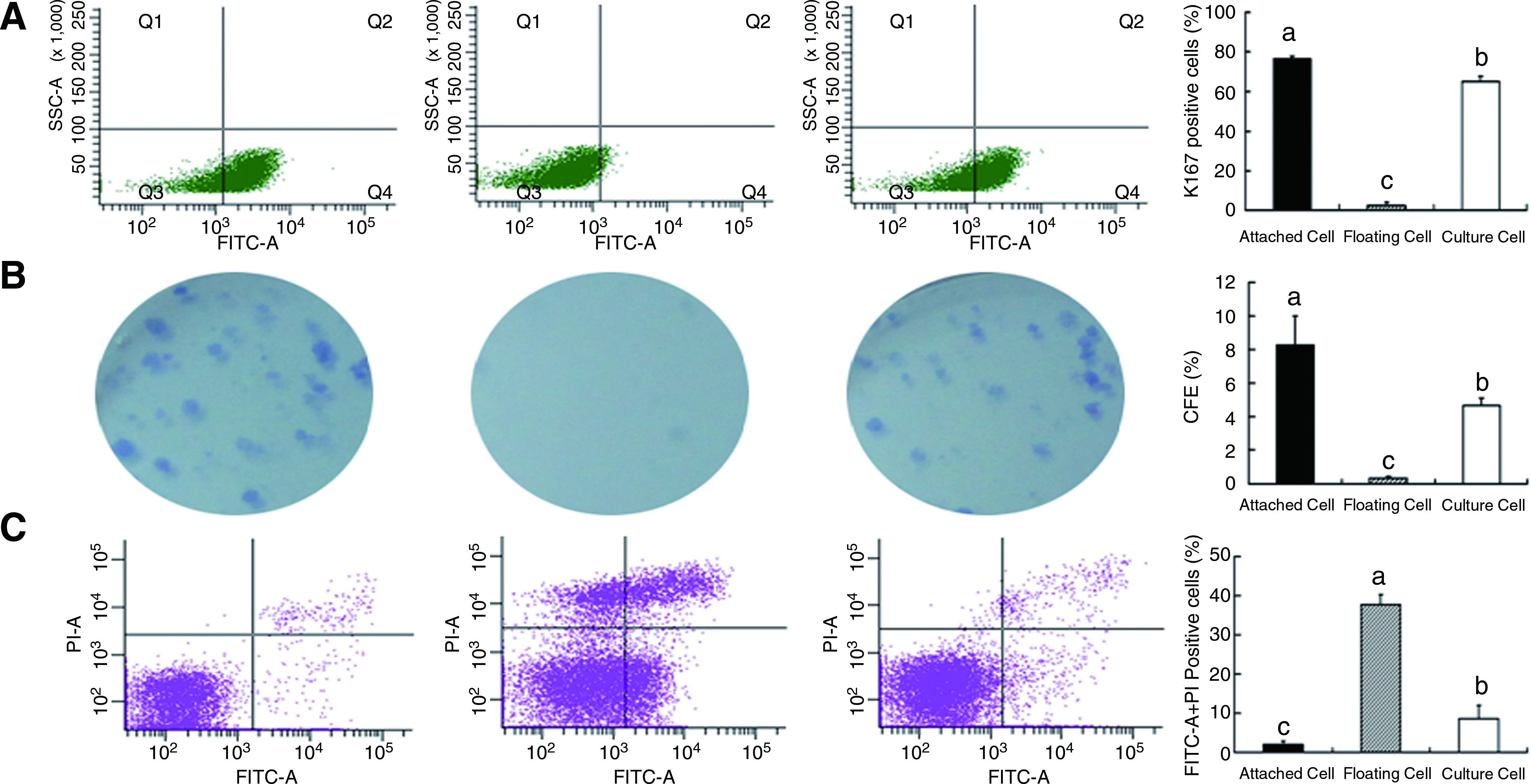
Centrifugal seeding cells have good survival and proliferation.
Properties of CCCES in vitro
Centrifugation force formed monolayer sheets in 4 min. Likewise, different cell layers of CCCES were formed in several minutes (Fig. 3A). The cell sheet exhibited a cobblestone-like morphology after 24 h (Fig. 3B). After being submerged and cultured in Transwell culture inserts for 3 days, CCCES from one layer to four multilayers were formed (Fig. 3D). After being cultured for 7 days, three-layer CCCES showed tight junction formation in apical cells (Fig. 3C). The labeled cells were present in CCCES (Fig. 3E). The CCCES was positive to K3 and ZO-1, but negative to p63 and ABCG2, similar phenotype to the cornea in vivo. Basement membrane components such as type 4 collagen, laminin, and integrin β1 were also expressed in basal cells (Fig. 3F).
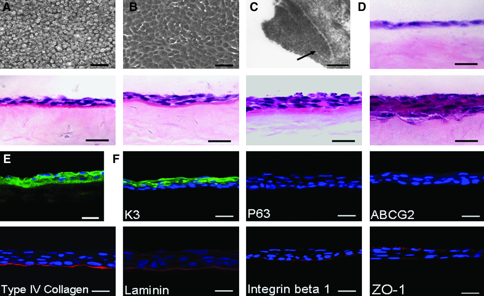
In vitro characteristics of centrifuge-constructed cornea epithelial sheets.
CCCES reconstructed rabbit cornea epithelia with limbal stem cell deficiency
As shown in Figure 4A, the corneas of CCCES group kept optical clarity for 30 days. Some eyes of AM dissolved before 10 days, and all AM was removed at 10 days. The corneas of CCCES group were smooth and transparent, and iris vessels could be observed underneath the cornea. The entire corneal surfaces were completely covered with epithelium, and most of the corneas neovascularization and inflammation were barely detected on the corneal surface for up to 30 days. The corneal surfaces of animals with no transplant became a white haze and showed partial epithelialization at 10 days, and they showed almost epithelialization and new blood vessel hyperplasia at 30 days.
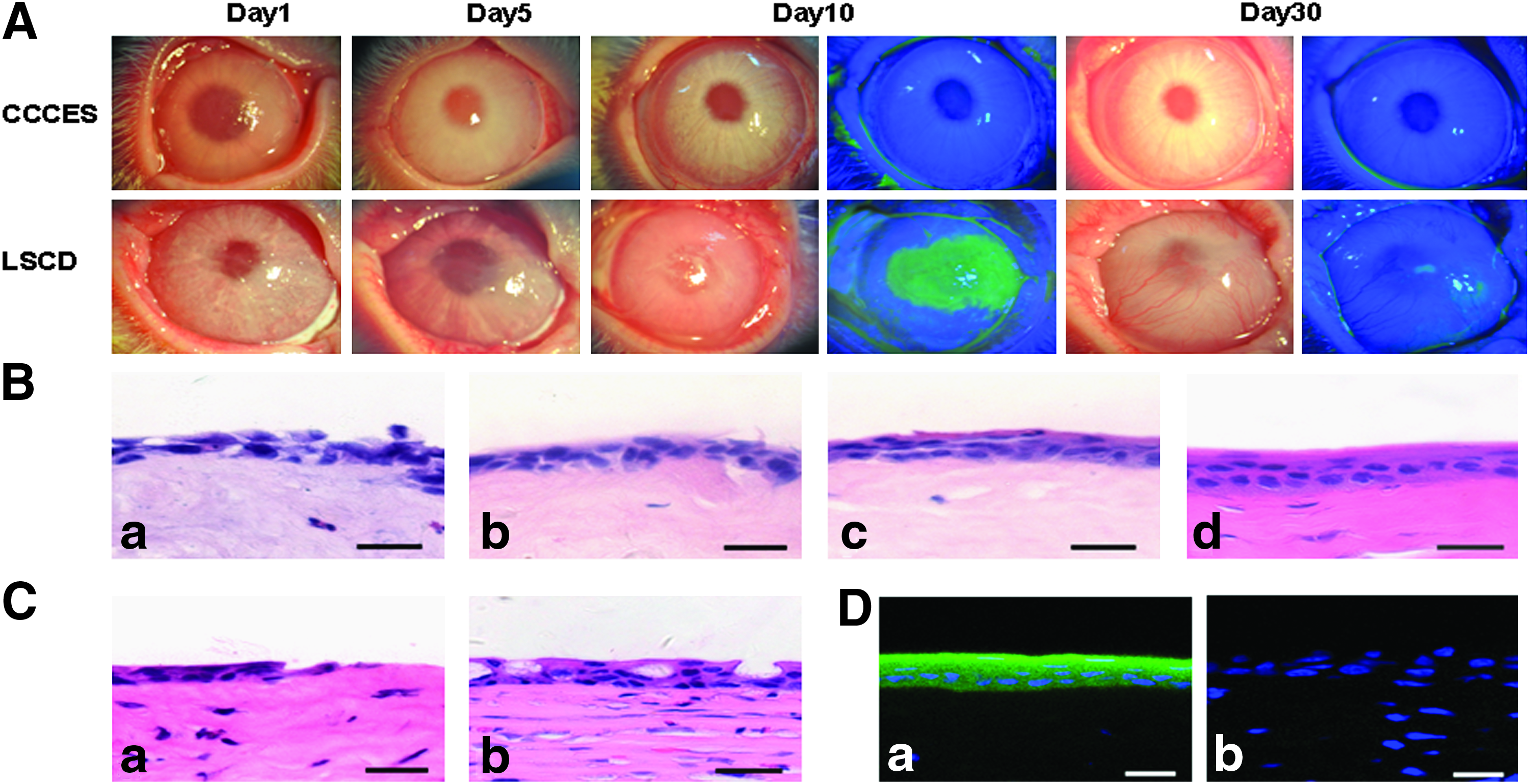
CCCES reconstructed rabbit cornea epithelia with limbal stem cell deficiency (LSCD).
HE histology examination of CCCES for 1 day (Fig. 4Ba), 5 days (Fig. 4Bb), 10 days (Fig. 4Bc), and 30 days (Fig. 4Bd) showed that materials between cells increased and fast coherence of cells was formed. CCCES molding was produced according to the requirements of stroma. Finally, it adhered well to the host corneal stroma with little subepithelial cell infiltration. The eyes received no transplant showed partial epithelialization in the periphery and host corneal stroma had large subepithelial cell infiltration (Fig. 4Ca) at 10 days. The eyes received no transplant showed epithelialization, new blood vessel hyperplasia, and goblet cells, as well as inflammatory cell infiltration in the ocular surface at 30 days (Fig. 4Cb). The labeled cells were present in CCCES group (Fig. 4Da) and no labeled cells were present in the eyes received no transplant (Fig. 4Db) up to 30 days.
As shown in Figure 5, immunohistochemistry of CCCES group showed K3 expression but no k4 and Muc5AC expression. The control animals with no transplant showed k4 and Muc5AC expression but no K3 expression, exhibiting epithelialization of conjunctival origin.

Representative immunohistochemical staining of keratin-3, keratin-4, and Muc5AC in CCCES group and no transplant group for 30 days. Only keratin-3 was expressed in CCCES group. In contrast, keratin-4 and Muc5AC were only expressed in no transplant group. Scale bar = 20 μm.
Discussion
Joseph Mazar et al. allowed organotypic cultures comprised entirely of stratified KCs creating epidermal equivalents rise from a submerged state to an air–liquid interface. 11 From the gene level, they found air–liquid interface culture promoting not only cell proliferation, differentiation, and cell junction but also cell senescence and death. Temperature-responsive polymer, degradation of fibrin glue, and collagenase can produce carrier-free sheets, but they all need submerging and air–liquid interface culture 2∼3 weeks. To harvest cell sheets from the dish, blunt mechanical separation or chemical separation will damage the cell sheets. The long culture time, culture method, and harvest way will decrease sheet viability. Lee W et al. worked hard to solve the problems, using cell print method to construct tissues.12,13
To avoid these shortcomings, we designed the centrifugal seeding method. Using the optimum viability cells, a high seeding efficiency of 80.04% can be achieved at a short seeding time of 4 min, as demonstrated in this study. The centrifugal force applied to the epithelia cells imposed an external force on the cells. However, it was demonstrated that this force not only caused no damage to the cells but also could choose good viability cells, and the viability of attached cells could reach 98.03%. The centrifugal seeding method could construct a multilayer epithelium sheets in several minutes. The sheet was cultured on AM for 24 h and then transplanted to the keratectomized cornea. The results show that it is a simple, fast, and effective method of reconstructing the corneal surface.
Optimization centrifugation parameters could form cell sheets rapidly. Ouyang A et al. applied centrifugal force to increase cell seeding in scaffolds, but they did not attempt to optimize the process. 14 Robin Ng studied cell density, centrifugation time, and centrifugation speed in centrifugal seeding and thought they all play important parts. 15 Optimization of centrifugation parameters should let cells attach quickly and have no damage to them. According to bionomics of living cells and when cell type is fixed, cell density, centrifugation time, and centrifugation speed may play important parts in the centrifugal force formula. In preliminary experiments, cell seeding efficiency increased from 500 to 2000 rpm and then decreased from 2000 to 2500 rpm. Floating death cells and cell debris increased in the medium when speed was higher than 2500 rpm. It was thought that too much centrifugal force caused cell death. To living cells, centrifugation time should be as short as possible. On the basis of above-mentioned data, orthogonal design experiments were performed and optimization parameters were produced. Cell monolayers could be formed in 4 min. Repetitive operation could construct multilayer sheets. Cells in different growth degrees were stained with trypan blue to analyze viability, and 80% confluent cells had the highest viability. So, we used 80% confluent cells to construct sheets to gain the highest viability CCCES.
Lee W et al. designed a cell print method to quickly construct tissues and avoid cell senescence.12,13 They used collagen to make cohesive cells and thus could avoid tissues broken to pieces when moving. With the same aim, Nishida also used a polyvinylidene fluoride membrane to transfer thermo-sensitive cell sheets when applied. We used two ways to keep cell sheets integrity. According to studies of Song and Manon Gaudreault, the formation of cell adherence between cornea epithelia is connected with laminin, fibronectin, heparin-binding peptide, and conglutinin famility.16,17 The factors express mostly in the first 24 h after cell seeding, so we cultured multilayer sheets for 24 h before use to gain sheet integrity. Three days after construction, HE showed that the sheets had integrity and were in good condition. First, cells were driven by physical forces to attach to each other. In the next stage, they attached to each other by extracellular materials that were secretive by themselves. AM was used as a temporary transfer vector to keep integrity.
Proper increasing of cell surface tension could raise cell density in the sheet. Adherent cells are spreading to the utmost before coming to confluence in the culture. Cell surface areas decrease after confluence, but the smallest surface area exists in suspension cells. At this time, cell sheets with maximum cell density can be achieved. Surface tension and osmotic pressure usually make suspension cell volume smaller. Using glycerin as an additive to enhance surface tension and osmotic pressure could make cell surface area decreased. Higher cell adherence ratio and high cell density could be gained and our experiment proved this. Glycerin as a lubricator lets cells distribute evenly and avoids cell agglomerating, which is important to subsequent cell culture and tissue development. Glycerin has no toxicity to cells and is used generally as a protectant in cell cryopreservation, but cells would die when glycerin concentration exceeded 1% in case volume obviously smaller. It is considered that high osmotic pressure surpassed cell tolerance limit.
Cell viability and proliferation show that optimization centrifugal force and 0.4% glycerin applied to the cells in centrifugal seeding did not cause any apparent damage to the cells. Contrast experiments showed that attached cells express more colonies, higher Ki67-positive and lower apoptosis than suspension cells and population culture cells. That is to say, healthy cells could be chosen through the centrifugal method. This is a very good advantage because postseeding viability of the cells is very important to ensure the tissue activity. It established a foundation for next tissue engineering. Robin et al. use centrifugal speed with 1000, 1500, and 1800 rpm, and found there was no damage to human colon cancer HT-29 cells. 15 Using low stir speed to culture cells for a long time also can promote cell proliferation.18–21 However, the centrifugal force method just needs several minutes for seeding and does not need a special stirring system. It is a good method to solve the problem of low cell seeding efficiency, speed, and cell viability.
Cell sheets were separated by thermo-sensitive material PIPAAm expansion, which is equivalent to blunt mechanical separation of cell sheet and will hurt the cell sheets. Degradation of glue by enzyme also would chemically damage cells. They used polyvinylidene fluoride membrane to transfer cell sheets, whereas we used AM as a transfer vector. Transplantation with AM outside the cornea has many virtues, such as to protect the transplant sheet when eye blinking, to avoid AM persistence in the cornea and avoid damage of harvest sheets. AM is translucent and only 0.02–0.05 mm thick. It often degrades in 1 week in clinical amniotic membrane grafting transplants, but this time could be long enough to protect sheet stable conjunction to the stroma.
The CCCES could repair cornea epithelia successfully. Centrifugal sheets could renew molding to repair tissue according to the need of the organism and form stratified epithelium that are similar to normal corneal epithelium. CCCES group showed K3, specificity maker of cornea epithelia, but no k4 and Muc5AC expression. The labeled cells were also present in CCCES group. These exhibited the cells that repair cornea were SDHCEC1 source. According to studies of Antonio Haddad, the corneal epithelium is renewing with a vertical turnover of 7–14 days in many mammals. 22 So, eyes were observed for 30 days and the period could show the stable long-term outcome. To avoid immune rejection, 0.15% cyclosporine A eye drops were applied to the eyes in the CCCES group.
The method has limitations: it requires large amount of cells. This centrifuging may be used in cell lines such as skin and other tissues. Using primary cells might improve outcome, it is true too difficult to obtain large amount of cells to construct tissues with the method. We had already done much work on how to gain large quantity of cells through small pieces of cornea tissue. Such as using embryonic stem cell conditioned medium to enhancement of long-term proliferative capacity of corneal epithelial cells 23 and cornea endothelial cells. 24 We injected single cells solution of SDHCEC1 cell line into the space between the limbal stem cell deficiency rabbits' ocular surface and the membrane-fixing device (BMFD-AM) contact lens in the experimental group and the cells successfully repaired the cornea. 25 In the future, cell sources may be not a big problem when using the method. Now, we do the experiments on using embryonic stem cell-conditioned medium to enhancement long-term proliferative of small biopsy of patients' cornea epithelia and the experiments show some good results. We believe that the two experiments can be benefit to each other when combined. This is the next step we will work but it will need a long time. Perhaps it will be another way to deal with limbal stem cell deficiency patients in the future.
This method can mix different type cells and construct multicellular tissues. The sandwich method can be used to solve nutritional ingredient and oxygen problem in case of thick tissue. That is to say, different tissues can construct vitro and then these tissues will be assembled and installed in vivo according to anatomy.
Many advanced techniques such as magnetic field,26,27 contact guidance, 28 and electrospinning29–31 can combine with this method to produce better tissue and organs that fit physiological functions.
Conclusion
In summary, the centrifugal force method could construct good viability in a multilayer cell sheet and the sheet could repair cornea epithelia successfully. The method is fast and easy. It may reach the aim of sandwich tissue engineering so that different tissues will be constructed vitro and assembled and installed in vivo according to anatomy for large tissue construction and application.
Footnotes
Acknowledgments
This work was supported by the National High Technology Research and Development Program of China (863 Project; Grant no. 2006AA02A133).
Disclosure Statement
No competing financial interests exist.