
Abstract
Engineering three-dimensional (3D) cell-dense tissues with a well-organized structure remains a challenge in tissue engineering. In this study, highly oriented fibrous bundles, consisted of composite fibers of poly(L-lactide-co-glycolide)/superparamagnetic iron oxide nanoparticles, were fabricated using an electrospinning technique. The magnetic properties of the fabricated fibrous bundles were examined by a vibrating sample magnetometer and a superconducting quantum interference device; the results demonstrate that the fabricated fibrous bundles revealed superparamagnetic behavior without magnetic hysteresis. After seeding C2C12 myoblasts on the fibrous bundles, cells were grown along the direction of the underlying fibers (cell rods), an aligned pattern similar to those in native skeletal muscle tissues. When treated with the differentiation medium, myoblasts were fused together and formed multinucleated myotubes. As soon as applying an external magnetic field, the cell rods can spontaneously response to the magnetic control and self-assemble into 3D tissues with a highly ordered architecture. These findings demonstrate that the magnetically susceptible fibrous bundles not only can serve as a functional unit providing the topographic cue for cell orientation, but also can be magnetically manipulated for the creation of 3D cell-dense constructs. This technique may be applied to various cell types and scaffold configurations, thus advancing the design of engineered tissues that more closely replicate native tissues.
Introduction
While scaffolds with oriented patterns show promise in engineering well-organized tissues, several limitations remain. In highly aligned dense scaffolds in particular, fiber packing increases density and reduces pore size, thus limiting the transport of oxygen and nutrients and the infiltration of cells. 14 For these reasons, cell growth and tissue formation are generally restricted to the surface layers of the scaffolds. Previous studies have shown that increasing the porosity within fibrous scaffolds improves cellular infiltration15–17 ; however, this may hinder the fiber alignment. To address these issues, we propose an approach to engineer three-dimensional (3D) cell-dense tissues via the magnetic actuation of superparamagnetic fibrous bundles and allow for the complete preservation of their structural orientation.
Over the past decade, manipulation of cells through magnetic actuation has received much attention in tissue engineering. Super paramagnetic iron oxide nanoparticles (SPION) have been used to manipulate the formation of cell sheets and tubular tissue structures as skin substitutes and blood vessels, respectively.18−20 Additionally, SPION have been employed to direct the differentiation of stem cells via the magnetic actuation of their specific ion channels and surface receptors. 21 These reported magnetic actuation techniques were realized by loading SPION onto or into the cells; however, their long-term effects on the cell physiology remain unknown.
To avoid the direct contact with SPION in the manipulation of cells, superparamagnetic fibrous bundles, consisted of highly oriented poly(L-lactide-co-glycolide) (PLGA) fibers embedded with SPION, are developed in the present research. Unlike ferromagnetic materials, superparamagnetic scaffolds do not retain any significant amount of magnetization in the absence of an externally applied magnetic field, and thus do not form aggregates. 22 When a magnetic field is applied, the magnetically susceptible bundles can be oriented along the magnetic moment of embedded SPION and create a desired construct.
The highly oriented PLGA/SPION fibrous bundles were fabricated by an electrospinning technique. To reduce the mass transfer barrier during cell culture, the diameters of the electrospun fibrous bundles were restricted to less than 200 μm for cell seeding. The oriented fibers within the bundle can guide the cell growth and then form a cell rod with an aligned cell orientation. Superparamagnetic scaffolds are of great interest in the present research because of their unique magnetic feature. In the absence of a magnetic field, these superparamagnetic fibrous bundles are free of magnetism and can be well dispersed in the culture dish to ensure the seeded cells obtaining sufficient oxygen and nutrients. After cells are confluent on the fibrous bundles, the bundles covered with cells can be magnetized and manipulated by an external magnetic field for the construction of a 3D cell-dense engineered tissue with a highly oriented architecture (Fig. 1).
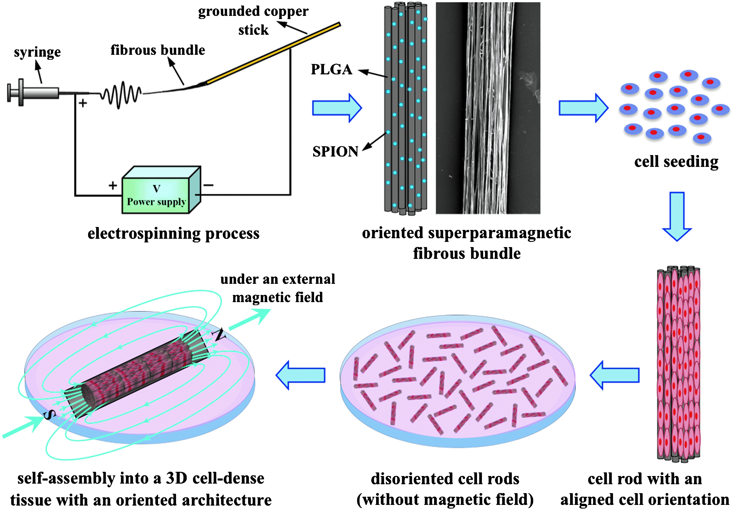
Schematic illustrations of the fabrication process of the electrospun superparamagnetic fibrous bundles and the concept of magnetically directed self-assembly of the magnetically susceptible bundles to form 3D tissue constructs with a highly ordered architecture. 3D, three-dimensional; PLGA, poly(L-lactide-co-glycolide); SPION, super paramagnetic iron oxide nanoparticles. Color images available online at
Materials and Methods
Materials
PLGA (L-LA/GA=85/15, intrinsic viscosity=2.39 dL/g) was obtained from Purac (Gorinchem). Hexafluoroisopropanol and dichloromethane (DCM) were acquired from Sigma-Aldrich. All other chemicals and reagents used were of analytical grade.
Preparation of PLGA/SPION fibrous bundles
The SPION used in the study were prepared according to a previously reported thermal decomposition method. 23 In brief, Iron(III) acetylacetonate (2 mmol), 1,2-hexadecanediol (10 mmol), oleic acid (6 mmol), oleylamine (6 mmol), and benzyl ether (20 mL) were mixed together and magnetically stirred under a flow of nitrogen. The mixture was heated to 200°C for 30 min and then, under a blanket of nitrogen, heated to reflux (∼300°C) for 30 min. The black-colored mixture was cooled to room temperature. Under ambient conditions, ethanol (40 mL) was added to the mixture, and a black material was precipitated and separated via centrifugation. The black product was dispersed in hexane, and centrifugation (6000 rpm, 10 min) was applied to remove any undispersed residues. The final step was repeated three times for further purification of SPION. Finally, the product was then redispersed into DCM.
X-ray diffraction measurements of the prepared SPION were performed with a powder diffractometer (Rigaku D/max-RB). Further, the morphology of SPION was examined by a transmission electron microscope (JEM-2010F, JEOL). The transmission electron microscopy (TEM) sample was prepared by placing a drop of SPION suspension onto a 400 mesh copper grid coated with carbon. Approximately 2 min after deposition, the grid was tapped with a filter paper to remove surface water and then dried in vacuum.
PLGA/SPION fibrous bundles were fabricated using an electrospinning setup equipped with a copper stick as a collector (Fig. 1). To obtain a homogeneous solution for electrospinning, a DCM dispersion of SPION (13 mg in 0.25 mL) was added to a PLGA solution in Hexafluoroisopropanol (130 mg in 0.75 mL) and then sonicated for 1 h. The composition of the blend of SPION/PLGA was 1/10 by w/w. The mixture was fed into a 1 mL standard glass syringe attached to a 18G stainless steel needle using a syringe pump (KDS 100, KD Scientific) at a flow rate of 1 mL/h with an applied voltage of 20 kV (CM-30P, SIMCO). The distance between the needle tip and the collector was 8 cm.
On application of high voltage, the polymer solution was drawn into fibers and self-bundled to the grounded copper stick collector. The collection time was set to 30 s to obtain a fibrous bundle with a diameter less than 200 μm. The obtained fibrous bundles were dried overnight under vacuum and used for characterization and cell culture studies.
Characterization of PLGA/SPION fibrous bundles
Test samples were sputter-coated with gold, and then their morphology was observed under a scanning electron microscope (SEM, Model JSM-5600, JEOL). After test samples were examined by SEM, an Image-Pro Plus 6.0 analysis software was used to measure the diameter of each fiber and its angle distribution with respect to the major axis of the fibrous bundle. 24 The dispersibility of SPION in the composite fiber was examined by a TEM. TEM samples were simply prepared by placing the composite fiber onto a 300 mesh copper grid coated with carbon and then inspected without staining. The composition of electrospun fibrous bundles was investigated by the energy dispersive X-ray analysis. Elemental maps of iron (L edge: 708 eV, ΔE=20 eV), oxygen (K edge: 532 eV, ΔE=20 eV), and carbon (K edge: 284 eV, ΔE=20 eV) were acquired and processed by the GIF with a Digital MicrographTM software (Gatan Inc.). 25
Magnetic properties of PLGA/SPION fibrous bundles
The saturation magnetization of fibrous bundles was determined using a vibrating sample magnetometer (VSM, Digital Measurement System). The temperature-dependent magnetization of test samples was measured by a superconducting quantum interference device (Quantum Design). The blocking temperature of test samples was read from the zero-field-cooled (ZFC) and field-cooled (FC) curves taken under an applied magnetic field of 100 Oe between 2 and 350 K. 26
Cell culture
Mouse C2C12 myoblasts (ATCC CRL-1772) were used to study cell alignment and differentiation on the fabricated fibrous bundles. Cells were cultured in the growth medium (Dulbecco's modified Eagle's medium supplemented with 10% fetal bovine serum and 1% penicillin-streptomycin) under standard culture conditions (37°C, 5% carbon-dioxide). For cell culture, fibrous bundle samples were cut into small pieces with a length of 1 cm and sterilized with UV overnight. Cells were grown onto the fibrous bundles in 24-well plates at 1×10⁵ cells per well and cultured in the growth medium for 3 days to form cell rods. To induce myotube formation, the medium was then replaced with Dulbecco's modified Eagle's medium containing 2% heat-inactivated horse serum (differentiation media) and cultured for an additional 9 days. The viability of cells was evaluated according to a live/dead assay using calcein-AM and ethidium homodimer (Molecular Probes # L3224; Eugene) 27 and the 3-(4,5-dimethylthiazol-2-yl)-2,5-diphenyl tetrazolium bromide (MTT) assay. 28
Immunofluorescence analysis
At 3 days post seeding, the cells on fibrous bundles were immunostained for endogenous extracellular matrix (ECM) molecules. The harvested cell rods were fixed in 4% paraformaldehyde in phosphate-buffered saline and then stained with a mouse monoclonal antibody against collagen type III (clone 3G4, Chemicon, Temecula), fibronectin (clone IST-9, Abcam), or laminin (clone 2E8, Chemicon). A Cy5-conjugated goat antimouse IgG (Jackson ImmunoResearch Laboratories) was then used as the secondary antibody. Cell rods were costained to observe nuclei by propidium iodide (P4864; Sigma-Aldrich). The stained cell rods were examined using an inverted confocal laser scanning microscope (TCS SL, Leica) with excitations at 543 and 633 nm. Fluorescence images were taken at 10 μm intervals and their 3D reconstruction images were created using an LCS Lite software (version 2.0).
For the in vitro differentiation study, cells were immunostained with phalloidin at 9 days after the induction of differentiation to observe their actin cytoskeleton. The harvested cells were treated with Alexa Fluor 488–conjugated phalloidin (A12379, Invitrogen) and propidium iodide and then examined under the 488 nm and 543 nm lasers of a confocal laser scanning microscope.
Magnetic-directed self-assembly of cell-dense rods
When C2C12 myoblasts were confluent on the fibrous bundles (about 3 days of culture), a cuboid magnet with a surface magnetic field of ∼1.0 T was applied to the incubated cell rods. The magnet was placed and fixed underneath the culture dish to drive the self-assembly of individual cell rods. The assembled cell rods were reincubated in the fresh growth medium under the external magnetic field for an additional 3 days. Subsequently, the magnet was removed and the morphology and cell viability of the engineered tissue were examined by SEM and fluorescence microscopy.
Results and Discussion
Engineering highly ordered constructs with a high cell density to mimic native tissues is essential for tissue engineering. It has been reported that cells cultured on the electrospun fibrous scaffolds showed excellent cell adhesion and spreading but poor cellular infiltration, due to their limited porosity. 29 To address this concern, highly oriented fibrous bundles with superparamagnetic properties were fabricated in this study. We demonstrated that the magnetically susceptible fibrous bundles not only can serve as a functional unit providing the topographic cue for cell alignment, but also can be specifically manipulated by an external magnetic field for the construction of 3D cell-dense tissues with a highly organized architecture. Mimicking the nature architecture of a specific tissue is significant for fabricating tissue-engineered constructs.
Characterization of PLGA/SPION fibrous bundles
The X-ray diffraction pattern of the prepared SPION matched well with that of the standard Fe3O4 powders (Fig. 2A). 30 The magnetically susceptible fibrous bundles were fabricated by electrospinning of a PLGA/SPION blend solution. To manipulate the aligned orientation, a grounded copper stick was used as a collector to induce the self-bundling of PLGA/SPION composite fibers during the electrospinning process (Fig. 1). A fibrous bundle with a diameter less than 200 μm can thus be obtained by controlling the electrospinning time for ∼30 s.

Figure 2B shows a representative SEM image of an obtained fibrous bundle. It was found that the diameter of the fibers within the bundle was 1.26±0.42 μm. The angle of most fibers ranged from 0° to 10° (Fig. 2C), indicating that nearly all fibers were oriented along the major axis of the bundle, a unique aligned topography. To examine the dispersion of SPION within the composite fibers, TEM images were taken. As shown in Figure 2D, the SPION were well dispersed within the electrospun fibers without significant aggregation. The characteristic energy dispersive X-ray spectrum of composite fibers further validated the presence of SPION (Fe) (Fig. 2E).
Magnetic properties of PLGA/SPION fibrous bundles
Figure 3A shows the ZFC and FC curves of the fibrous bundles measured using a superconducting quantum interference device magnetometer. As shown, the divergence between the susceptibility in the ZFC and FC processes was a typical feature of superparamagnetic materials, indicating that the superparamagnetism of SPION was preserved after the fabrication of composite fibers. The ZFC curve exhibited a broad peak at about 80 K, indicative of a characteristic blocking temperature, which is an important parameter in the study of a magnetic fibrous scaffold because the encapsulated SPION would exhibit superparamagnetic properties above the blocking temperature. 31

Results of
The magnetic hysteresis loop measured by a VSM is shown in Figure 3B. The results demonstrate that the fabricated fibrous bundles revealed superparamagnetic behavior without magnetic hysteresis. Magnetization measured using VSM provides a quantitative evaluation for assessing the ability to respond to an external magnetic field. It was found that the saturation magnetization of the fibrous bundles was 2.6 emu/g, which was sufficient to allow the bundles to be influenced by a magnet with a surface magnetic field of ∼1.0 T (as discussed in the next section).
Magnetic manipulation of PLGA/SPION fibrous bundles
To evaluate the response of the superparamagnetic bundles to magnetic manipulation, an external magnetic field was applied to the bundles that were originally suspended in phosphate-buffered saline randomly in a cell-culture plate. The time course of their self-assembly was recorded by photography. As shown in Figure 4 and Supplementary Video S1 (Supplementary Data are available online at

Photographs of the time course of self-assembly of the superparamagnetic fibrous bundles (indicated by the white arrows). Color images available online at
Effect of fiber alignment on cell growth and differentiation
Alignment of cells plays a critical role in the skeletal muscle tissue engineering because the natural skeletal muscle has a well-oriented architecture comprising long, parallel multinucleated myotubes formed through differentiation and fusion of myoblasts. 32 To examine the effect of fiber alignment on cell orientation and proliferation, mouse C2C12 myoblasts were used as a model cell line and seeded onto the fibrous bundles. Figure 5A shows an SEM image of the morphology of the grown myoblasts after 3 days in culture. As shown, C2C12 myoblasts were confluent on the oriented fibrous bundles and proliferated along the direction of the underlying fibers (cell rods), an aligned pattern similar to those in native skeletal muscle tissues.
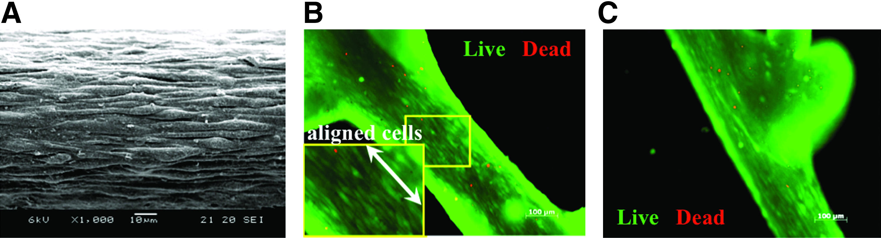
The cytotoxicity of test bundles was qualitatively evaluated according to a live/dead cell viability assay using calcein-AM and ethidium homodimer and quantitatively measured by the MTT assay. Calcein-AM hydrolysis in live cells produces a green fluorescent signal, whereas ethidium homodimer is excluded from live cells and produces a red fluorescent signal only in dead cells. As shown in Figure 5B, the alignment of C2C12 myoblasts along the axis of bundles was evident. The cell viability obtained by the MTT assay was expressed as a fraction of viable cells on the PLGA/SPION fibrous bundles and normalized to that of cells on the PLGA fibrous bundles in the absence of SPION (control). It was found that the viability of cells on the PLGA/SPION fibrous bundles (94.3±4.5%) was comparable to their control counterpart (Fig. 5C; 100.0±7.1%). These results suggest that the superparamagnetic fibrous bundles developed were cytocompatible and can indeed provide topographical cues to induce cell orientation.
The incubated cell rods were harvested and then fixed in 4% paraformaldehyde for immunofluorescence analyses. It was found that ECM molecules (collagen type III [Fig. 6A]) and integrative adhesive agents (fibronectin [Fig. 6B] and laminin [Fig. 6C]) were clearly identified on the cell rods. The reconstructed 3D confocal images further validated that the surface of the cell rods was covered by collagen type III (Fig. 6A), fibronectin (Fig. 6B), and laminin (Fig. 6C). It is known that ECM provides not only a physical support but also outside-in signals that regulate many cellular functions such as adhesion, migration, and proliferation and must be maintained for optimal cellular benefits. 33
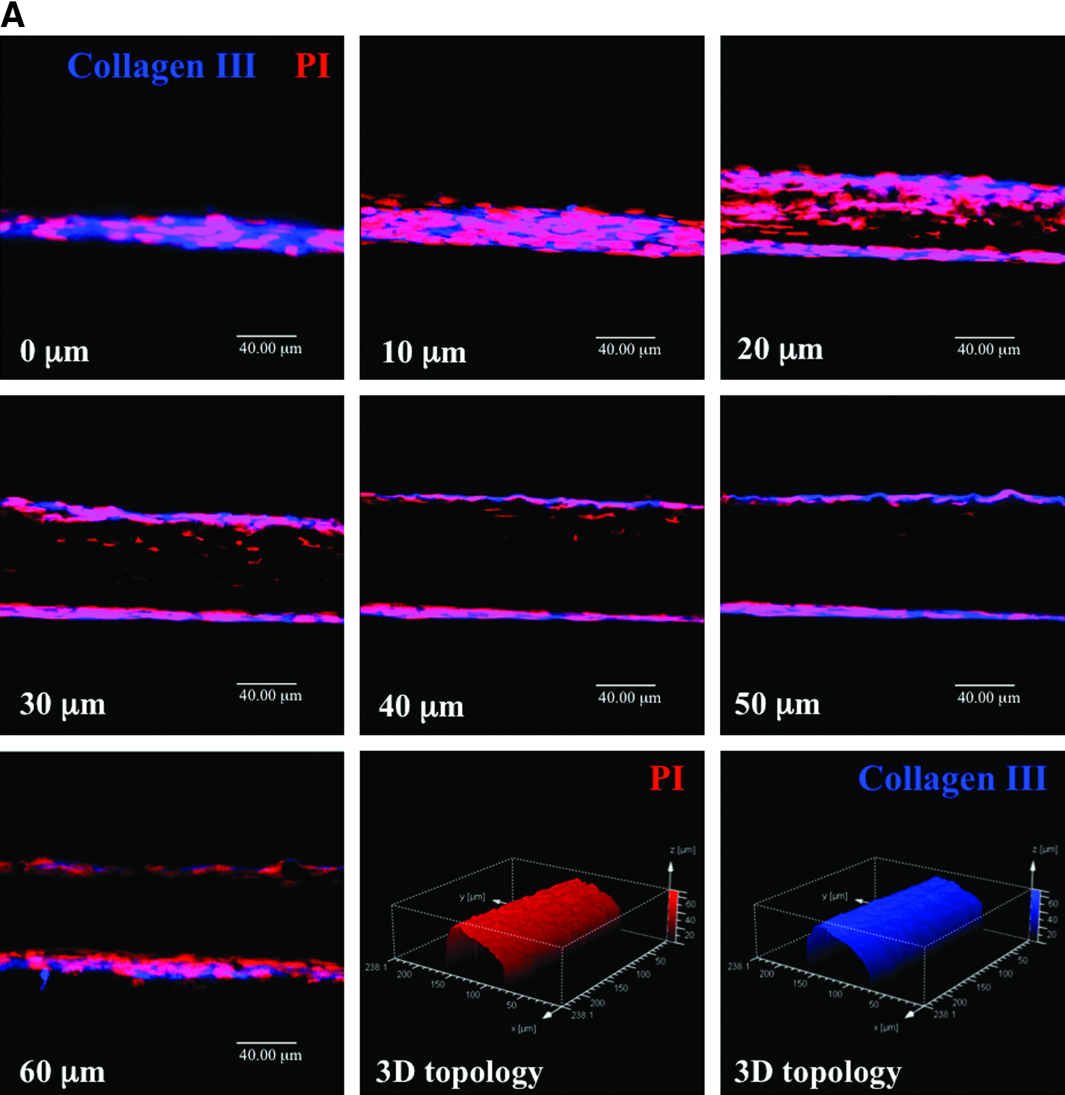
Confocal images of seven optical sections (and those after 3D reconstruction) of the cell rod using the immunofluorescence staining for
Fusion of undifferentiated myoblasts into multinucleated myotubes is a prerequisite for the developmental myogenesis and postnatal muscle growth. 34 To induce the differentiation of myoblasts in vitro, nearly confluent C2C12 cells in a culture dish were incubated in the differentiation medium for 9 days. Figure 7A shows the microscopic and immunofluorescence images of the time course of the formed multinucleated myotubes.

It has been reported that the cell–scaffold interaction may influence cell functions such as proliferation, differentiation, and cell death. 35 To evaluate the differentiation ability of myoblasts grown on the fibrous bundles, confluent C2C12 cells were treated with the differentiation medium and then stained with the live/dead fluorescent dye for observation. As shown in Figure 7B, C2C12 myoblasts fused together, and tube formation was clearly observed at day 9 after the induction of differentiation. Additionally, the nucleus number, length, and width of myotubes on the PLGA/SPION fibrous bundles (25±4, 1.1±0.3 mm, and 16.5±0.8 μm, respectively) were comparable to their control counterparts (the fibrous bundles without SPION: 26±5, 1.2±0.4 mm, and 15.8±0.9 μm, respectively). The above-mentioned results demonstrate that the prepared fibrous bundles did not cause adverse effects on C2C12 cell differentiation.
Technical feasibility for forming 3D cell-dense tissues
To further test our approach in forming 3D engineered tissue, an external magnetic field was utilized to drive the self-assembly of cell rods. As expected, the incubated cell rods can be magnetically manipulated and self-assemble to form a well-organized structure. After reculturing for 3 days, the external magnetic field was removed and the morphology of the assembled structure was examined by SEM. As shown in Figure 8A, 3D cell-dense tissues with a highly ordered architecture were successfully obtained. This observation can be attributed to the fact that the neighboring cell rods adhered to each other via their integrative adhesive agents (e.g., fibronectin [Fig. 6B] and laminin [Fig. 6C]) on endogenous ECM and then became an integrated engineered tissue during reculture. Additionally, the results of the live/dead assay demonstrate that most cells on the engineered tissue were viable (Fig. 8B).

An ideal tissue-engineering scaffold must be designed from biodegradable polymers and their degradation products should not provoke significant inflammation or toxicity when implanted in vivo. 36 PLGA can be degraded into lactic acid and glycolic acid through hydrolysis of its ester bond; the degradation products are then removed from the body through the citric acid cycle. 37 SPION have been clinically used in magnetic resonance imaging for the detection and delineation of focal liver lesions 38 ; they are eventually phagocytosed or endocytosed by the reticuloendothelial system in vivo.39,40 Once compartmentalized within the lysosomes of reticuloendothelial system cells, the iron oxide particles are digested, with the majority of the SPION iron stored as ferritin and/or hemosiderin. 40
The architecture and characteristics of biomaterial scaffolds play an important role in affecting cell behavior and guiding the regeneration of functional tissues. Recent studies emphasize on the use of scaffolds with various advanced techniques to form guidance patterns or channels, which accurately mimic a natural repairing process in the human body.41–44 Reconstruction of well-organized 3D tissues is vital to tissue engineering. Compared to the traditional scaffolds, which provide only the topographical signal for cells, our fibrous bundles with unique superparamagnetic properties can be magnetically directed to construct a highly ordered 3D architecture. A 3D culture environment may re-establish the physiological cell–cell and cell–ECM interactions that can mimic the specificity of native tissues.
Conclusions
In this study, aligned electrospun fibrous bundles composed of PLGA/SPION were fabricated as a biomaterial scaffold. The results demonstrate that the topographical features on the surface of the electrospun fibrous bundles can spontaneously induce cell alignment and formed cell rods. When treated with the differentiation medium, C2C12 myoblasts grown on the fibrous bundles can be fused together and differentiated into multinucleated myotubes. Given an external magnetic field, the cell rods can self-assemble into 3D cell-dense tissues with a highly oriented structure. Mimicking the nature architecture of specific tissues is significant for fabricating tissue-engineered constructs.
Footnotes
Acknowledgments
This work was supported by grants from the National Science Council (NSC 98-2120-M-007-007) and the Center for Frontier Materials and Micro/Nano Science and Technology (CFMMNST), National Cheng Kung University (D99-2700), Taiwan.
Disclosure Statement
No competing financial interests exist.
References
Supplementary Material
Please find the following supplemental material available below.
For Open Access articles published under a Creative Commons License, all supplemental material carries the same license as the article it is associated with.
For non-Open Access articles published, all supplemental material carries a non-exclusive license, and permission requests for re-use of supplemental material or any part of supplemental material shall be sent directly to the copyright owner as specified in the copyright notice associated with the article.