
Abstract
Previous studies have shown that novel scaffold-free self-assembled constructs can be an ideal alternative for cartilage tissue engineered based on scaffolds, which has many limitations. However, many questions remain, including the choice of seeding cells and the role of growth differentiation factor 5 (GDF-5) in constructing self-assembled engineered cartilages. Moreover, whether the optimum construct is effective in human chondral defect repair is still unknown. In this study, we generated self-assembled constructs of human mesenchymal stem cells (hMSCs) using four different approaches: direct self-assembly of hMSCs with or without GDF-5, and predifferentiated hMSCs self-assembly with or without GDF-5. Histological, immunohistochemical, and biochemistry analyses indicated that the constructs generated from predifferentiated hMSCs induced by GDF-5 (Group D2) exhibited up-regulated glycosaminoglycan (GAG) and type II collagen expression and contained higher amounts of GAG and total collagen than any other group. After 3-weeks of in vitro culturing of the constructs in a chondral defects explant culture system, the contructs from Group D2 were stably adhered to the surface of the cartilage matrix. Immunohistochemically, the repair tissue was positive for type II collagen, toluidine blue, and safranin O. These data demonstrated that the generation of self-assembled tissue-engineered cartilage from chondrogenically differentiated hMSCs induced by GDF-5 is a promising therapeutic strategy for cartilage repair.
Introduction
Particular research efforts have been drawn continually to improve cartilage healing, such as autologous chondrocyte implantation (ACI), microfracture techniques, and cartilage tissue-engineering strategies.4–9 However, questions remain about the limited proliferative capacities of chondrocytes, the de-differentiation of chondrocytes during expansion, and the sacrifice of neighboring normal articular cartilage; and the use of scaffolds has been associated with potential problems including the phenotypic alteration of cells, stress-shielding, poor engraftment rates after implantation, and degradation product toxicity.10–13
Previous studies have successfully constructed tissue-engineered cartilage using chondrocytes as seeding cells to repair cartilage defects with hyaline articular cartilage without employing any scaffold.14–17 However, the use of chondrocytes as seeding cells also suffers from the aforementioned concerns and disadvantages of using scaffolds. On the other hand, human bone marrow-derived mesenchymal stem cells (hMSCs) have shown their multiple differentiation potential, rapid proliferation and easy availability, which makes them a promising candidate for cartilage tissue engineering.18–21 In addition, hMSCs can be isolated through a noninvasive procedure and have an ample source.
In this study, we evaluated the feasibility of constructing scaffold-free tissue-engineered cartilage by self-assembly with hMSCs as the seeding cells and further optimized the construction procedure. We generated self-assembled constructs in four ways: directly self-assembled hMSCs with or without growth differentiation factor 5 (GDF-5), and predifferentiated hMSCs self-assembled with or without GDF-5. Further, we assessed the effect of GDF-5 on the chondrogenesis of self-assembled constructs and compared the effect of different construction procedure on the chondrogenesis of self-assembled constructs. At last, we determined an optimum construction method to effectively repair in vitro chondral defects.
Materials and Methods
Isolation of hMSCs
Bone marrow samples were derived from the iliac crests of eight healthy volunteer donors (three men and five women) following a protocol approved by the Ethical Committee of Tongji Medical College, Huazhong University of Science and Technology, China. The median age of volunteers was 36 years old (ranging from 24 to 48 years old). Bone-marrow aspirates were separated by density gradient centrifugation at 900 g for 20 min; mononucleated cells were collected and resuspended in hMSC growth medium consisting of low glucose Dulbecco's modified Eagle's medium (DMEM; HyClone) complemented by 10% fetal bovine serum (FBS; Gibco-BRL), 100 U penicillin-streptomycin (Gibco-BRL), and 2 mM L-glutamine (Gibco-BRL). Cells were cultivated in T25 culture flasks at a density of 2×10⁵ cells/cm2 and cultured for 12–14 days (Passage 1) at 37°C in a humidified, 5% CO2 atmosphere. The cells were passaged at 1:2 dilutions after reaching 90% confluency under the same culture condition. The experiments described next were performed using cells from passages 4–7.
Identification of hMSCs
Fluorescein isothiocyanate (FITC)-labeled anti-human CD44, CD45, phycoerythrin (PE)-labeled anti-human CD14, CD29 antibodies were purchased from BD Biosciences, and FITC-labeled anti-human CD105 was purchased from R&D Systems Inc. A total of 1×10⁶ hMSCs (Passage 4) were harvested, resuspended in 1 mL of phosphate-balanced solution (PBS; Sigma), and incubated with FITC - or PE - conjugated anti-human antibodies, which had been diluted with PBS containing 2% bovine serum albumin (BSA), for 20 min at room temperature in the darkness. Cells released by the cell dissociation buffer were phenotyped using a Coulter FC500 flow cytometer. The Macintosh CELL Quest software program (BD) was employed to analyze the data.
In vitro multi-differentiation potential of hMSCs
Adipogenesis and osteogenesis
The hMSCs (Passage 4) were plated onto six-well plates at a density of 1×10⁴ cells/cm2 and cultured for 24 h in hMSC growth medium, which was then changed to adipogenic or osteogenic medium for an additional 21-day of culturing. The adipogenic medium consisted of DMEM-H containing 10% FBS, 0.5 mM isobutylmethylxanthine (Sigma), 100 nM dexamethasone, and 10 μg/mL insulin. The osteogenic medium was composed of DMEM-L supplemented with 10% FBS, 10 mM β-glycerophosphate, 100 nM dexamethasone, and 0.1 mM ascorbate-2-phosphate. Adipogenesis was evaluated by Oil Red O staining, whereas osteogenesis was assessed by Alizarin Red staining.
Chondrogenesis
The hMSCs (Passage 4) were cultivated on 12-well tissue culture plates at a density of 2×10⁴ cells/cm2 in chondrogenic media (CM). The CM was composed of DMEM-L medium (Invitrogen), 40 μg/mL L-Proline, 1% ITS+1 (Sigma), 100 μg/mL sodium pyruvate, 100 nM dexamethasone, 1% FBS, and 50 μg/mL ascorbate 2-phosphate. CM supplemented with GDF-5 (100 ng/mL) or not were added to the 12-well tissue culture plates. The medium was changed every 2–3 days. After 21 days of induction, the newly synthesized proteoglycan was detected by alcian blue staining; and the Col II mRNA level of the induced hMSCs from both groups was assessed by reverse transcription–polymerase chain reaction (PCR).
Reverse transcription–polymerase chain reaction
Total RNAs from each group of chondrogenically differentiated hMSC were extracted using a TRIZOL® Reagent kit (Invitrogen). Complementary DNAs (cDNAs) were produced by reverse transcription reaction of 1 μg total RNA with oligo (dT) 20 as the primer. Polymerase chain reaction were carried out using a 25 μL reaction volume consisting of 1 μL of cDNAs, 12.5 μL of 2×Taq PCR MasterMix, 3 μL of primer-probe mix, and 8.5 μL of RNase-free water. Polymerase chain reactions were performed in a thermocycler (Biometra) for 40 cycles according to the following procedure: 94°C for 2 min; 94°C for 30 s; 52°C for 30 s; 72°C for 1 min, and followed by extension of 5 min at 72°C. The primers were used as following: collagen II sense 5′-TTCA GCTATGGAGATGACAATC-3′ and antisense 5′-AGAGT CCTAGAGTGACTGAG-3′; GAPDH sense 5′-ACCACAGT CCATGCCATCAC-3′ and antisense 5′-TCCACCACCCTGT TGCTGTA-3′.
Self-assembly of hMSCs
To prepare the agarose-coated 24-well plates, 2% molecular biology grade agarose solutions were added into each well, and then plates were gently shaken to ensure that agarose evenly covered the plate wall. The hMSCs (Passage 4) were cultivated in T75 culture flasks and divided into four groups until cells reached 80%–90% confluence. The hMSCs were digested and resuspended in CM at a density of 2.5×10⁷ cells/mL, and 400 μL of cell suspension was then transferred to the agarose-coated 24-well plates and self-assembled with CM for 6 weeks (CM1 group). The hMSCs were resuspended in CM supplemented with 100 ng/mL GDF-5 at the same concentration and then self-assembled with CM containing 100 ng/mL GDF-5 for 6 weeks (D1 group). In the other two groups, the hMSCs were first induced using CM supplemented with 100 ng/mL GDF-5 in monolayer cultures for 3 weeks. Then, the predifferentiated hMSCs were digested and resuspended in CM at a concentration of 2.5×10⁷ cells/mL; the predifferentiated hMSCs suspensions were added to agarose-coated 24-well plates and self-assembled using CM for 6 weeks (CM2 group). The predifferentiated hMSCs were suspended in CM supplemented with 100 ng/mL GDF-5 at the same concentration and then self-assembled using CM containing 100 ng/mL GDF-5 for 6 weeks (D2 group). The media in these 6-week self-assembled cultures were changed once per day.
Construct processing
The time point at which the hMSCs suspension was seeded into the agarose-coated 24-well plate was defined as t=0 weeks. Constructs harvested at t=3 weeks were weighed to obtain wet weights (WW) and dry weights (DW), photographed, and then measured to determine the construct diameters by digital calipers with 0.02 mm accuracy. Samples harvested at t=1, 3, and 6 weeks were used for histological, immunohistochemical, and biochemical analyses.
Histological and immunohistochemical analysis of the self-assembled constructs
The constructs were fixed with 4% paraformaldehyde for 24 h, embedded in paraffin, and then sectioned at a thickness of 5 μm. Toluidine blue (Sigma) staining was performed to detect extracellular sulfated glycosaminoglycan (GAG) deposition, whereas immunostaining was employed to detect the expression of type II collagen and type X collagen, respectively. After deparaffinization and hydration, sections were predigested with pepsin at 37°C for 30 min for optimal antigen retrieval and then treated with 3% H2O2 in PBS for 20 min at room temperature to quench endogenous peroxidases. The sections were incubated with 10% goat serum at 37°C for 30 min for blocking nonspecific antibody binding sites. The primary antibodies, consisting of rabbit anti-human collagen II and rabbit anti-human collagen X, were diluted at a ratio of 1:100 with PBS containing 2% BSA, and then applied to the sections and incubated overnight at 4°C. After being rinsed with PBS, the sections were incubated with the secondary antibody, goat anti-rabbit immunoglobulin (Vectastain ABC kit), for 20 min at 37°C followed by incubations with an avidin-biotin-peroxidase complex (Amresco) for 20 min at 37°C and 3,3-diaminobenzidine (Amresco) for 3–5 min. Finally, the sections were counterstained with hematoxylin.
Biochemical analyses
The dimethylmethylene blue, PicoGreen, and hydroxyproline biochemical assays were used. Constructs harvested at t=1, 3, and 6 weeks were digested with 125 mg/mL papain (Sigma) in 50 mM phosphate buffer supplemented with 2 mM N-acetyl cysteine and 2 mM ethylenediaminetetraacetic acid (EDTA) at 65°C overnight. Sulfated GAG was assessed by the Blyscan™ Sulfated Glycosaminoglycan Assay kit (Biocolor) following the manufacturer's instructions. Total DNA contents were measured by the PicoGreen® Cell Proliferation Assay kit (Invitrogen). The Sircol Collagen Assay kit (Biocolor) was used to measure the total collagen contents following the manufacturer's instructions.
Implantation of self-assembled constructs into full-thickness cartilage defects
The whole-thickness of articular cartilage plate was aseptically harvested from the knee joints of two 16-year-old patients who suffered from osteosarcoma and required amputation. To test the cell viability of the constructs at 6 weeks, the predifferentiated hMSCs were incubated with 4 μM calcein-AM (37°C, 30 min) in the incubator for live cell labeling, and then self-assembled with CM+100 ng/mL GDF-5 for 6 weeks (D2 group) in darkness. Constructs harvested at t=6 weeks were used to make frozen sections, and then observed under a fluorescence microscope. Full-thickness concentric circular cartilage defects (2 mm in diameter) were made by a biopsy punch, and the height was 2 mm. The constructs were implanted into cartilage defects that had been previously created, which were then transferred to six-well plates with culture medium. The thicknesses of the repair cartilage were maintained for 3 weeks in CM, and the medium was changed each day. The implants were also processed by standard histological methods (Combined hematoxylin and eosin, type II collagen immunohistochemical, safranin O, and Alcian blue).
Statistical analysis
The data are presented as the mean±standard deviation, and the statistical analysis was performed using SPSS 12.0. The intergroup differences were assessed by Student's t-test or one-way analysis of variance. Significance was defined as p<0.05. All experiments were repeated thrice or more times.
Results
Distinctive morphology and surface antigens of the hMSCs
The hMSCs obtained from aspirated iliac crest marrow using plastic adherence in a complete medium looked similar to spindle-shaped fibroblasts. In the first passage of the culture, the cells expanded slowly and reached confluency in 2–3 weeks. At passage 3, a homogeneous population containing flat and fibroblast-like cells was obtained (Fig. 1A). To characterize the cell-surface markers of the hMSCs, flow cytometry was employed to analyze these cells at passage 4. These cells experienced a high level of expression of CD29 (87.16%±4.3%), CD44 (87.26%±2.9%), and CD105 (89.4%±7.1%) and a negative expression of CD14 (0.64%±0.13%) and CD45 (0.45%±0.09%) (Fig. 1B–G), which was consistent with previous reports.22–24
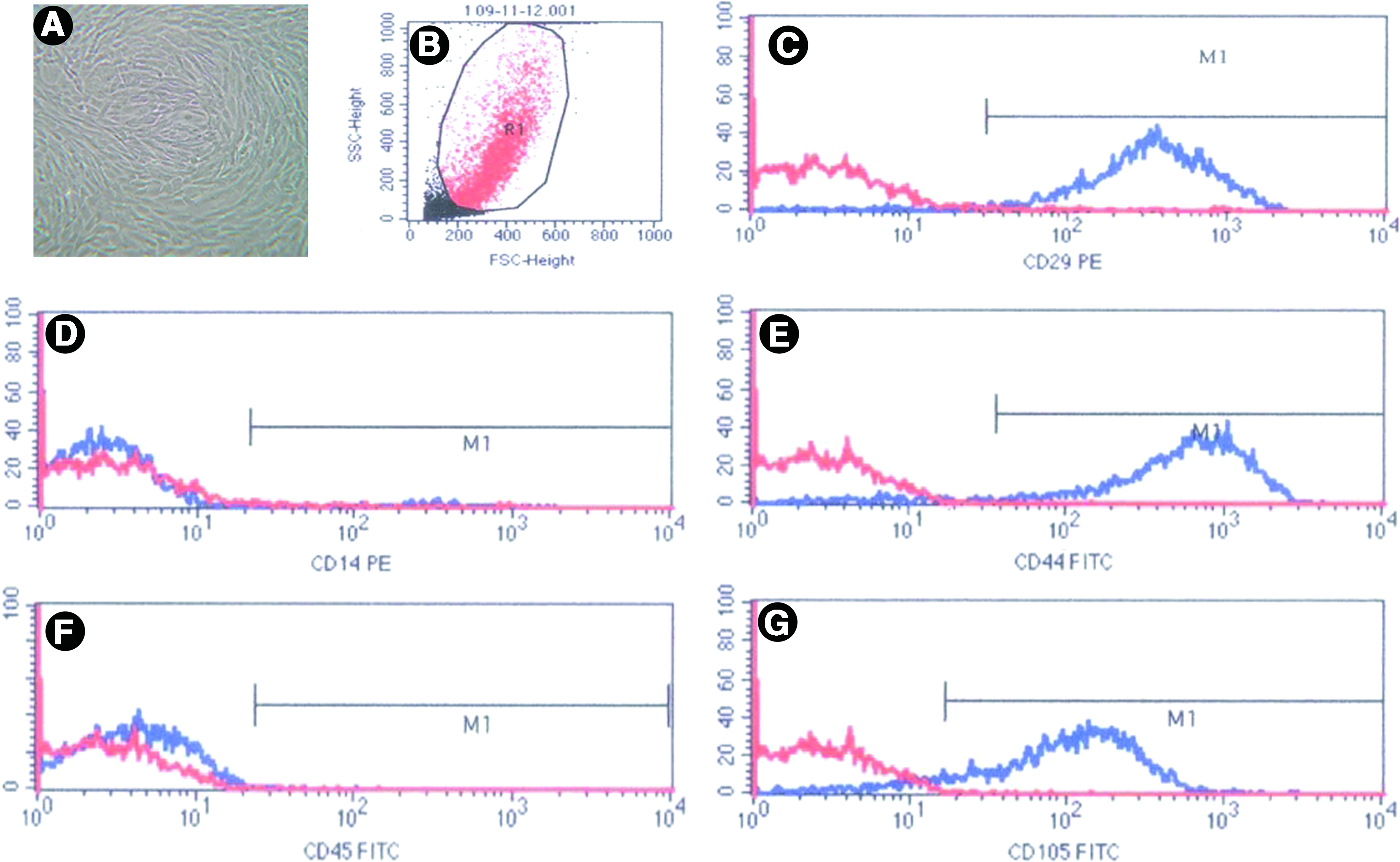
Identification of hMSCs using flow cytometry.
In vitro multi-differentiation potential of hMSCs
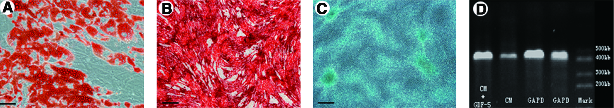
After 21 days of adipogenic induction, several clusters of adipocytes and intracellular lipid vacuoles were observed, which were positively stained by 1% Oil Red O (Fig. 2A). The osteogenic potential of the hMSCs was confirmed by the positive alizarin red staining of the colonies (Fig. 2B). After 21 days of chondrogenic induction, the cell colonies were positively stained by alcian blue (Fig. 2C) and showed positive expression of type II collagen mRNA (a marker of chondrogenic differentiation, Fig. 2D). These results demonstrated that the hMSCs obtained in this study had a multi-differentiation potential.
In vivo multi-differentiation potential of hMSCs.
Gross appearance of the self-assembled constructs
When a cell suspension was seeded onto an agarose-coated 24-well plate (defined as t=0 weeks), the cells from all four groups began to aggregate. During the first 6 h, the cells gradually sank into the surface of the agarose (Fig. 3A). Within 24 h, the cells aggregated into a transparent membrane that was detached from the substratum (Fig. 3B). Subsequently, the membrane gradually shrank into a round or oval construct within 4 days (Fig. 3C). During the next weeks, the constructs that were cultured over agarose showed a smooth, spherical, and cartilage-like appearance. This process applied to all of the groups.
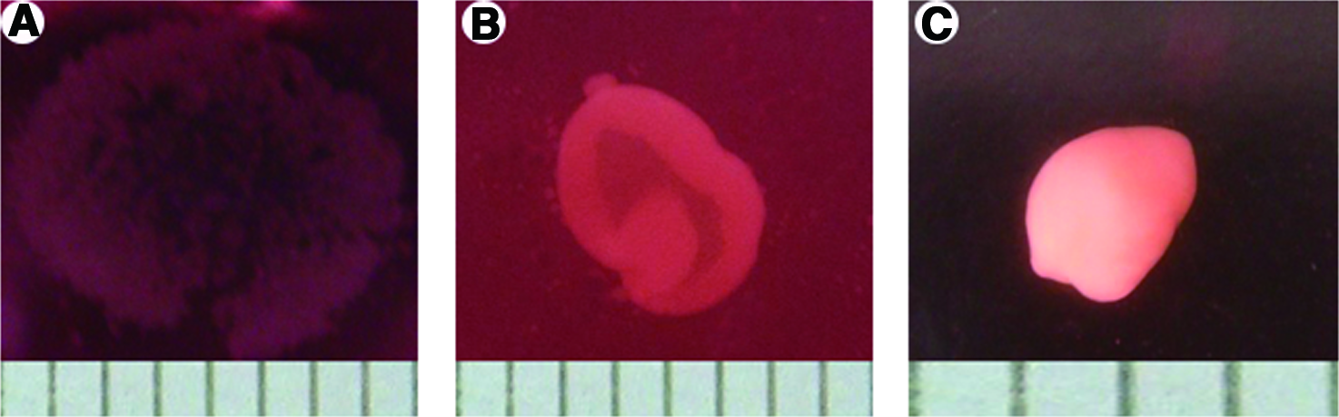
The process of self-assembly.
After 3 weeks of culturing under the same conditions, the diameters of the constructs generated from the predifferentiated hMSCs (Groups D2 and CM2) were significantly larger than those of the constructs generated from unpredifferentiated hMSCs (Groups D1 and CM1; p<0.05). For the same cell type, D1 constructs had an obviously larger diameter than that of CM1 constructs (p<0.05), whereas there was no significant difference between the CM2 and D2 constructs. Constructs generated from the predifferentiated hMSCs (Groups D2 and CM2) had significantly higher WW and DW compared with those generated from unpredifferentiated hMSCs (Groups D1 and CM1; p<0.05). The WW (4.39±0.50 mg) and DW (1.64±0.42 mg) of the CM2 constructs were greater than 3.1- and 2.2-fold higher, respectively, than the CM1 constructs (1.39±0.11 mg WW and 0.75±0.04 mg DW). The D2 constructs showed a similar trend in the WW and DW (3.2- and 2.6-fold higher, respectively) relative to the D1 constructs. Moreover, the D2 constructs had significantly higher WW and DW compared with the CM2 constructs (p<0.05), whereas there was no significant difference in weight between the D1 and CM1 constructs (Table 1).
Values are reported as the mean±standard deviation. Data separated by different superscript letters indicate significant differences; p<0.05.
Histological and immunohistochemical analysis of the self-assembled constructs
All of the self-assembled constructs appeared to have a spherical shape after 3 weeks of culturing (Fig. 4A–D). Hematoxylin and eosin staining showed that the matrix surrounding the chondrocytes was similar to cartilage lacunae found in normal cartilage (Fig. 4E–H). Collagen X, which is a marker of hypertrophic chondrocytes, was positively stained in all the groups. However, the expression of Collagen X was significantly higher in the CM2 constructs that were cultured in the chondrogenic medium lacking GDF-5 relative to the other constructs (Fig. 4M–P). Expression of type II collagen was uniformly stronger in both CM2 and D2 constructs; and negative and marginally positive staining was observed in the CM1 and D1 constructs, respectively (Fig. 4I–L). Similar trends were observed for the toluidine blue staining. Strong toluidine blue staining in the D1 and D2 constructs reflected the extent of chondrogenic differentiation of the constructs treated with GDF-5. Conversely, the CM1 and CM2 constructs showed weak toluidine blue staining (Fig. 4Q–T).
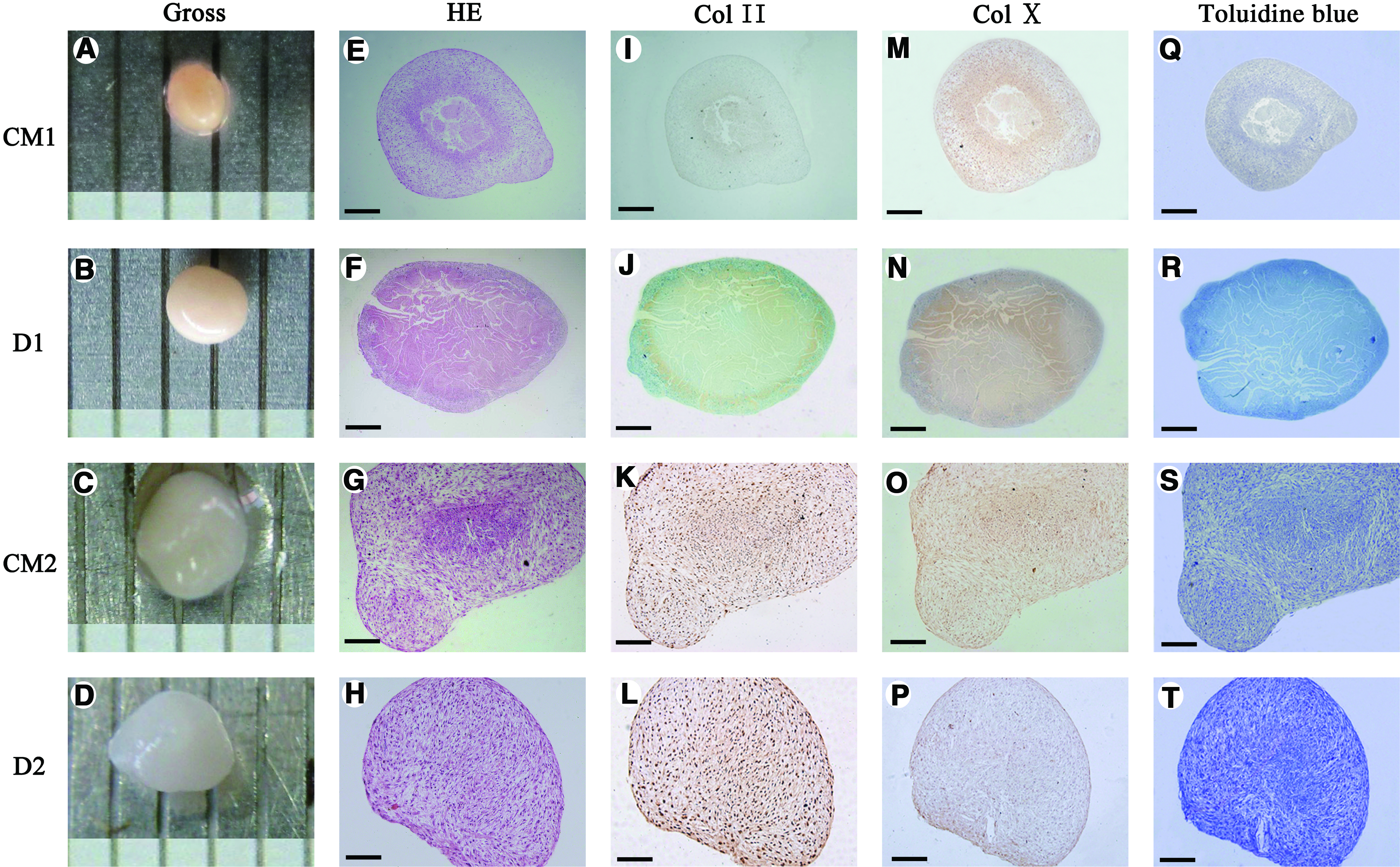
The gross appearance, histological structure, and staining of self-assembled constructs.
Biochemical analyses
DWs were measured to normalize the biochemical contents of the self-assembled constructs from different groups. The constructs generated from the predifferentiated hMSCs (Groups D2 and CM2) displayed significantly higher GAG and collagen per DW at any given time point relative to those generated from unpredifferentiated hMSCs (Groups D1 and CM1, p<0.05). For the CM1 and D1 constructs, the GAG/DW increased within the first 3 weeks and then decreased during the last 3 weeks. To be exact, the GAG/DW of the CM1 constructs increased from 2.13±0.40 at t=1 week to 2.65±0.52 at t=3 weeks and then decreased slightly to 1.95±0.36 during the last 3 weeks (p>0.05). The GAG/DW of the D1 constructs increased from 3.54±0.34 at t=1 week to 5.91±0.46 at t=3 weeks (p<0.05) and then decreased slightly to 4.93±0.23 during the last 3 weeks (p>0.05). However, the GAG/DW of the CM2 and D2 constructs continued to increase during the 6 weeks of self-assembly. To be exact, the GAG/DW of the CM2 constructs increased significantly from 6.03±0.88 to 9.28±0.95 within the first 3 weeks (p<0.05) and then showed a slight increase to 10.68±0.70 during the last 3 weeks (p>0.05). The GAG/DW of the D2 constructs increased significantly from 7.91±0.59 to 13.24±0.92 within the first 3 weeks (p<0.05) and achieved a final GAG/DW of 14.42±1.15 at t=6 weeks (p>0.05, Fig. 5 and Supplementary Fig. S2).

GAG contents of self-assembled constructs from all groups at t=1, 3, and 6 weeks. Data are presented as the means±SD (n=6 per group). *p<0.05 versus CM1 group at the same time point; **p<0.05 versus D1 group at the same time point; #p<0.05 versus D1 group at t=1 week; ##p<0.05 versus CM2 group at t=1 week; ▴p<0.05 versus D2 group at t=1 week. GAG, glycosaminoglycan; SD, standard deviation; DW, dry weight.
Likewise, the Collagen/DW of the CM1 constructs increased from 4.15±0.56 at t=1 week to 5.31±0.85 at t=3 weeks (p<0.05) and then decreased slightly during the last 3 weeks (p>0.05). The Collagen/DW of the D1 constructs showed a slightly increasing trend over time (p>0.05). The Collagen/DW of the CM2 constructs increased significantly from 10.71±1.24 to 16.50±1.90 within the first 3 weeks (p<0.05), and this level was maintained throughout the last 3 weeks. The Collagen/DW of the D2 constructs increased significantly from 15.46±0.96 at t=1 week to 21.38±1.34 at t=3 weeks (p<0.05) and reached a final value of 23.48±1.45 at t=6 weeks (p<0.05) (Fig. 6 and Supplementary Fig. S3).

Total collagen contents of self-assembled constructs from all groups at t=1, 3, and 6 weeks. Data are presented as the means±SD (n=6 per group). *p<0.05 versus CM1 group at the same time point; **p<0.05 versus D1 group at the same time point; #p<0.05 versus CM1 group at t=1 week; ##p<0.05 versus CM2 group at t=1 week; ▴p<0.05 versus D2 group at t=1 week.
There was no significant difference in the cell number among the four groups at t=1 week (Fig. 6). The number of cell in the CM1 constructs decreased significantly from 5.5±0.8 million at t=1 week to 2.2±0.5 million at t=6 weeks (p<0.05); the number of cells in the D1 constructs also decreased significantly from 5.1±0.7 million at t=1 week to 2.4±0.5 million at t=6 weeks (p<0.05). The number of cells in constructs CM2 and D2 generated from the predifferentiated hMSCs did not fluctuate significantly during 6-weeks of culture self-assembly and ranged from 5.3±0.9 million to 6.4±0.7 million cells per construct (Fig. 7). Moreover, we have compared the biochemical composition of tissue-engineered cartilage from this study with previous reports (Supplementary Table S4).
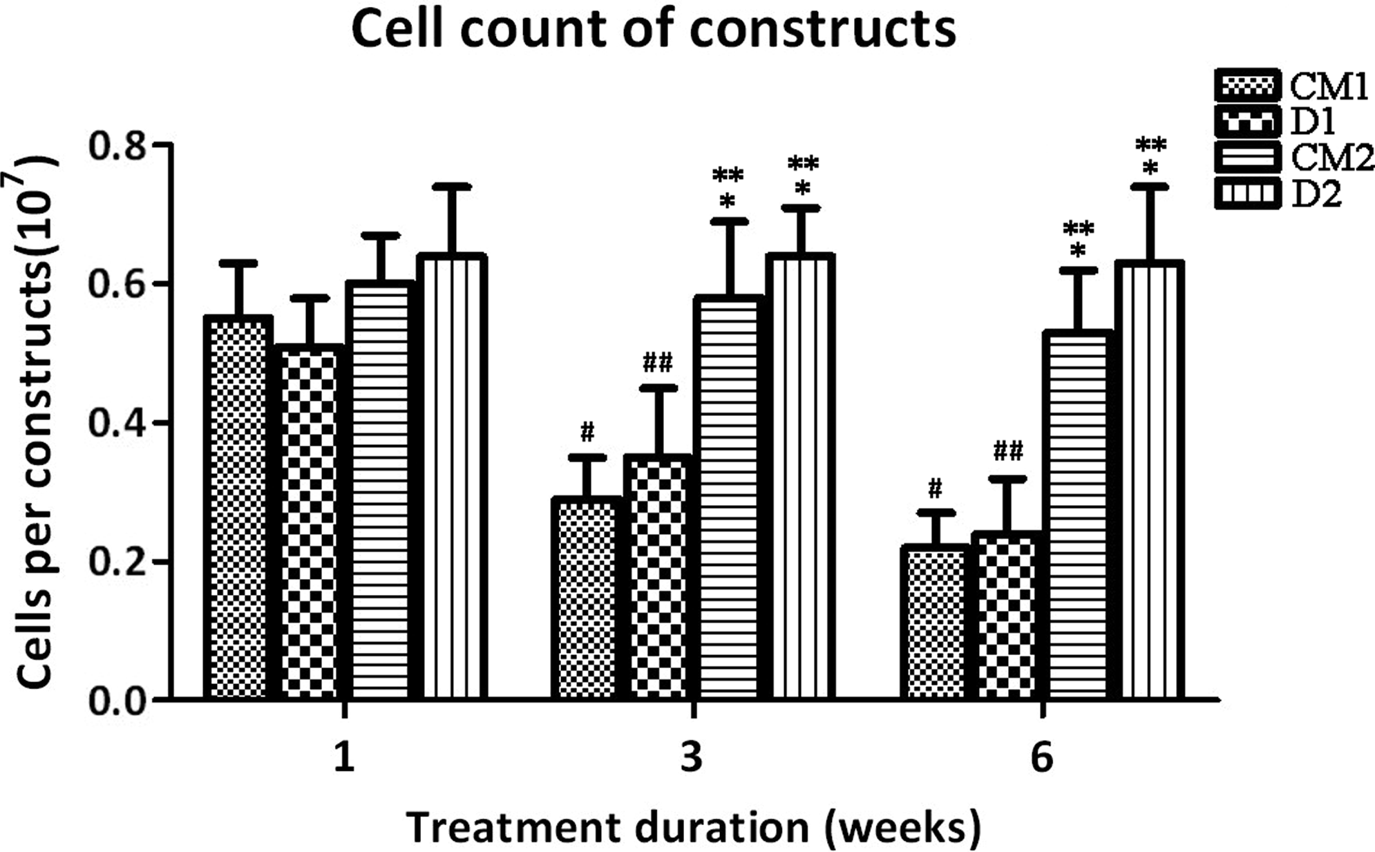
Cell counts of self-assembled constructs from all groups at t=1, 3, and 6 weeks. Data are presented as the means±SD (n=6 per group). *p<0.05 versus CM1 group at the same time point; **p<0.05 versus D1 group at the same time point; #p<0.05 versus CM1 group at t=1 week; ##p<0.05 versus D1 group at t=1 week.
Implantation of self-assembly constructs into chondral defects in vitro
As we all know, the most important indicator of the success of tissue-engineering methods to repair cartilage defects is the integration of native tissue and repair tissue. The repair in this study was regarded as a smooth, stable repair tissue that filled the defect and appeared to be adhering to the surrounding cartilage according to the International Cartilage Repair Society (ICRS) -Histological Visual Scale (See Supplementary Table S3; Supplementary Data are available online at
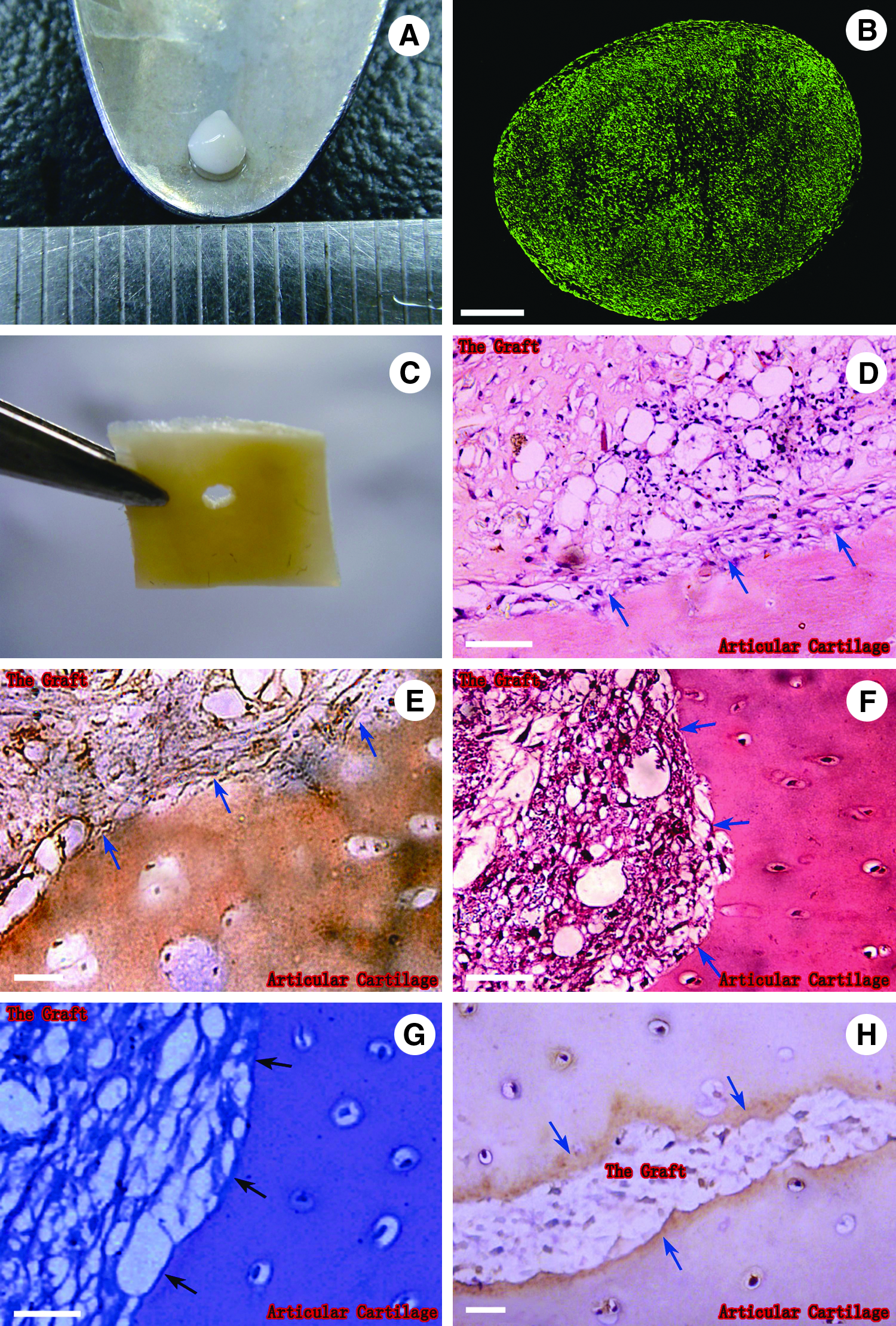
Macroscopic and histological assessment of the cartilage defect repair model constructed using D2 constructs and human cartilage explants.
Discussion
The most common approach to cartilage tissue engineering involves a biocompatible and structurally and mechanically sound scaffold seeded with either chondrocytes or undifferentiated MSCs. However, scaffolds-based cartilage tissue engineering is prone to a number of limitations including poor adhesiveness, immune rejection of the materials used, a material-induced inflammatory response, the toxicity of degradation products, the mechanical properties of the scaffolds, and the biocompatibility between materials and the implanted cells.10,12,14,25,26 Therefore, the development of an improved tissue-engineered alternative has been a recent research focus. Currently, scaffold-free tissue-engineered constructs with a hyaline-like appearance, excellent adhesiveness, and histological, biochemical, and biomechanical properties similar to those of native articular cartilage have been harvested by a self-assembly process and used to successfully repair chondral defects in vivo.14,27 The self-assembly technique has been shown to have the most potential for the development of tissue-engineered cartilage. However, an optimum construction method for self-assembled constructs is still under investigation. Hence, we tested four different construction methods in an attempt to develop an optimum scaffold-free tissue-engineering alternative and to repair in vitro chondral defects.
The scaffold-free method could be a viable alternative for avoiding unknown risks from the application of scaffolds. The self-assembly approach used in this study is feasible, because when cells are seeded at a high density over the substratum coated by agarose, they are inhibited by the agarose substratum from attaching and flattening. Moreover, since the only substrate of these cells for adhesion is other cells, this may reinforce the intercellular communication essential to the maintenance of chondrocytic phenotype.14,28,29 In this case, cartilage-like constructs self-formed during the process termed “self-assembly.” In this study, the self-assembly tissue-engineering method can attain similar effects that are based on the cell-seeded scaffold, but prevent the potential problems caused by application of scaffold materials.
Another interesting finding was the use of hMSCs as the seeding cells in this study. Autologous chondrocytes have previously been chosen as the seeding cells for cartilage tissue engineering. However, limitations regarding the use of chondrocytes as the seeding cells also suffers from the aforementioned concerns and disadvantages of using scaffolds.16,24,30 In contrast, hMSCs derived from bone marrow have been considered to possess the capacity of proliferating and maintaining their multipotent differentiation in vitro.31,32 Moreover, hMSCs are easily accessible after a minimally invasive procedure. Therefore, hMSCs is an ideal cell type for cartilage tissue engineering. In this study, hMSCs were isolated from iliac crest bone marrows aspiration of 8 healthy volunteer donors. We investigated the effect of aging on the pluripotent differentiation capacity of hMSCs, and whether the number of hMSCs decreases with age (see Supplementary Tables S1 and S2). The results indicated that age did not exert an influence on the pluripotent differentiation potential of hMSCs, and no significant difference was found in the phenotype of the hMSCs (data not shown). However, we found that the number of hMSCs harvested by bone marrow declined with age, and the hMSCs from younger volunteers had a stronger proliferative capacity than those from older individuals. Therefore, we chose bone marrow from younger volunteer donors (ranging from 24 to 48 years; median age 36 years). Whether or not the number of hMSCs decline with age is still unknown. Roura et al. found that age does not affect the adipogenic and myogenic differentiation potential of human CD105 (+) MSCs. 33 Oreffo et al. found that aging did not appear to alter several other cell phenotypes expressed by MSC. 34
The third important issue is the optimization of differentiation-inducing methods and agents. GDF-5 is a recently discovered growth factor belonging to the bone morphogenetic protein family and the transforming growth factor-β (TGF-β) superfamily. GDF-5 can regulate the bone growth of endogenous cartilage by acting on the duration of the growth of the plate chondrocytes, which is very important for skeletal development and joint formation.35,36 Since GDF-5 also is a part of the TGF-β superfamily, we investigated whether GDF-5 contributed to the regulation of the differentiation of MSCs into chondrocytes by a self-assembling tissue-engineering method. A previous study demonstrated that 100 ng/mL of GDF-5 is the most effective concentration of inducing the differentiation of fibroblasts,37,38 periosteum-derived cells,39,40 and stem cells41,42 into chondrocytes, and it promotes the secretion of a chondrocyte-specific collagen and proteoglycan matrix. Moreover, GDF-5 can enhance the GAG production of chondrocytes.43,44 Further, our data indicated that GDF-5 could effectively induce hMSCs to form cartilage tissue. Since the main aim of this study was to examine the effect of GDF-5 on the chondrogenesis of self-assembled constructs rather than exploring the difference of GDF-5 and TGF-β on the chondrogenic inductive effect of hMSCs, we failed to compare our results with the more common chondrogenic differentiation using TGF. However, Bai et al. showed that higher doses of GDF-5 or TGF-β1 stimulated the differentiation of MSCs into chondrocytes, whereas low doses of GDF-5 combined with TGF-β1 could manifest obvious chondrogenic effects on MSCs. 41 Feng et al. also demonstrated that exogenous GDF-5 (100 ng/mL) had a much greater effect on chondrogenesis than TGF- β1 (10 ng/mL). 42
Our data further demonstrated that GDF-5 (100 ng/mL) could stimulate cells or self-assembled constructs to secrete specific sulfated proteoglycans and collagen type II cartilaginous matrices. In our study, the gross appearance, histological and biochemical properties of self-assembled constructs illustrated that D2 constructs generated from chondrogenically predifferentiated hMSCs treated with GDF-5 outperformed the other constructs. The D2 constructs, which contained the highest GAG and collagen contents and number of living cells, were most similar to native hyaline cartilage in appearance, histology, and biochemistry. Moreover, GDF-5 may, to some extent, inhibit self-assembled engineered cartilages from undergoing hypertrophy. This inhibition was inferred from the fact that the CM2 constructs without GDF-5 treatment strongly expressed Collagen X (a specific marker of hypertrophic chondrocytes). Therefore, the use of chondrogenically pre-differentiated hMSCs treated with GDF-5 as seeding cells represents another improvement in cartilage tissue engineering.
The utilization of hMSCs and a unique scaffold-free technique of tissue engineering could minimize the limitations that are commonly related to the use of scaffolds and ACI. Our data showed that the D2 constructs based on chondrogenically pre-differentiated hMSCs contained higher GAG/DW and collagen/DW than those based on fibrochondrocytes. In addition, GAG/DW values reached just one tenth the level of those based on hyaline chondrocytes, whereas collagen/DW values were more than half of those based on hyaline chondrocytes.29,45 The difference in GAG and collagen contents may result from the fact that the chondrogenically pre-differentiated hMSCs with GDF-5 had a greater capacity for the synthesis and secretion of GAG and collagen than fibrochondrocytes but a weaker capacity than hyaline chondrocytes. On the basis of these observations, hMSCs may be a viable alternative to chondrocytes in cartilage tissue engineering.
To further study whether the self-assembled constructs generated from chondrogenically pre-differentiated hMSCs treated with GDF-5 could effectively repair chondral defects, an in vitro cartilage defects model was constructed. Previous studies have found that articular cartilage has anti-adhesive properties due to proteoglycans, decorin, and fibromodulin, which can inhibit the adhesion of cells to cartilage. 46 Therefore, the integration of implanted constructs into adjacent cartilage is also a concern. Several approaches have been proposed to overcome this problem, including enzymatic treatment of the surface of the cartilage matrix and the strengthening of the initial fixation.12,47 However, these measures cannot solve the fundamental problem. Therefore, it is urgent to develop an optimum tissue-engineered cartilage that possesses excellent adhesive properties for the repair of cartilage defects. On the basis of the histological and immunohistochemical results showing that the D2 constructs was integrated with the explant at 3 weeks post-implantation, we could infer that the D2 constructs also had ideal adhesive properties, and FN may be related to the adhesive properties of the D2 constructs as reported elsewhere, to some extent. 27 However, the key factors that determine the adhesiveness have not been identified and still remain to be determined. The histological results also indicated that the tissues exhibited plasticity at the interior of the constructs, which may result from the adhesiveness that improved signal transduction between the implanted constructs and the cartilage explants. Due to these excellent adhesive properties, we speculate that a self-assembled construct could be developed that matches the different size and shape to repair the cartilage defects in clinic that do not reach the subchondral plate.
Conclusions
This study has demonstrated that it is feasible to repair injured cartilage in an allograft model using scaffold-free self-assembled constructs generated from chondrogenically pre-differentiated hMSCs, and the pre-differentiation of hMSCs using GDF-5 is an improved method for self-assembled tissue-engineered constructs. In addition to the excellent adhesive properties, the D2 constructs from pre-differentiated hMSCs with GDF-5 showed increased GAG contents, collagen contents, and cell numbers relative to other groups, thereby indicating that this approach is a more promising therapeutic alternative for cartilage repair. In the future, our constructing method may be further improved by modifying the induction time and standardizing the geometric environment.
Footnotes
Acknowledgments
This study was supported by the National Natural Science Foundation of China (No.30800654).
Disclosure Statement
No competing financial interests exist.
References
Supplementary Material
Please find the following supplemental material available below.
For Open Access articles published under a Creative Commons License, all supplemental material carries the same license as the article it is associated with.
For non-Open Access articles published, all supplemental material carries a non-exclusive license, and permission requests for re-use of supplemental material or any part of supplemental material shall be sent directly to the copyright owner as specified in the copyright notice associated with the article.