
Abstract
This study describes an injectable, thermo-responsive hyaluronic acid-g-chitosan-g-poly(N-isopropylacrylamide) (HA-CPN) copolymer for bone tissue engineering. The wettability, temperature-dependent change of water content, and volume of HA-CPN hydrogel were measured, together with its biocompatibility in vitro and in vivo. The dried hydrogel morphology shows a three-dimensional, porous structure with interconnected pores. Canine bone marrow-derived mesenchymal stem cells (cBMSCs) were encapsulated in HA-CPN hydrogel and osteoinduction was assessed by comparing samples with different osteogenic differentiation induction times but with the same total cell culture time. Cell proliferation and time-dependent osteogenic differentiation, evident from secretion of extracellular matrix and formation of mineral deposits, were observed. The cells showed better proliferation in HA-CPN hydrogel than on tissue culture polystyrene after osteo-induced for 21 days and higher alkaline phosphatase activity regardless of osteo-induction times. Mineralization extent of cBMSCs in HA-CPN followed by Alizarin red stains showed positive stained nodules after osteo-induced longer than 7 days. The cells/hydrogel construct also showed increased mechanical strength and elasticity after osteogenic differentiation, and the increase could be correlated with osteo-induction time. In vivo studies confirmed the biocompatibility and bioresorption of the HA-CPN hydrogel and ectopic bone formation when the hydrogel was used as a cell carrier for osteo-induced cBMSCs and implanted in nude mice subcutaneously. Taken together, the results indicate the feasibility and efficacy of HA-CPN hydrogel as an injectable bone tissue engineering scaffold with cBMSCs.
Introduction
An in situ forming hydrogel from a polymer solution is one of the most popular injectable scaffolds.2–4 For a polymer solution to perform sol-to-gel phase transition, the gelling mechanism could be photo-polymerization, ionic crosslinking, chemical crosslinking, or environmental stimuli including temperature, pH, or ionic strength change of the surrounding medium.5,6 Nonetheless, among all in situ forming hydrogels, a thermo-responsive hydrogel, which could be formed spontaneously from a polymer solution in response to a temperature change around the physiological temperature, might be the easiest to apply for tissue engineering purpose. The polymer usually shows sol-gel phase transition above a certain temperature and the threshold temperature is called the lower critical solution temperature (LCST). 7 The polymer is soluble in an aqueous solution below the LCST with the solution being in a free-flowing state to facilitate the scaffold to be mixed evenly with cells and growth factors. The polymer becomes increasingly hydrophobic and insoluble in an aqueous solution above the LCST, which leads to gel formation.
Poly(N-isopropylacrylamide) (PNIPAM) and its related copolymers are widely investigated thermo-responsive hydrogel polymers, which have been applied for drug delivery and cell therapy.8–12 Recently, those gels have also been proven to be useful in cell encapsulation for bone and cartilage tissue engineering.13–18 However, with major limitations such as nonbiodegradability and relatively low biocompatibility, 19 PNIPAM have been modified with many natural polymers, including chitosan, collagen, hyaluronic acid, or arginine-glycine-aspartate (RGD) peptide, to increase cell attachment, strengthen mechanical properties, and improve cell proliferation.13,15–17 Such modifications also render PNIPAM-based copolymers more biodegradable without changing their reversible sol-gel transition characteristics. 16
Being the second most abundant natural biopolymer, chitosan is commonly found in cell walls of fungi and shells of marine crustaceans. Chitosan is a polysaccharide derived from chitin and is with a linear structure composed of glucosamine and N-acetyl glucosamine-linked by β(1→4) covalent bonds. In recent years, biomaterials based on chitosan have been applied in bone tissue engineering due to its osteo-conductive ability, minimal foreign body reaction, and antibacterial nature. 20 When used in osteoblasts culture, chitosan can promote cell proliferation and mineral-rich matrix deposition.2,21–23 Many studies also reported that chitosan-modified calcium phosphate, biphasic calcium phosphate, or hydroxyapatite could enhance the mechanical strength of the inorganic phase and be developed for bone tissue engineering applications.24–29
Hyaluronic acid (HA) is a linear polysaccharide with repeated disaccharide units of N-acetyl-D-glucosamine-D-glucuronate linked by β(1→4) and β(1→3) linkages. It is a nonsulfated anionic glycosaminoglycan widely distributed in connective, epithelial, and neural tissues. Large amounts of HA are synthesized during bone morphogenesis in bone matrix implants in rats. 30 During the healing process of fractured bones, HA is secreted in early stages of callus formation. As scaffolds for bone tissue engineering, HA-based materials were demonstrated in several studies to be suitable for bone regeneraton.31–33 Kim et al. found that a HA-based hydrogel could be a scaffold for treating rat calvarial bone defect by mixing with BMP-2 and human bone marrow stem cells. 34 Jang et al. addressed that a HA gel can deliver a demineralized bone matrix loaded with mesenchymal stem cells for new bone formation in obliteration of mastoid cavity. 35
Therefore, we synthesized a PNIPAM-base copolymer, hyaluronic acid-g-chitosan-g-poly(N-isopropylacrylamide) (HA-CPN), from those natural bone-enhancing biomaterials (chitosan and HA). In our previous study, the copolymer hydrogel was demonstrated to be a good cell carrier for chondrocytes and meniscus cells for tissue engineering of cartilage and meniscus. 36 In this study, we hypothesize that HA-CPN could also be a good bone tissue engineering scaffold using canine bone marrow-derived mesenchymal stem cells (cBMSCs). The cells were encapsulated in the hydrogel and studied for osteogenic differentiation in vitro and ectopic bone formation in vivo.
Materials and Methods
Isolation, cultivation, and characterization of cBMSCs
cBMSCs were harvested and isolated according to the procedure previously described. 37 All experiments were approved by Chang Gung University's Institutional Animal Care and Use Committee and follow the guidelines of experimental animal care. Briefly, we aspirated bone marrow from a dog's iliac crest by using an aspiration syringe filled with heparin (1000 U/mL bone marrow) to prevent clotting. Retrieved bone marrow was plated in cell culture flasks containing the normal growth medium (Dulbecco's modified Eagle's medium [DMEM] supplemented with 100 U/mL penicillin, 100 μg/mL streptomycin, and 10% fetal bovine serum [FBS]). cBMSCs attached to the culture flask were passaged when cells were 80% confluent and expanded for 10–12 days with medium change every 3 days.
To prove the multilineage differentiation potential of cBMSCs, cells at passage 2 were tested for differentiation into osteogenic, chondrogenic, and adipogenic lineages. After cells reaching 60% confluence in the culture flask, the control medium was changed to an osteogenic, adipogenic, or chondrogenic induction medium. The osteogenic medium contained DMEM/10% FBS supplemented with 10 mM β-glycerolphosphate, 50 μg/mL ascorbic acid, and 100 nM dexamethasone. Alizarin red and alkaline phosphatase (ALP) stains were used to confirm the success of osteogenic differentiation. The adipogenic medium contained DMEM/10% FBS supplemented with 0.5 mM isobutyl methylxanthine, 200 μM indomethacin, 10 μM insulin, and 1 μM dexamethasone. Adipogenesis was evaluated by oil red O stain to show the intracellular lipid accumulation. The chondrogenic medium contained DMEM/10% FBS supplemented with 6.25 mg/mL insulin, 50 nM ascorbate-2-phosphate, and 10 ng/mL TGF-β1. Chondrogenesis was evaluated by Alcian blue stain to demonstrate cartilage matrix production.
Preparation of HA-CPN polymer hydrogel
The HA-CPN copolymer was synthesized as described previously. 36 Briefly, N-isopropylacrylamide and 2-mercaptoacetic acid was first reacted by free radical polymerization in benzene with azobisisobutyronitrile as an initiator to form end-capped PNIPAM-COOH. To synthesize chitosan-g-poly(N-isopropylacrylamide) copolymer (CPN), 0.5 g chitosan and 5 g PNIPAM-COOH were dissolved in 50 mL 0.1 M, pH 5.0 MES buffer containing 1-ethyl-3-[3-dimethylaminopropyl] carbodiimide hydrochloride (EDC) and N-hydroxysuccinimide (NHS). After polymerization for 12 h at 25°C by mixing at 180 rpm, the solution temperature was raised to 50°C to completely recover the copolymer by thermal precipitation. For HA-CPN synthesis, the CPN obtained above was mixed with 0.25 g HA in 100 mL of 0.1 M, pH 5.0 MES buffer in the presence of 0.46 g EDC and 1.37 g NHS. The grafting reaction was carried out for 12 h at 25°C by mixing at 180 rpm. Impurities and residual HA after the reaction were removed simultaneously by thermal precipitation at 50°C. Purified HA-CPN copolymer was obtained by dialysis against de-ionized water at 4°C for 4 days (300K MWCO dialysis tubings) followed by lyophilization.
Properties of HA-CPN hydrogel
Influence of temperature change on water content and volume shrinkage
To analyze the effect of temperature change on water content of hydrogel, 1 mL 10% (w/v) PNIPAM-COOH or HA-CPN solution in phosphate-buffered saline (PBS) was placed in a preweighted 4 mL sample vial and incubated at a constant temperature from 25°C to 75°C. After equilibrated at each temperature for 2 h, the weight of the polymer solution or hydrogel was determined. For hydrogel above LCST, the surface water above the wet gel was first carefully removed using filter papers before weight determination (Eq. 1).
where Whydrogel is the weight of the hydrogel, and Wpolymer is the dry weight of the polymer.
The volume shrinkage (%) of hydrogel at different temperature was defined as the water volume loss percentage after the polymer solution was gelled (Eq. 2).
where Vi is the volume of initial polymer solution at 25°C,
Contact angle measurement
The surface wettability of hydrogel was characterized by static contact angle measurements using a sessile drop method. A 1×1 cm polyethylene terephthalate (PET) film was used as the control surface. The film was coated with cold (4°C) 10% (w/v) PNIPAM-COOH or HA-CPN solution prepared in PBS and dried in a 37°C oven to obtain a fully-covered hydrogel layer. Static contact angle was measured at 37°C, using a 5 μL water droplet in a telescopic goniometer. A×23 magnification telescope was equipped with a protractor of 1 graduation. For each value reported, at least six measurements at different locations were averaged.
Scanning electronic microscope examination
To prepare a hydogel sample for scanning electron microscopic (SEM) observations without disrupting its original porous structure, the hydrogel was first frozen instantaneously by immersing in liquid nitrogen and followed by lyophilization in a freeze dryer. The dried samples were fractured, sputter-coated with gold, and examined with a Hitachi S-3000N SEM.
Evaluation of in vitro biocompatibility of HA-CPN hydrogel
To form a uniform hydrogel layer at the bottom of a 12-well cell culture plate, 10% (w/v) sterilized HA-CPN hydrogel solution was placed in each well and the plate was incubated at 37°C for gel forming. cBMSCs were seeded on top of the hydrogel layer in each well at 1×10⁵ cells/well and cultured in DMEM/10% FBS medium at 37°C in a CO2 incubator. After 0, 7, and 14 days, the cultured cells were assayed for viability by the LIVE/DEAD Viability/Cytotoxicity Assay Kit (Invitrogen). The kit provides two molecular probes, calcein AM, and ethidium homodimer-1 (EthD-1) for simultaneous visualization of live cells and dead cells. Live cells emit green fluorescence when calcein AM enters cells and is hydrolyzed to calcein by intracellular esterase. EthD-1 enters dead cells that have damaged membranes and binds to nucleic acids to produce a bright red fluorescence. A Leica DMIL inverted fluorescence microscope was used for observation and imaging. The excitation and emission wavelengths for calcein AM are 494 nm and 517 nm, respectively. The excitation and emission wavelengths for EthD-1 are 528 nm and 617 nm, respectively.
Osteogenic differentiation of cBMSCs in HA-CPN hydrogel
To determine the osteogenic differentiation potential of cBMSCs in HA-CPN hydrogel, 1×10⁵ cells were mixed uniformly with 250 μL sterilized HA-CPN polymer solution (10%, w/v) at 25°C. The cell-hydrogel solution was injected into each well of a 24-well culture plate and incubated in a 37°C CO2 incubator. The mixture turned into a solid within 5 min and the solidified hydrogel provided a porous three-dimensional (3D) cell culture environment. Samples were divided into five groups to compare the osteogenic differentiation of cBMSCs in 3D hydrogels with different osteogenic differentiation induction times but with the same total cell culture time (21 days). Group I (n=6): cultured the cell-hydrogel mixture in control culture medium (DMEM/10% FBS) for 21 days. Group II (n=6): cultured the cell-hydrogel mixture in control culture medium for 18 days followed by in osteogenic medium for 3 days. Group III (n=6): cultured the cell-hydrogel mixture in control culture medium for 14 days followed by in osteogenic medium for 7 days. Group IV (n=6): cultured the cell-hydrogel mixture in control culture medium for 7 days followed by in osteogenic medium for 14 days. Group V (n=6): cultured the cell-hydrogel mixture in osteogenic medium for 21 days. After cultivation for 21 days in each group, the specimen was harvested, and the cell proliferation and osteogenic differentiation were determined by Trypan blue stains and ALP activity measurements. Cell proliferation and ALP activity of 1×10⁵ cBMSCs grown on tissue culture polystyrene (TCPS) under the same condition in each experimental group were used as controls. Histology by Alizarin red stain and ALP stain were used to confirm the calcium deposition and ALP activity of osteo-induced cBMSCs. Mineral deposits and interactions between cells and hydrogel were observed with an SEM (Hitachi S-3000N).
Cell proliferation
To determine cell proliferation in HA-CPN hydrogel, cells were stained with Trypan blue and the number of viable cells was counted by a hemocytometer. Generally speaking, the chromophore of Trypan blue is negatively charged and only interacts with cells with damaged cell membrane. Cells with intact cell membranes that exclude the dye can therefore be deemed as viable. The cells/hydrogel constructs were cooled down to 4°C to reverse the sample from a gel state to a solution form before measurements. Two hundred fifty microliters of cells/hydrogel solution was mixed with 50 μL of 0.4% Trypan blue solution and the number of viable cells was determined after 5 min. The control was cells seeded on TCPS and cultured under the same condition. Cell numbers were reported as relative values compared to the initial seeding cell number (1×10⁵ cells/well).
ALP activity
To analyze intracellular ALP activity, each specimen was suspended in 500 μL cell lysis solution containing 0.1% Triton X-100 detergent and 5 mM MgCl2, and centrifuged at 13,000 rpm for 10 min at 4°C. The supernatant was assayed for ALP activity with the substrate p-nitrophenyl phosphate. Briefly, 80 μL supernatant was mixed with 100 μL p-nitrophenyl phosphate (5 mM) in 150 mM 2-amino-2-methyl-1-propanol buffer solution and reacted for 15–45 min at 37°C. After the incubation period, 100 μL 0.2 N NaOH was used to stop the reaction by denaturing ALP. The optical density (OD) was determined with a multi-mode microplate reader (BioTek Synergy HT) at 405 nm. ALP activity was normalized to cell number for each sample and reported as enzyme activity (ΔOD405/h) per 105 cells.
Histology
Cells/hydrogel constructs were fixed with 4% formaldehyde, washed with PBS, and incubated with 1% Alizarin red S (Sigma) solution for 10 min at room temperature to detect mineralized nodules. For ALP stain, the sample was fixed with 4% formaldehyde, washed with PBS, and stained with 1% ALP substrate solution (1 mg/mL Fast blue RR and 1% naphthol ABSI phosphate; Sigma) for 30 min at 37°C.
SEM observation
cBMSCs (1×10⁵ cells) cultured in HA-CPN hydrogel for 21 days with 7 and 14 days osteo-induction were fixed with 4% formaldehyde, washed with PBS, and dehydrated in ethanol (stepwise increasing concentrations from 50% to 100%). The fixed cells/hydrogel samples were dried in a critical point dryer (Balzers CPD030), and images were taken with a JEOL JSM 5410 SEM.
Rheological properties of cells/hydrogel constructs
To understand the change of rheological behavior of cells/hydrogel constructs with osteogenic induction times, 1×10⁵ cBMSCs were cultured in 0.5 mL HA-CPN hydrogels (10%, w/v) for 21 days with 0, 7, and 14 days osteo-induction, respectively, followed by rheological analysis. The rheological properties of all cells/hydrogel samples were obtained from a Carri-Med CSL
2
100 controlled stress rheometer (TA Instruments). The storage modulus (G′), loss modulus (G′′), complex shear modulus (G*), and loss tangent (tan δ) were determined at different frequencies. G′ provides information about the elastic property of the sample or the energy stored during deformation, whereas G′′ describes the viscous property of the sample or the energy dissipated as heat during deformation. The complex shear modulus (G*) was calculated as
In vivo nude mice experiments
Animal procedures were approved by the ethical committee of Chang Gung Memorial Hospital. Male athymic nude mice (3 months old) weigh between 250 and 300 g were used. Animals were anesthetized with 7 mg/kg xylazine (Rompum, Bayer) and 140 mg/kg ketamine (Ketalar, Roche) and the backs were sterilized with 75% alcohol solution. The animals were fed with standard animal feed. A 0.5 mL sterilized HA-CPN polymer solution (10%, w/v) was injected into the subcutaneous pocket of nude mice (n=4) for evaluating the biocompatibility and biodegradability in vivo. The skin was examined by direct observation every week for 2 months to check for any inflammation or necrosis. The resorption time of the hydrogel was recorded once the subcutaneous mass totally disappeared and no residual hydrogels were confirmed after opening the skin.
For assessment of ectopic bone formation, 0.5 mL sterilized HA-CPN polymer solution (10%, w/v) was mixed with 1×10⁶ osteo-induced cBMSCs (cultured in osteogenic medium for 14 days on TCPS) and injected into the subcutaneous pocket of nude mice (n=4). Bone formation was analyzed by an animal microcomputed tomography (μ-CT; BioScan) 4 months after implantation. The helical CT data were acquired using a high-resolution frame as a setup in the system, with tube voltage=55 KeV, pitch=1.0, and projection=180. The axial scanning range was set as 3 cm with the subcutaneous mass at the center field of view. The CT images with the matrix size of 280×280×300, and an isotropic voxel size of 0.1 mm were reconstructed. The 3D surface rendering images were generated by Osirix software. The animals were sacrificed 4 months after implantation under anesthesia with overdosed pentobarbital and the implants were harvested. Samples examined by histology were fixed in 4% paraformaldehyde, dehydrated, and embedded in paraffin. Four-micrometer-thick serial sections were obtained and subject to hematoxylin and eosin (H&E), Masson's Trichrome, and von Kossa stains.
Statistical analysis
All data are reported as mean±standard deviation. Statistics among multiple groups on cell proliferations and biochemical assays were carried out using one-way analysis of variance test to determine significant differences. Turkey's post hoc test was used to determine the difference between any two groups with p<0.05 considered statistically significant.
Results and Discussion
Characteristics of cBMSCs
Compared with rabbit or human BMSCs,17,18,38,39 cBMSCs have been less often reported as a cell source for tissue engineering.40–42 cBMSCs harvested and purified from dog iliac bone marrow could adhere to culture dishes and had the appearance of spindle shape as previously reported. 38 Since cBMSCs by definition must have the ability of multilineage differentiation, the cells were first tested for their differentiation abilities. The positive Alcian blue stain confirms cBMSCs could differentiate into chondrocytes after subcultured in chondrogenic medium (Fig. 1). The positive oil red stain indicates the intracellular lipid accumulation in cBMSCs after induced in the adipogenic medium (Fig. 1). The positive Alizarin red stain confirms the minerals produced by osteo-induced cBMSCs and the positive ALP stain demonstrates the elevated ALP activity after the cells was induced into the osteoblast lineage (Fig. 1). All those data supported the multilineage differentiation potential of cBMSCs isolated from dog iliac bone marrow.
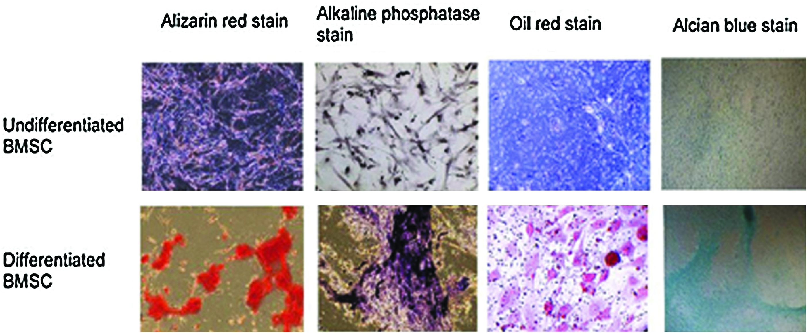
Differentiation potentials of cBMSCs into osteoblasts, adipocytes, and chondrocytes. Positive Alizarin red stain and ALP stain prove the osteogenic differentiation. Positive oil red stain verifies the adipogenic differentiation. Positive Alcian blue stain confirms the chondrogenic differentiation. Controls with undifferentiated cBMSCs show negative stains in all cases. cBMSCs, canine bone marrow-derived mesenchymal stem cells; ALP, alkaline phosphatase. Color images available online at
Characteristics of HA-CPN hydrogel
As tissue engineering scaffolds, both natural-based and synthetic thermo-gelling polymers have been developed and tested in vitro and in vivo. PNIPAM is one of the thermo-sensitive polymers that have been explored widely for biomedical applications. However, pure PNIPAM polymer was reported to be cytotoxic at 37°C due to the hydrophobic property above the LCST. 19 To improve cell viability, and to enhance cell proliferation and differentiation, we chose to modify PNIPAM by grafting bone-enhancing natural materials (HA and chitosan) onto it.
We controlled the degree of grafting (percentage of amino groups in chitosan conjugated with PNIPAM-COOH) to be 5% during the synthesis of CPN. 31 This strategy results in CPN polymer with sufficient amino groups in its chitosan backbone to facilitate subsequent HA conjugation and synthesis of HA-CPN hydrogel with sufficient mechanical strength. The synthesized HA-CPN copolymer contains 27 CPN polymer chains grafted onto a HA molecule and the molecular weight of HA-CPN was 3.22×10⁷/g/mol with 94% yield. The LCST measurement confirmed that HA-CPN still possessed sol-to-gel and gel-to-sol reversible phase transition property after modifying PNIPAM with HA and chitosan. The LCST of HA-CPN was 30.3°C during the heating cycle and 27.8°C during the cooling cycle. 36
The water content in respond to temperature change showed sharp decrease from 25°C to 35°C (around LCST) and reached a plateau after 45°C for both HA-CPN and PNIPAM-COOH (Fig. 2a). However, there is a distinctive difference for the extent of decrease. For HA-CPN the water content decreases from 9.96±0.25 to 8.29±0.22, whereas the value is from 9.99±0.23 to 1.83±0.23 for PNIPAM-COOH. These results demonstrate that HA-CPN have about 4.53 times water retention ability compared to PNIPAM-COOH near the physiological temperature. Similar trends are also observed from the volume shrinkage in Figure 2b. HA-CPN could significantly improve structure collapse by preventing excessive volume contraction during gel formation. The volume shrinkages are 21% and 95% for HA-CPN and PNIPAM-COOH, respectively. The water content plays an important role in application of hydrogels as scaffolds for cells delivery. 43 Higher water content raises the transport efficiency of nutrients into and wastes out of the hydrogel. Due to the higher water absorption and retention ability of HA and chitosan, HA-CPN retains significantly more water than PNIPAM-COOH after gel formation.
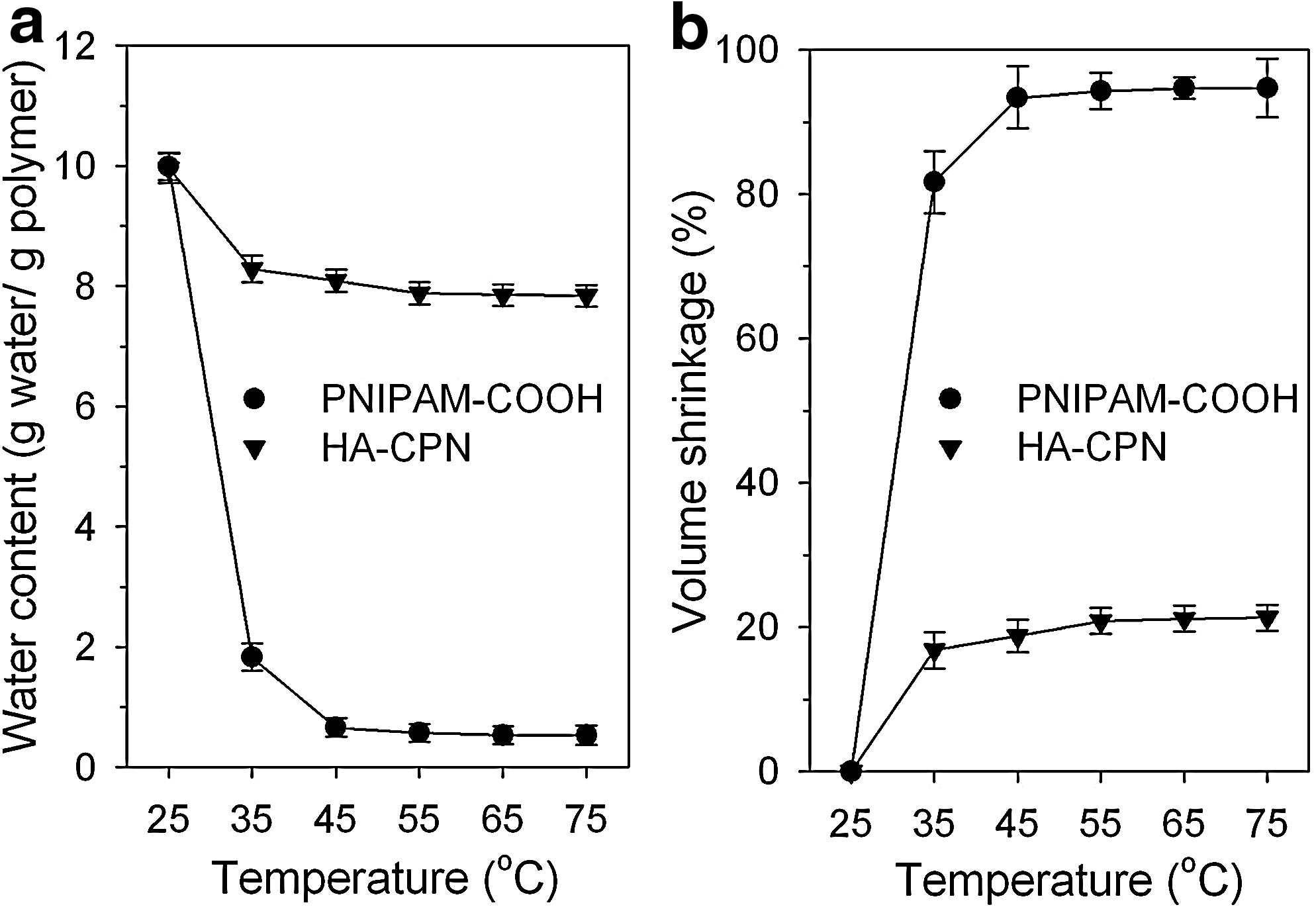
The effects of temperature change on
Hydrophilicity of polymer is another important factor for cell proliferation. PNIPAM was reported to be cytotoxic due to reduced hydrophilicity at 37°C. 14 The hydrophilicity was significantly improved after conjugating PNIPAM with chitosan and HA. The water contact angle of PET film decreased from 66.7±3.8° to 53.3±2.9° (PET film coated with PNIPAM-COOH hydrogel), and further to 38.5±2.5° (PET film coated with HA-CPN hydrogel). Thus, incorporating chitosan and HA into PNIPAM-based copolymer can increase the hydrophilicity of the hydrogel scaffold at 37°C. At 25°C, a 10% (w/v) HA-CPN polymer solution is clear and free-flowing and injectable through a syringe needle. At 37°C, the polymer solution turns into a rigid hydrogel by compelling water and the gel sticks firmly to the vial holding the polymer solution (Fig. 3a). The SEM images in Figure 3b show the 3D porous structure of HA-CPN hydrogel that contains interconnected pores with an average pore size around 20 to 30 μm (Fig. 3b), which is suitable as an injectable cell carrier for tissue engineering applications.

The phase transition behavior of HA-CPN polymer hydrogel.
For in vitro biocompatibility of HA-CPN hydrogel, cBMSCs survived well within the hydrogel according to the LIVE/DEAD test where increase of green fluorescence (live cells) with time was evident with no red fluorescence (dead cells) observed throughout the culture period (Fig. 4).
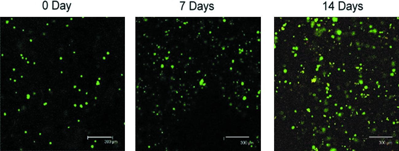
Live/dead fluorescence image of cBMSCs after cultured in HA-CPN hydrogel (10%, w/v) for 0, 7, and 14 days in the normal growth medium. Bar=300 μm. Color images available online at
Cell proliferation
Cartilage- or bone-derived cells are believed to have better proliferation and differentiation capabilities in a 3D environment than in a 2D environment owning to the 3D architecture in bone and cartilage tissue.44–46 The cells cultured in 3D hydrogels may mimic the in vivo condition more closely. Therefore, the osteogenic potential of cBMSCs was compared between 3D hydrogel and 2D TCPS environment in this study to further prove the advantage of HA-CPN hydrogel. The relative cell number increases of cBMSCs cultured on TCPS were similar for induction time up to 7 days but were significantly reduced for 14 and 21 days osteo-induction groups (Fig. 5). It could be realized that when cBMSCs are induced into osteoblast lineage, cells will become more mature and cell growth will decrease due to limited cell proliferation of matured stem cells. Compared to cells on TCPS, cBMSCs in HA-CPN hydrogel had a similar relative number increase for induction time up to 7 days (2.5 to 3 times), but showed significantly higher cell number increase for 14 and 21 days osteo-induction groups (p<0.05; Fig. 5), indicating the proliferation of osteo-induced cBMSCs was better in 3D hydrogel than on 2D TCPS surface. Taken together, it proves that HA-CPN is not only nontoxic to cBMSCs but also provides a suitable 3D environment to enhance proliferation of osteo-induced cBMSCs. Previous papers also address that BMSCs grow better in 3D scaffolds than on 2D TCPS.32,47
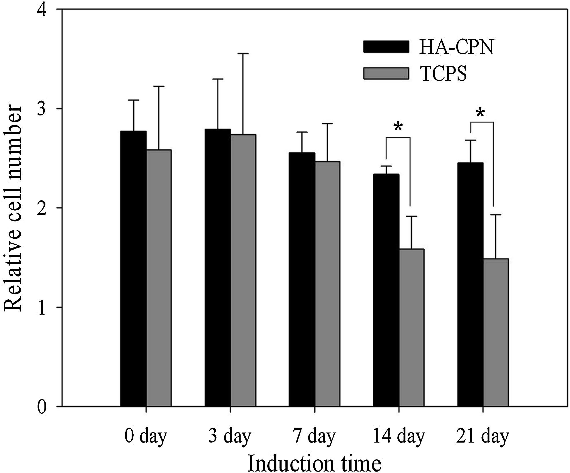
The relative cell number increase of cBMSCs cultured on TCPS and in HA-CPN hydrogel (10%, w/v) for 21 days and with different osteogenic differentiation induction times. Cell number at day 0 is taken as 1. Cell seeding density=1×10⁵ cells. *p<0.05. TCPS, tissue culture polystyrene.
AP activity and mineralization
Bone formation stages involve cell proliferation at first followed by extracellular matrix (ECM) maturation and mineralization. During the differentiation stage of bone cells, a high level expression of ALP by the cells will occur first followed by ECM maturation. The matured ECM will mineralize at the end. Therefore, elevation of ALP activity and production of mineralized matrix are two major specific events during osteogenic differentiation of cBMSCs. As shown in Figure 6, cells in HA-CPN hydrogel show increasing ALP activity with induction time and the ALP activity is significantly different from that of noninduced group (0 day) when the induction time is longer than 3 days (p<0.05). The ALP activity was also significantly higher in HA-CPN hydrogel than on TCPS regardless of the length of induction time (p<0.05). It further implied that cBMSCs presented better ability of osteogenic differentiation in 3D porous environment than on 2D surface. 48 Positive ALP stain (violet color) in HA-CPN hydrogel also proved the increase of intracellular ALP activity with induction time, especially for groups osteo-induced longer than 3 days (Fig. 7), which is consistent with the trend observed for quantitative measurement of ALP activity by biochemical assays (Fig. 6). In contrast, negative ALP stain was observed for culture in normal growth medium only (0 day). Mineralized matrix was also confirmed by Alizarin red stain with positive stained nodules in red appearing after osteo-induced longer than 7 days (Fig. 7). Taken together, cBMSCs can differentiate into osteogenic lineage well in the 3D HA-CPN hydrogel by production of intracellular ALP and mineralization of ECM. The SEM images in Figure 8 confirm that cBMSCs subject to osteo-induction for 14 days in HA-CPN are embedded within more abundant mineral-rich ECM than 7 days where mineralized particles could be observed.

The ALP activity of cBMSCs cultured on TCPS and in HA-CPN hydrogel (10%, w/v) for 21 days with different osteogenic differentiation induction times. Cell seeding density=1.0×10⁵ cells. *p<0.05. OD, optical density.

Alizarin red stains for detection of mineralization and ALP stains for detection of intracellular ALP activity of cBMSCs after cultured in HA-CPN hydrogel (10%, w/v) for 21 days with different osteogenic differentiation induction times. Color images available online at

Scanning electron micrograph of cBMSCs cultured in HA-CPN hydrogel (10%, w/v) for 21 days with 7
Mechanical properties of cells/hydrogel construct
To ascertain the enhanced mineral-rich ECM production and mineral deposits of cBMSCs after osteogenic differentiation, the mechanical properties of cells/hydrogel constructs with different osteo-induction times were subject to rheological studies. Both the storage modulus (G′) and the loss modulus (G′′) increased with induction time (Fig. 9a, b). However, the extent of increase of G′ with induction time was more pronounced than that of G′′ over the whole frequency range under study. That the value of G′ dominated over G′′ indicates the construct will show predominantly an elastic behavior. The complex shear modulus (G*) and mechanical strength also increased substantially with induction time (Fig. 9c). The G* value at 10 HZ for the construct subject to 14 days induction increased 3.1 times compared to that of the noninduced one, indicating the construct will be a much stronger injectable bone after osteogenic differentiation of cBMSCs. In contrast, the loss tangent (tan δ) decreased with induction time (Fig. 9d). Indeed, the abundance of mineralized ECM secreted by well differentiated cBMSCs will make the cells/hydrogel construct more elastic (G′>G′′) and also enhance its mechanical strength.
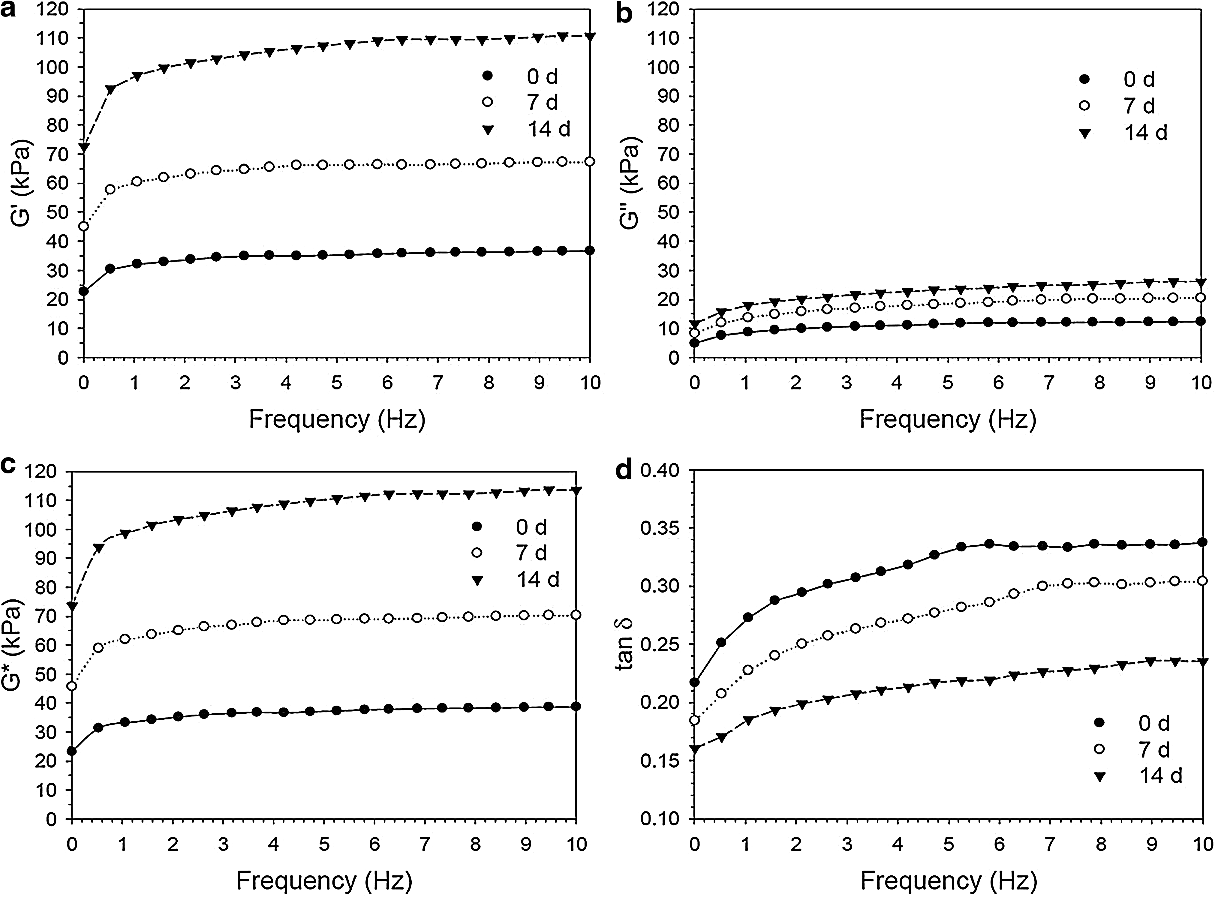
Previous studies of the mechanical properties of bone found that a less mineralized bone was softer in nature and endowed with lower elasticity. 49 The storage modulus and loss modulus of bone decreased, whereas the loss tangent increased with decreasing bone mineral contents. 50 Les et al. reported that the storage modulus of bone was positively correlated with its mineralization extent, whereas the loss tangent was negatively correlated with its mineralization. 51 An increase in the mineral content of bone's ECM also increased both the hardness and the indentation modulus. 52 For bone, mineral crystals can be viewed as a rigid filling agent for the matrix collagen fibers. Being with much higher elastic modulus than collagen, minerals can provide bone with elasticity and the ability to store and recover most energy under loading. 50 The increase of G′ with increasing induction time shown in Figure 9a therefore supports time-dependent mineralization of cBMSCs during cell differentiation. Indeed, the progressive reinforcement from the mineral phase will lead to a gain in storage modulus. For G′′, results from a previous study using hydroxyapatite reinforced starch composite indicates the increase of G′′ of bone can be related to increased energy dissipation during collagen fibers movement as more mineral phase can lead to more interactions between mineral particles and collagen fibers. 53 The results that G′′ increases after osteo-induction (Fig. 9b) therefore also supports successful time-dependent mineralization of ECM. However, since the extent of the increase of G′ is greater than that of G′′, tan δ (=G′′/G′) decreases upon increase of mineral content under osteogenic differentiation condition.
In vivo nude mice experiment
Experiments with nude mice were first used to confirm in situ gel formation after subcutaneous injection (Fig. 10A) and to test in vivo biocompatibility and bioresorption. In general, if a material is toxic when tested in vivo, the animal will show mild to moderate signs like erythematous change around subcutaneous mass, skin necrosis, or even pus formation to severe symptoms of death. 54 In our study, no skin necrosis or any inflammatory changes such as erythema, local heat, or infected wounds were found in the subcutaneous mass of nude mice during follow-up periods. Also, no nude mice were dead during the follow-up periods. The subcutaneous mass completely disappeared after 1 month (Fig. 10B) and no residual hydrogel was found after the skin was opened (Fig. 10C). The thermo-gelling HA-CPN hydrogel is thus a biocompatible and bioresorbable material, and nontoxic to the animals.
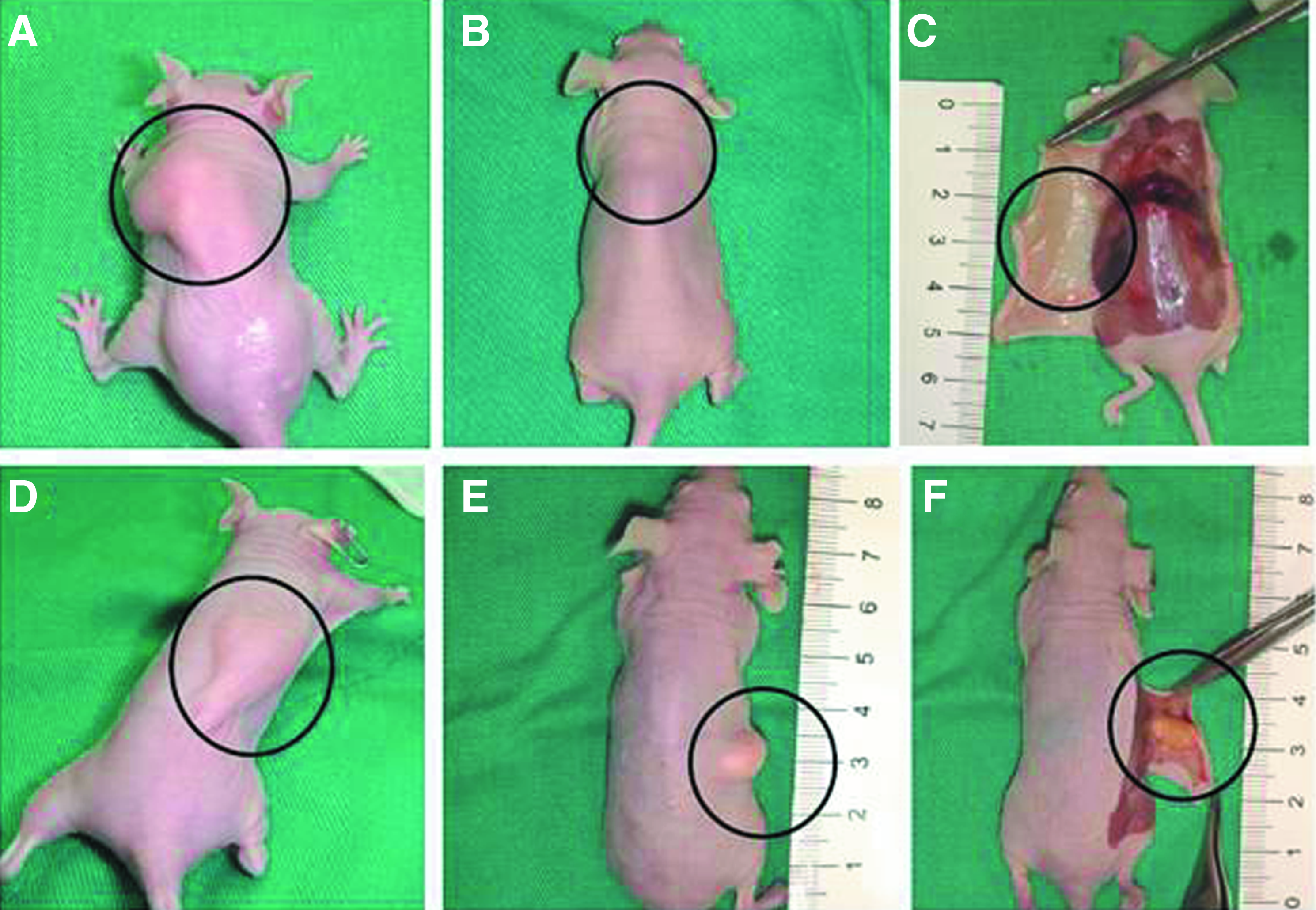
When the HA-CPN polymer solution was mixed with osteo-induced cBMSCs and injected subcutaneously on the back of nude mice (Fig. 10D), the implants were not totally degraded as was found for HA-CPN hydrogel alone and the mass sustained the shape even after 4 months (Fig. 10E). It implies that osteo-induced cBMSCs grew well within the hydrogel in vivo to form ectopic bone. The nude mice were subject to μ-CT analysis and the mass in the back was shown in soft tissue window (Fig. 11A). The bone density mass was demonstrated when it was adjusted to bone window in 2D axial view (Fig. 11B) and in 3D view (Fig. 11C). The specimens were further harvested (Fig. 10F) and examined by histology. H&E (Fig. 11D) and Manson's Trichrome (Fig. 11E) stains all showed osteoid formation with osteoblasts in the bone matrix. von Kossa stain (Fig. 11F) also proved the mineralization on the ground of matrix. Taken together, the data supported that the injected cell mass has formed ectopic bone tissue in HA-CPN hydrogel.

The mass of the HA-CPN/cBMSCs implant over the back of nude mice in Figure 10 after 4 months was analyzed by μ-CT in soft tissue window
Conclusions
Thermo-responsive HA-CPN hydrogel was found to be suitable as an injectable cell carrier for cBMSCs. The hydrophilic copolymer has high water retention ability and low volume shrinkage after gel formation, to give a 3D hydrogel that is porous and nontoxic to cBMSCs and nude mice. Overall, cBMSCs cultured in HA-CPN hydrogel showed better cell proliferation and much improved osteogenic differentiation than on TCPS. Microscopic images and staining results demonstrated cBMSCs live well in HA-CPN hydrogel with secretion of mineralized ECM when in an osteo-induction environment. The HA-CPN hydrogel was strengthened with time due to secretion and mineralization of ECM by cBMSCs after osteogenic differentiation. In vivo study also demonstrated ectopic bone formation when HA-CPN hydrogel was used as a carrier for osteo-induced cBMSCs. This study suggests HA-CPN hydrogel will be feasible as an injectable scaffold for future bone tissue engineering applications using BMSCs.
Footnotes
Acknowledgments
This work was supported by the National Science Council of the Republic of China (NSC-96-2221-E-182-028-MY2 and NSC-96-2314-B-182A-076-MY3) and Chang Gung Memorial Hospital (CMRPD290101 and CMRPD170292). Technical supports from the Animal Molecular Imaging Center of Gung Memorial Hospital for μ-CT experiments and images reconstruction are highly appreciated.
Disclosure Statement
The authors have declared that no conflict of interest exists.