
Abstract
Hepatocytes derived from embryonic stem cells (ESCs) are expected to be useful for basic research and clinical applications. However, in several studies, genetic methods used to detect and obtain them are difficult and pose major safety problems. Therefore, in this study, we established a novel detection system for hepatocytes by using indocyanine green (ICG), which is selectively taken up by hepatocytes, based on nongenetic manipulation. ICG has maximum light absorption near 780 nm, and it fluoresces between 800 and 900 nm. Making use of these properties, we developed flow cytometry equipped with an excitation lazer of 785 nm and specific bandpass filters and successfully detected ESC-derived ICG-positive cells that were periodic acid-Schiff positive and expressed hepatocyte phenotypic mRNAs. These results demonstrate that this detection system based on nongenetic manipulation with ICG will lead to isolate hepatocytes generated from ESCs and provide the appropriate levels of stability, quality, and safety required for cell source for cell-based therapy and pharmaceutical studies such as toxicology.
Introduction
One of the major metabolic functions of the liver is the elimination of various endogenous and exogenous compounds such as bile acids, organic anions, and organic cations.14,15 Indocyanine green (ICG) is a nontoxic organic anion that is exclusively eliminated by hepatocytes and is clinically used as a test substance to evaluate liver function. 16 In a large number of reports, the uptake and release of ICG is frequently used to identify ESC-derived hepatocytes.17–19 Moreover, ICG is not only selectively taken into hepatocytes, but it also has a wide optical absorption band, extending from 600 nm to above 800 nm. The maximum absorption is near 780 nm and has been used for the fluorescence diagnosis in ophthalmology for dye-enhanced photocoagulation. The fluorescence of ICG can be observed at 800–900 nm with appropriate excitation. 20 Therefore, making use of these properties, we developed a flow cytometer equipped with an excitation lazer of 785 nm with specific bandpass filters and attempted to detect ICG-positive cells.
We show here that rat primary hepatocytes and ESC-derived hepatocyte-like cells selectively took up ICG and could be detected by flow cytometry. Our approach will lead to the development of an effective system for purifying ESC-derived hepatocytes without genetic modification.
Materials and Methods
Isolation and culture of rat primary hepatocytes
All experimental procedures and protocols were approved by the animal care and use committees of Shinshu University. Primary hepatocytes were isolated from 8 week-old Wistar rats (Japan SLC, Inc.) by the collagenase perfusion method. Hepatocytes were separated from the resulting cell suspension by centrifugation. The isolated hepatocytes were plated onto gelatin-coated dishes (12-wells) at a density of 1×10⁵ cells/well and cultured in Lanford medium (Nissui Pharmaceutical Co.).
Cultured cell lines
AR42J cells of a rat pancreatic adenocarcinoma cell line, MCF7 cells of a human breast adenocarcinoma cell line, HEK 293T cells of a human embryonic kidney cell line, and HepG2 cells of a human hepatocellular carcinoma cell line were obtained from RIKEN Bioresource Center and cultured in Dulbecco's modified Eagle's medium (DMEM; GIBCO), containing 10% fetal bovine serum and 100 μM nonessential amino acids (GIBCO).
ESC culture and induction for hepatocyte-like cells
Mouse ESC line R1, a kind gift of Dr. Andras Nagy, was used throughout this study. Frozen stock was thawed and seeded on SNL feeder cells, a kind gift of Dr. Yoh-ichi Tagawa. ESCs were cultured in DMEM containing 15% knockout serum replacement (GIBCO), 100 μM nonessential amino acids, 1 mM sodium pyruvate (GIBCO), 100 μM 2-mercaptoethanol (Sigma-Aldrich Corp.), 103 units/mL of leukemia inhibitory factor (Chemicon), and antibiotics. Induction of hepatocyte-like cells was performed as previously described. 8
ICG staining of cultured cells and detection of ICG-positive cells by FISHMAN-R flow cytometry
A sterile stock solution of ICG (Daiichi-sankyou Pharmaceutical Co., Ltd) at 5 mg/mL was freshly diluted in culture medium. Primary hepatocytes, SNL, HepG2, and ESC-derived cells were incubated at 5% CO2 and 37°C in diluted ICG at specific concentrations and times. After washing, they were dispersed by using 0.1% collagenase and 0.1% trypsin. Single cells were separated from the nondigested cell clusters by using a 40 μm cell strainer (BD Biosciences). The viability of each cell population by trypan blue exclusion was typically >90%. All cells were resuspended in phosphate buffered saline and then analyzed by a FISHMAN-R flow cytometer (On-chip Biotechnologies Co., Ltd) with an excitation lazer of 785 nm and 815–850 nm bandpass filters to detect ICG-positive cells.
Isolation and culture of ESC-derived ICG positive-cells
ESC-derived cells at day 22 were dispersed by 0.1% trypsin, and a 40-μm cell strainer was used to separate single cells from the nondigested cell clusters. The harvested cells were re-plated and cultured on Type I collagen-coated dishes (BD Biosciences) with Lanford medium for 5 days. Then, they were incubated in 1 mg/mL ICG for 30 min at 37°C. After washing, ICG-positive and ICG-negative cells were picked up under light microscopy and cultured on a Type I collagen-coated dish, which has been extensively used for the proliferation and culture of mature and immature hepatocytes,21,22 with Lanford medium for 5 days.
Periodic acid-Schiff assay for glycogen
ESC-derived cells were incubated in 1 mg/mL ICG for 30 min at 37°C. After washing, cellular uptake of ICG was recorded by light microscopy. Then, the cells were returned to culture medium and incubated for 6 h, whereon the release of the ICG stain 18 was documented by light microscopy. The cells were then fixed in 10% formalin in methanol for 30 min, and subjected to periodic acid-Schiff (PAS) staining (Polysciences) according to the manufacturer's instructions.
Reverse transcription–polymerase chain reaction
Total RNA was extracted by using Trizol reagent (Invitrogen) according to the manufacturer's protocol. First-strand cDNA was synthesized from 1 μg total RNA by using Primescript reverse transcriptase (TaKaRa Bio Inc.). cDNA samples were subjected to polymerase chain reaction (PCR) amplification with a thermal cycler (MyCycler; Bio-Rad). The PCR cycling conditions were as follows: cycle of 94°C for 4 min; 30 cycles of 94°C for 30 s, annealing at the temperatures specified for each of the primer sets for 30 s, 72°C for 30 s; and 1 cycle of 72°C for 7 min. The primer sets and the predicted size of the PCR product are shown in Table 1.
Quantitative real-time RT-PCR
Real-time PCR analysis was performed by using the Thermal Cycler Dice Real Time System (TaKaRa Bio Inc.). Cycling was performed for 10 min at 95°C, followed by 40 cycles of 5 s at 95°C and 30 s at 60°C, which were the default conditions of the Thermal Cycler Dice Real-Time System software (TP 800 ver. 3.0). The threshold cycle method was used to analyze the data, with gene expression levels calibrated to those of the housekeeping gene β-actin. The primer sets were as shown in Table 2.
Results
Detection of ICG-positive cells by flow cytometry
For positive controls, primary adult hepatocytes were stained with ICG at 1 mg/mL. Although they took up some ICG (Fig. 1A), analysis by flow cytometry indicated that only 8% of the cells were ICG positive (Fig. 1Ba, b). The absorption wavelength of ICG changes depended on the concentration of ICG. 23 We assumed that primary hepatocytes took up so much ICG that they could not be easily detected by FISHMAN-R flow cytometry, because the absorption wavelength was changed by the high concentration. Therefore, we assessed ICG uptake at incubation concentrations between 50 ng/mL and 1 mg/mL for 30 min. ICG-positive cells were somewhat detectable at 20 μg/mL (Fig. 1Bc, d) and readily detected, about 90% of the cells, at 5 μg/mL (Fig. 1Be, f). At 50 ng/mL ICG, they were not detectable (Fig. 1Bg, h). These results showed that an appropriate concentration of ICG, one that does not change the absorption wavelength, is required for detecting ICG-positive cells.
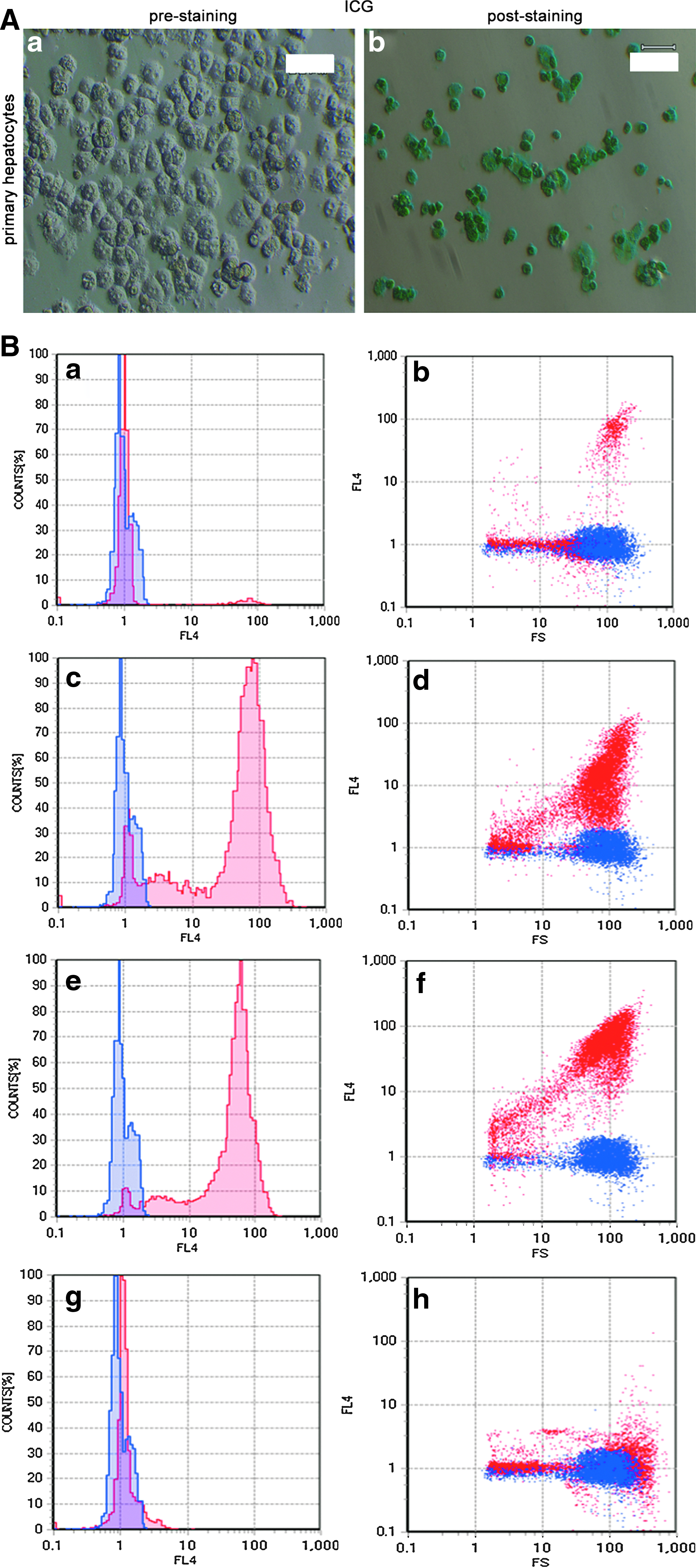
Detection of ICG-positive cells.
Time-dependent, nonspecific ICG staining of nonhepatocytes
We next examined the optimal time of ICG staining. We stained the nonhepatocytes such as SNL feeder cells, AR42J cells, MCF7 cells, and HEK 293T cells. After 30 min, there was almost no detectable staining with 5 μg/mL ICG (Fig. 2Aa, Ba, Ca, Da). Between 2 and 24 h, there was a detectable presence of ICG in the SNLs, AR42Js, MCF7s, and HEK 293Ts (Fig. 2Ab–d, Bb–d, Cb–d, Db-d). However, the levels were 10-fold lower than for primary hepatocytes (Fig. 1B). Thus, nonhepatocytes nonspecifically took up ICG when incubated for a sufficiently long time.
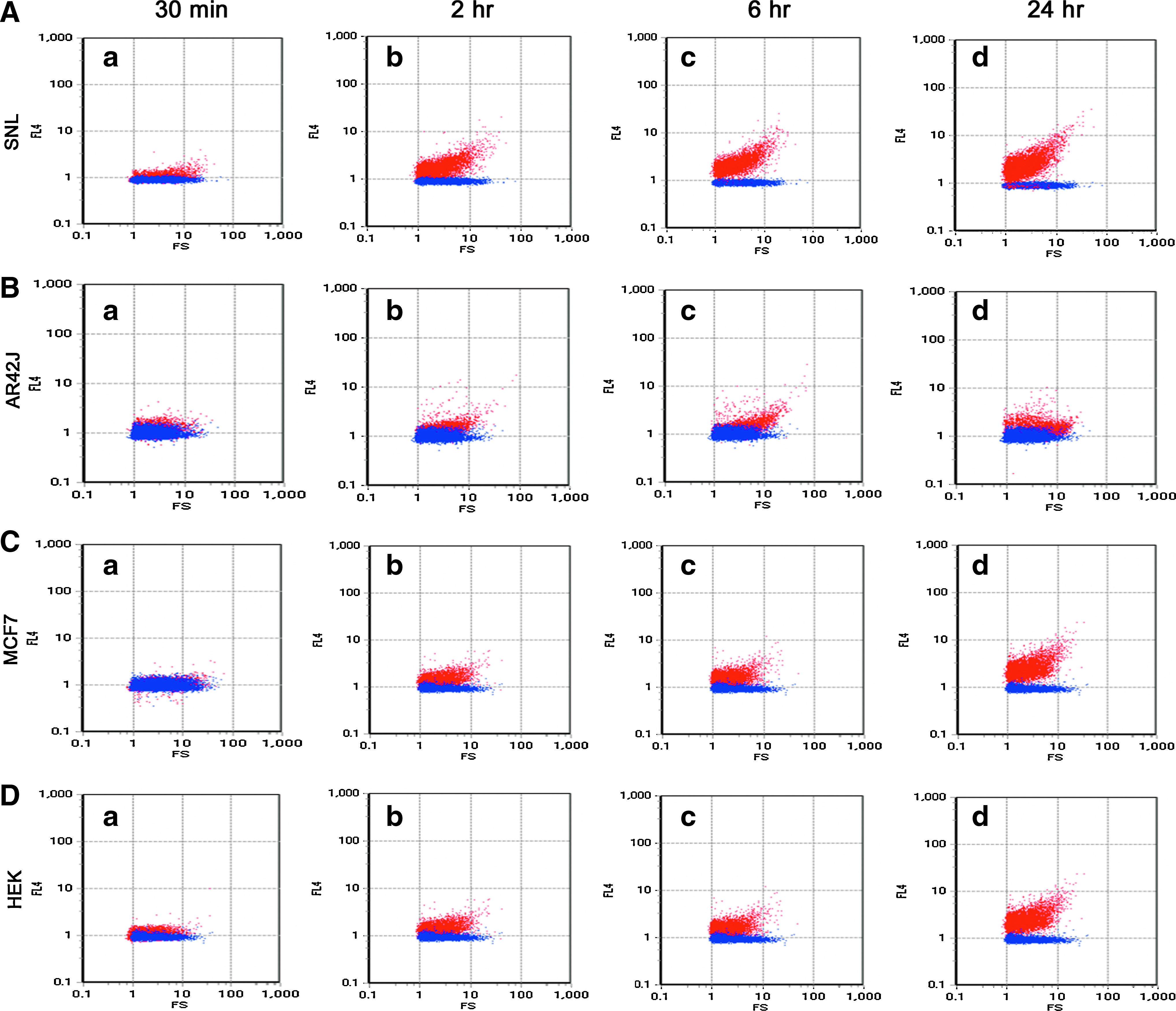
Time-dependent nonspecific uptake of ICG by nonhepatocytes.
Detection and characterization of ESC-derived ICG-positive cells
We then determined whether ESC-derived ICG-positive cells could be detected by using the FISHMAN-R flow cytometer. ESC-derived hepatocyte-like cells 8 were incubated in 5 μg/mL ICG for 30 min, which was the optimal time and concentration for staining of the primary hepatocytes. Based on FISHMAN-R flow cytometry, ESC-derived ICG-positive cells increased in a time-dependent manner (Fig. 3Aa–d). In particular, about 10% of the ESC-derived cells at day 22 were ICG-positive cells (Fig. 3Ad). Likewise, the expression of hepatocyte marker alpha-fetoprotein (AFP) and albumin (ALB) increased over time (Fig. 3Ba, b), and the expression of undifferentiated ESC-marker Nanog gradually decreased (Fig. 3Bc). Further, about 40% of human hepatocellular carcinoma HepG2 cells, which have some characteristics of hepatocytes, were also stained by ICG (Fig. 3C). HepG2 cells consisted of a relatively homogeneous population. It is possible that the phenotype and characteristics of these cells changed during the repeated passages. Taken together, these findings showed that we could detect ESC-derived ICG-positive cells by FISHMAN-R flow cytometry under appropriate conditions of ICG staining.
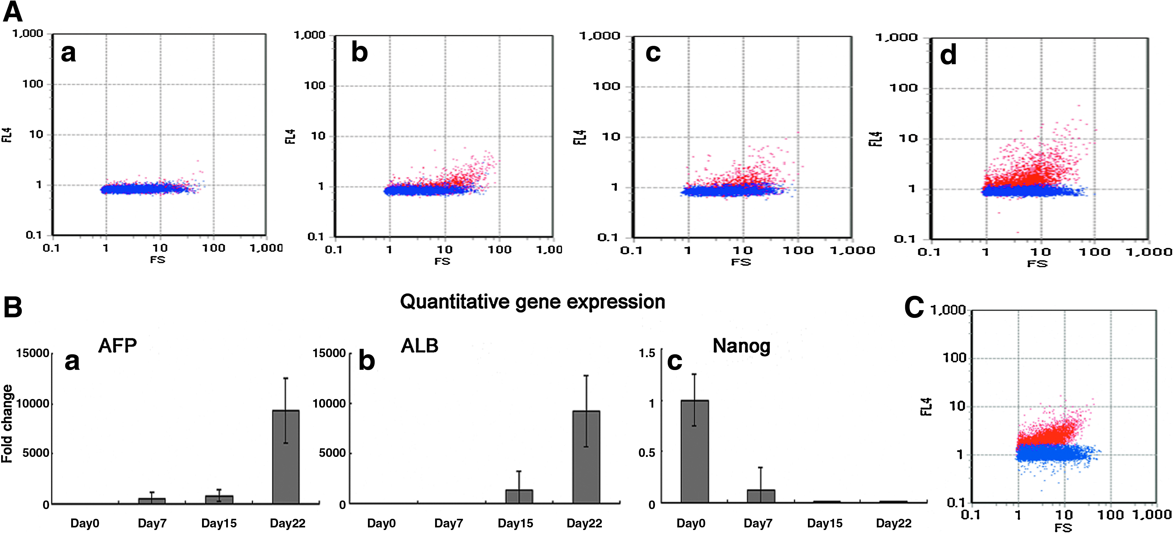
Detection of ESC-derived and HepG2 ICG-positive cells under optimal conditions.
The FISHMAN-R flow cytometer was not equipped with a cell sorting system to purify ICG-positive cells. Therefore, we could not determine whether the ESC-derived ICG-positive cells had the characteristics of hepatocyte-like cells. Hence, we examined them by ICG staining at a high concentration (1 mg/mL) for 30 min, after which we could visualize incorporation of ICG into the cells. The cells were then stained with PAS. A large number of the ESC-derived cells took up ICG (Fig. 4Aa) and then released it during the 6 h postuptake incubation (Fig. 4Ab). Further, the cells that took up and released ICG also stored glycogen as shown by PAS staining (Fig. 4Ac), which is a characteristic of hepatocyte-like cells.
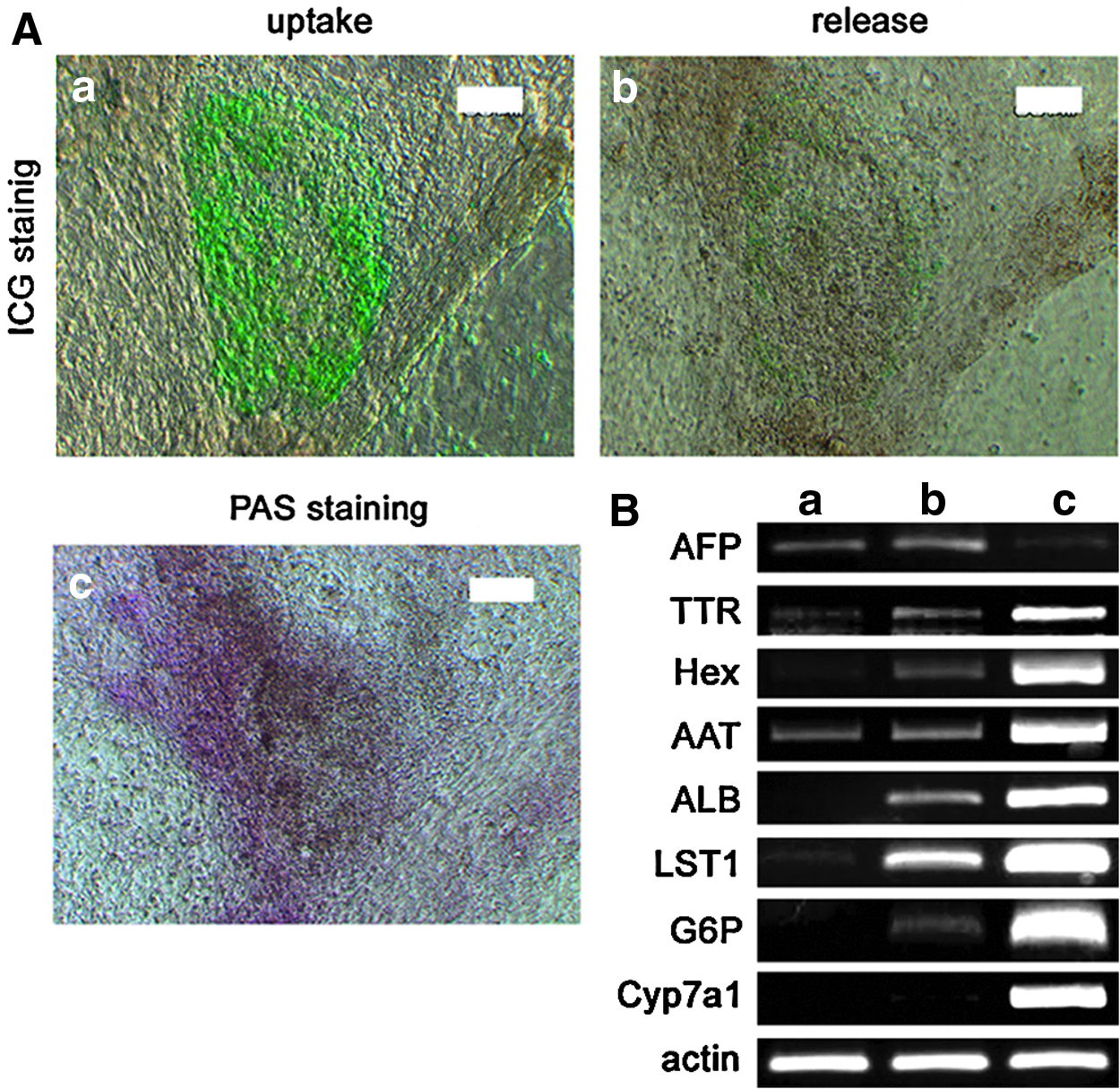
Characterization of ESC-derived ICG-positive cells.
To further analyze the properties of the ESC-derived ICG-positive cells, we examined the expression of hepatocyte-related genes by reverse transcription PCR. During the induction of hepatocyte differentiation and exposure to ICG, some of the cell clusters did not take up ICG. For these cells, the expression of hepatocyte phenotypic mRNAs for AFP, transthyretin (TTR), Hex, alpha 1-antitrypsin (AAT), ALB, and LST1 was weak or not detected (Fig. 4Ba). However, for all the ICG-positive cell clusters, the mRNAs were either weakly or strongly detected (Fig. 4Bb). The expressions of glucose-6-phosphatase (G6P) and cytochrome P450 7a1 (Cyp7a1) mRNAs, which are expressed by mature hepatocytes such as mouse primary hepatocytes (Fig. 4Bc), were only weakly detected in ICG-positive cell clusters (Fig. 4Bb). These results showed that cells within the ICG-positive ESC-derived cell clusters had the characteristics of hepatocyte-like cells.
Isolation and functional characterization of ESC-derived ICG-positive cells
Finally, we isolated ICG-negative cells and ICG-positive hepatocyte-like cells under light microscopy and enriched them on Type I collagen-coated dishes for functional characterization of the pure populations. Again, they were stained with ICG at 1 mg/mL. The population of ICG-negative cells remained ICG-negative (Fig. 5Aa), whereas the ICG-positive cells were nearly ICG-positive (Fig. 5Ab). The expression levels of AFP and ALB mRNAs were prominently higher in ICG-positive cells than in ICG-negative ones (Fig. 5B). Then, we quantified the expression of mRNAs for transferase, transporters, and cytochrome, which is a gene superfamily that catalyzes the oxidative metabolism of endogenous and exogenous compounds and plays a major role in the biotransformation of xenobiotics in the liver. The expression of Ugt1a1 (Fig. 5C), Sult2a1 (Fig. 5D), Slco1a4 (Fig. 5E), Cyp1a1 (Fig. 5F), Cyp2a10 (Fig. 5G), and Cyp2b10 (Fig. 5H) was obviously higher in ICG-positive cells (Fig. 5Cb–Hb) than in ICG-negative cells (Fig. 5Ca–Ha). Further, the expression levels of ICG-positive cells were similar to or higher than those in the fetal liver (Fig. 5Cc–Hc). However, the expression of these genes in ICG-positive cells was remarkably lower than that in the adult liver (Fig. 5Cd–Hd). These results indicated that the ICG-positive cells had functional characteristics of hepatocyte-like cells.
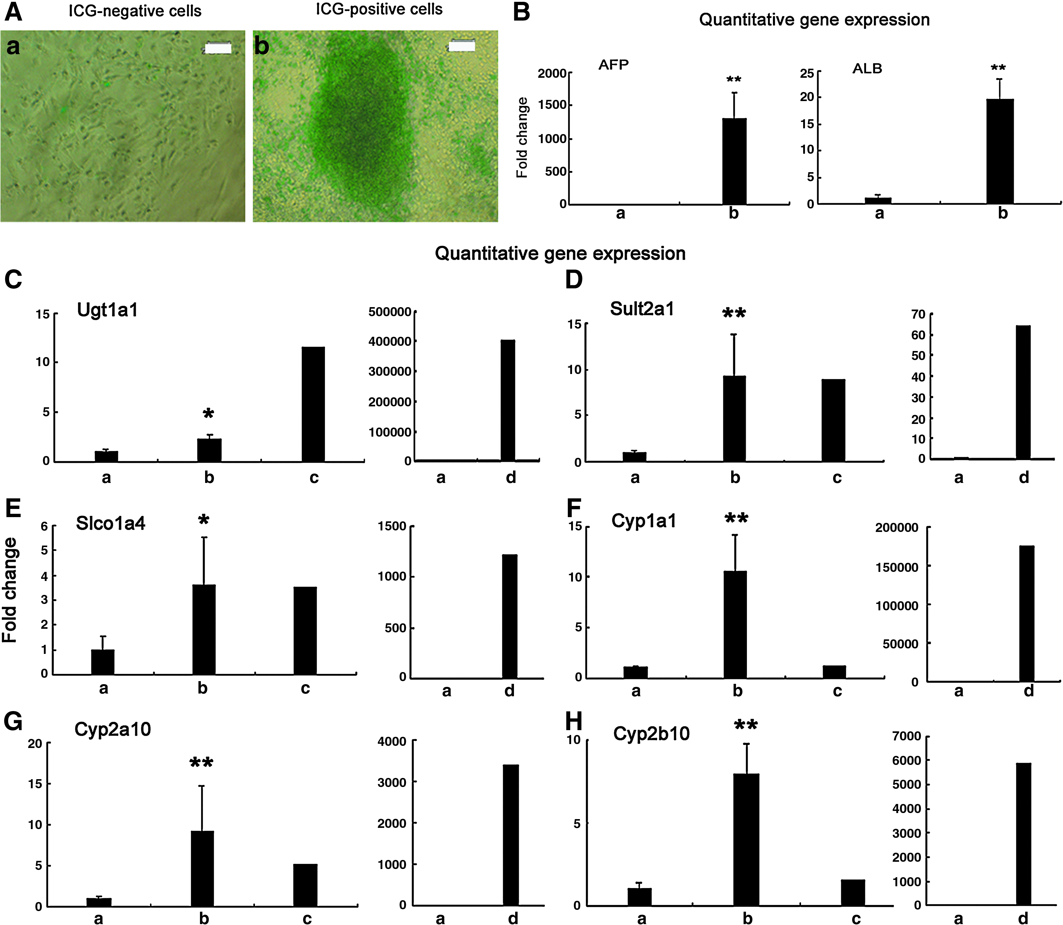
Isolation and functional characterization of ESC-derived ICG-positive cells.
Discussion
In the current study, we developed a new system for detecting hepatocytes that does not depend on any genetic modification. Rather, it utilizes the innate ability of hepatocytes to take up ICG, a nontoxic, fluorescent organic anion. Previous methods for detecting and/or isolating hepatocytes depended on transfer of reporter genes driven by liver-specific promoters9,10 or the use of antibodies that capture single or multiple surface antigens in the hepatic cell membrane.24,25 However, inserted exogenous genes that are randomly integrated into nuclear chromosomes can be silenced due to positional effects associated with local chromatin structure, especially in ESCs.26,27 Further, targeted integration is not optimal, because it is difficult and time consuming,26,27 and genetic modification may damage endogenous genes and increase the risk of tumorigenesis. 12 The use of antibodies also has drawbacks. For instance, the antibodies are expensive, and the activity may be inconsistent between batches and, thus, yield variable results. Our detection system overcomes these problems and has the advantage of being easy, inexpensive, and relies on the selective uptake by hepatocytes of ICG, a stable fluorescent chemical compound.
ICG absorbs light over a wide spectrum of wavelengths, and it has the unusual property that the absorption maximum is dependent on concentration. In solution, including plasma, it tends to aggregate, which changes its absorption properties. 23 Therefore, we could not detect ICG in primary hepatocytes when they were incubated with high concentrations. Moreover, cells other than hepatocytes can acquire it if incubated with ICG for long periods of time or at high concentrations (data not shown). Thus, the proper concentration and incubation time with ICG are required for detecting hepatocytes.
A previous study reported that irradiation of ICG with a lazer at 100 J/cm2 can generate singlet oxygen ( 1 O2), which is damaging to cells. 20 However, among the cells we analyzed by flow cytometry, most were alive as determined by trypan blue exclusion (data not shown). The power of the FISHMAN-R lazer was 5 mJ/cm2, which is considerably lower than the 100 J/cm2 lazer and much less likely to generate singlet oxygen. Thus, the ICG detection system for hepatocytes appears to do little or no damage to the cells.
The FISHMAN-R flow cytometer was successful in detecting ESC-derived ICG-positive cells. Interestingly, the fluorescence intensity of the ICG was similar to that of the HepG2 cells, but was lower than that of primary hepatocytes. These results indicate that our detection system can distinguish between mature hepatocytes and immature hepatocytes by the difference of fluorescence intensity. However, the FISHMAN-R cannot now isolate and purify ICG-positive cells. Therefore, we confirmed that ESC-derived ICG-positive cells had characteristics of hepatocytes by PAS staining of ESC-derived cell clusters which showed glycogen deposits. Additionally, reverse transcriptase PCR of ICG-positive ESC-derived cell clusters showed the expression of typical hepatocyte genes AFP, TTR, Hex, AAT, ALB, LST1, and G6P. Previous research reported that organic anion transporter, LST1, has been found to be exclusively expressed in the liver28,29 and is considered to play a role in the hepatocellular uptake of ICG. 17 Our results are also consistent with those reports.
Finally, we used light microscopy to identify and harvest individual ICG-positive cells that we then expanded in subsequent cultures. Using these isolated and expanded cells, we confirmed that expression of mRNAs for transferase, transporters, and major cytochrome P450s responsible for drug metabolism, such as Ugt1a1, Sult2a1, Slcola4, Cyp1a1, Cyp2a10, and Cyp2b10 was higher in ICG-positive cells than in ICG-negative cells. However, the expression levels of ICG-positive cells were lower than those of adult liver. Interestingly, these results were similar to those of flow cytometry, indicating that immature hepatocytes also have the ability to take up ICG. Further, a previous study reported that transplanted ESC-derived ICG-positive cells are normally incorporated into liver parenchymal structure. 17 Further, these cells are morphologically indistinguishable from host hepatocytes and do not promote development of teratomas. These results indicate that ESC-derived ICG-positive cells exhibit functional characteristics of hepatocytes.
In conclusion, this study demonstrates that the FISHMAN-R flow cytometer ICG detection system could detect ICG-positive hepatocyte-like cells. It has the potential to be developed for purifying hepatocytes produced from pluripotent stem cells such as ESCs, induced pluripotent stem cells, and mesenchymal stem cells. Since it is based on technology that does not depend on gene analysis or manipulation, the FISHMAN-R flow cytometer provides an appropriate level of stability and safety required for identifying cell sources for the treatment of hepatic diseases. Additionally, our method also has the ability to screen for differentiated hepatocytes derived from ESCs.
Footnotes
Acknowledgments
The authors thank Kayo Suzuki and Dr. Kiyokazu Kametani (Research Center for Instrumental Analysis, Shinshu University) for their excellent technical assistance.
Disclosure Statement
No competing financial interests exist.