
Abstract
Microscopy techniques based on laser-induced nonlinear optical processes allow for chemically specific imaging of unmodified samples at high spatial resolution in three dimensions and provide powerful tools for characterization of tissue-engineering constructs. This is highlighted by the simultaneous imaging of scaffold material, cells, and produced extracellular matrix collagen in samples consisting of osteoprogenitor MC3T3-E1 cells seeded on microporous bacterial cellulose (BC), a potential scaffold material for synthesis of osseous tissue. BC and collagen have been visualized by second harmonic generation (SHG) microscopy, and verification of collagen identification on cellulose scaffolds has been carried out on sectioned samples by comparison with the conventional histological staining technique. Both methods showed similar collagen distributions and a clear increase in the amount of collagen when comparing measurements from two time points during growth. For investigations of intact cellulose scaffolds seeded with cells, SHG was combined with simultaneous coherent anti-Stokes Raman scattering (CARS) microscopy for visualization of cell arrangement in three dimensions and to be correlated with the SHG data. Results showed that the osteoprogenitor cells were able to produce collagen already during the first days of growth. Further on, developed collagen fiber networks could be imaged inside compact regions of cells located in the cellulose micropores. Collagen production, the initial step of tissue mineralization, demonstrates the potential of BC as a scaffold material for bone tissue engineering. Furthermore, the noninvasive in situ monitoring of collagen inside compact tissue clearly manifests the benefits of nonlinear microscopy techniques, such as SHG and CARS, for use in tissue engineering.
Introduction
Thus, development of cell-containing materials to produce replacement tissue that remains interactive after implantation is of high interest. This requires a biomaterial scaffold acting as supporting structure to guide cell arrangement and initial tissue development 7 at the implant site for replacement and production of new extracellular matrix (ECM). A three-dimensional scaffold structure of interconnected pores facilitates cell migration and nutrient transport, 8 and a variety of materials have been examined as potential scaffolds for bone tissue engineering, including ceramics, composites, and polymers. 9
Bacterial cellulose (BC), consisting of a mesh of submicron fibers synthesized during fermentation of Gluconacetobacter xylinus, has emerged as a biomaterial with promising properties for use as tissue-engineering scaffolds. 10 The material has shown good biocompatibility, 11 has relatively high tensile strength, 12 and cellulose constructs can be manufactured into various sizes and shapes. Cellulose has been used as scaffold material for tissue-engineered blood vessel substitutes13,14 as well as cartilage replacements 15 ; and it has been shown that endothelial cells, 16 smooth muscle cells, 17 and chondrocytes 15 are able to adhere to the material. In addition, BC with interconnected micropores fabricated into the material 18 has been evaluated as a scaffold for engineered bone tissue. 19
The successful design of optimal scaffolds requires powerful methods for material characterization, and microscopy providing detailed structural information is of particular importance. Moreover, information on the early tissue formation during the first days of growth is highly relevant to gain insights in the process. Microscopy techniques based on laser-induced nonlinear optical processes, for example, multiphoton fluorescence, have been developed and increasingly used during the last decade. 20 Excitation using near-infrared radiation results in efficient sample penetration, and the strong signal dependence on laser intensity limits image contrast generation to the small focal volume of the focused excitation beam. Thus, these methods allows for confocal imaging with high spatial resolution in three dimensions. These are clearly favorable properties for the investigation of soft samples consisting of cells embedded in scaffold material combined with ECM components. Fluorescence techniques often require a fluorescent marker attached to the structure or cellular component to be imaged and depend on uptake or expression of the fluorophore in the material as well as on an often unknown fluorescence yield, all introducing measurement uncertainties. However, nonlinear processes that probe intrinsic properties, such as second harmonic generation (SHG) and coherent anti-Stokes Raman scattering (CARS), allow for noninvasive, label-free imaging. SHG is an optical frequency-doubling process generated in both BC and type I collagen via dipole interactions and is enhanced by their molecular chirality.21,22 In addition, symmetry conditions for the SHG process require a noncentrosymmetric arrangement. Overviews of SHG microscopy 23 have been presented as well as reviews addressing tissue engineering applications.24,25 The BC fiber network can be visualized by SHG microscopy, 22 and the cellulose synthesis as well as the structure of cellulose scaffolds intended as blood vessel substitutes have been investigated. 26 Collagen type I is the major component of ECM and a strongly SHG-active molecule 27 for which the SHG process has been characterized in detail. 21 Applications of SHG microscopy for collagen visualization include investigations of ECM production by mesenchymal stem cells differentiated into osteogenic cells28,29 and the arrangement of collagen fibers in osseous tissue with developed osteogenesis imperfecta.30,31 In this study, SHG has been combined with CARS microscopy, allowing for selective imaging by probing vibrations of molecular bonds. The main application of CARS microscopy is imaging of lipids probing acyl chain CH2 groups.32,33 However, probing other hydrocarbon bonds in polymers makes the method also useful for label-free confocal imaging of scaffold structure, which is valuable in tissue engineering. Both cellulose and collagen have molecular vibrations that in principle are possible to probe by CARS. However, here SHG microscopy is the preferred alternative, both for the hydrogel-like cellulose containing large amounts of bound water giving a CARS background signal, and for collagen due to its very strong SHG signal. In this study, CARS microscopy has instead been set to probe lipid structures and membranes for visualization of the cell arrangement in the scaffolds. Thus, the combined microscopy techniques allow cell integration, proliferation, and differentiation to be followed in the three-dimensional scaffold structure.
After a characterization of BC as a scaffold material for engineered bone tissue, 19 we have in this study investigated the development of ECM collagen on scaffolds seeded with osteoprogenitor cells. The applicability of SHG microscopy for monitoring collagen on cellulose-cell constructs has been verified by comparison with established histology staining. Furthermore, combined SHG and CARS microscopy have been carried out on intact cell-seeded scaffolds to monitor the production of collagen in the porous structure, the initial step of osteogenesis and a prerequisite to eventually obtain a functional graft for bone tissue.
Materials and Methods
Scaffold production
A protocol for production of microporous BC scaffolds based on particle leaching has been previously described in detail. 18 Briefly, paraffin wax microspheres having diameters in the range of 300–500 μm were added to the cellulose synthesis by G. Xylinus subsp. sucrofermentas BPR2001, trade number: 700178™ (LGC Promochem AB). A volume of 2.5 mL of 3.7×10⁶ cfu mL−1 bacteria in culture medium was seeded to each bioreactor. During fermentation, the bacteria extruded cellulose fibers into a mesh imbedding the wax microspheres. After 7 days of growth, the obtained cellulose structures were purified from bacteria, and the wax particles were leached out from the cellulose matrix as previously described. 18 The obtained microporous cellulose scaffolds were placed in deionized water and steam sterilized (1 bar, 121°C) for 20 min. After cooling to room temperature, the scaffolds were placed in an osteoprogenitor cell growth medium (details in the following text) for pretreatment over night to facilitate cell adhesion. The growth medium was removed just before cell seeding.
Cultivation of MC3T3-E1 osteoprogenitor cells
Cells of the MC3T3-El mouse calvaria-derived cell line (subclone 4. ATCC) were seeded onto the scaffolds (10 μL/scaffold at 2×10⁵ cells mL−1), and allowed to attach for 4 h in the incubator before the addition of growth medium containing alpha-minimum essential medium (αMEM) (Invitrogen), 10% fetal bovine serum (Gemini Bio-Products), and 1% antibiotic/antimycotic solution (Invitrogen). The next day, the growth medium was replaced with differentiation medium (growth medium supplemented with 0.13 mM L-ascorbic acid 2-phosphate, 2 mM β-glycerophosphate, and 10 nM dexamethasone (Sigma Aldrich)) for a set of eight samples, in the following text referred to as sample category I. A group of four samples were cultivated using only growth medium, in the following text referred to as sample category II. Before SHG/CARS microscopy, these samples were fixed in 3.7% formaldehyde (Polyscience) and stored in PBS medium (Fisher). Cells were grown in an incubator at 37°C in an atmosphere containing 5% CO2 at 95% relative humidity. Growth and differentiation media were changed every 2–3 days.
Histology
Four scaffold-cell samples cultivated in differentiation medium were harvested 1 and 7 days after cell seeding for histological analysis. The sample preparation followed by collagen type I staining was carried out at HistoCenter AB. The scaffold-cell constructs were placed in 4% buffered formaldehyde for dehydration, embedded in paraffin, and sliced along the vertical plane of the scaffolds into 5 μm thick sections. After deparaffinization, the sections to be stained for collagen type I were sequentially incubated in citric buffer pH 6.0 2×5 min, 3 vol% H2O2 for 5 min, 1% BSA (Sigma) for 20 min, streptavidin/biotin for 15 min, primary antibody (goat anti-collagen type I, Chemicon) diluted 1:20 in antibody dilutent (Dako) for 60 min, and Streptavidin HRP for 30 min. Incubated sections were then stained for collagen type I with DAB (Vector) for 5 min. Sample analysis was carried using an inverted microscope (Olympus CKX 41) at 20× magnification. Histology sections were also prepared without collagen staining to be used for comparative SHG microscopy.
Nonlinear microscopy
The setup for nonlinear microscopy has been previously described in detail. 26 Briefly, two laser beams of synchronized pico-second pulse trains with near-infrared wavelengths are coupled into an inverted microscope and focused on a mounted sample. Sample illumination with laser beams at wavelengths of 817 nm and 1064 nm simultaneously induces the SHG and CARS processes in the sample. This allows for combined imaging of SHG-active cellulose and collagen fibers simultaneously with the seeded cells (CARS), the latter by probing a vibration of hydrocarbon CH2 groups at frequency (wavenumber) 2845 cm−1.
Both fixated whole cellulose scaffolds and sectioned samples were investigated using nonlinear microscopy. The intact scaffolds were contained in glass-bottom Petri dishes (WillCo-dish® GWSt-5030, thickness 0.17 mm), whereas the sectioned scaffolds were mounted using a microscope slide and a 0.17 mm thick cover slip. The laser beams were focused on the sample (objective 40× Nikon Plan Fluor N.A. 1.3, working distance 0.21 mm), and forward propagating SHG and CARS signals transmitted through the sample were collected, separated by a dichroic beamsplitter, and registered by single-photon-counting photomultiplier tubes. Bandpass filters were used to isolate the SHG and CARS signals, generated at 409 nm and 663 nm, respectively, from background radiation. Typical average excitation powers at the sample were 40 mW per laser beam. SHG microscopy images were acquired in sectioned samples of category I for days 1 and 7, the image area was 200×200 μm2, and the acquisition time was 60 s. Image data from the intact scaffolds of sample category II were acquired as simultaneous SHG and CARS microscopy z-stacks covering horizontal planes of area 100×100 μm2 distributed over vertical ranges of 5–100 μm (vertical step size 0.5–2 μm). For the z-stack measurements, with multiple images collected at each lateral position, the image acquisition time was either 20 s or 60 s depending on the signal at the measurement location. Measurements were carried out on days 1, 4, 6, and 8 after cell seeding, one sample for each day. Merged SHG and CARS images and volume views showing the cell/scaffold system in three dimensions were generated using the software ImageJ. A measure on collagen content was evaluated from the image data as an area percentage of collagen fibers in the cell regions. The number of collagen signal pixels, isolated by manual thresholding of the SHG images, was divided by the area of the cell region identified from the corresponding CARS image. Collagen content was quantified for data measured at 1 (day 1), 5 (day 4), 2 (day 6), and 3 (day 8) different lateral positions and up to six slices, separated 5–10 μm, were evaluated in each z-stack.
Results and Discussion
Sample category I
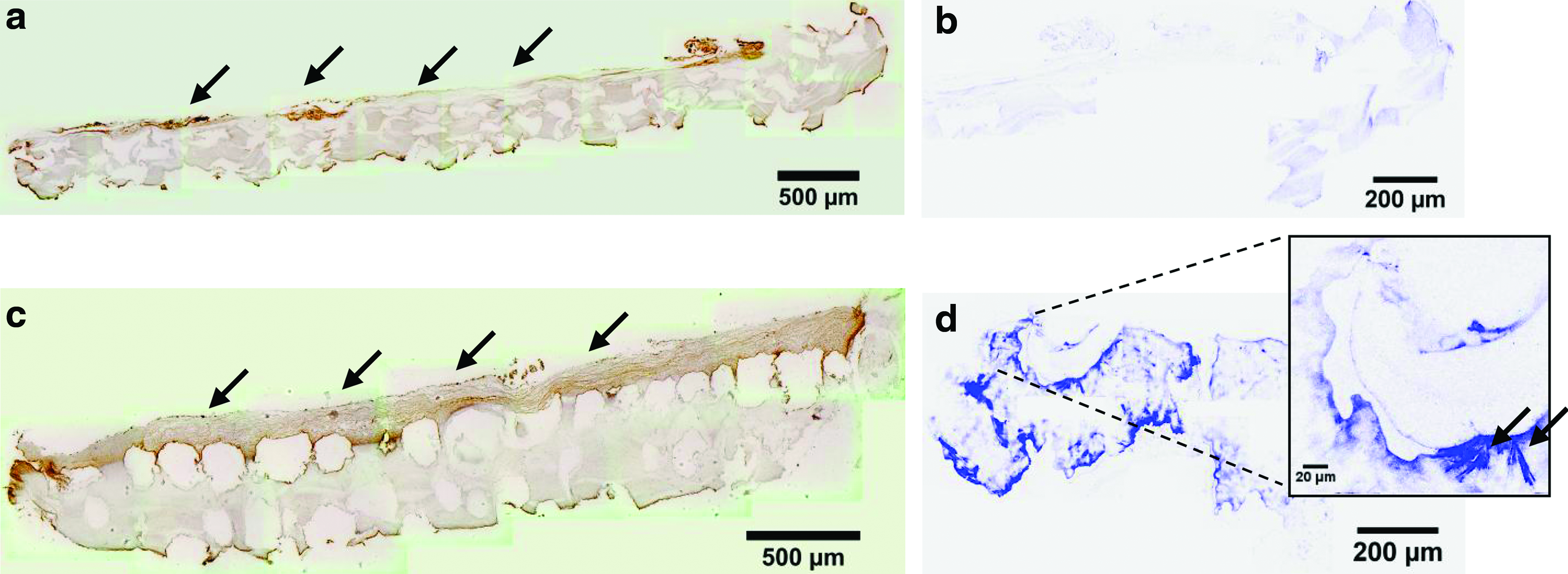
Histology sections of samples from category I, that is, cellulose scaffolds with osteoprogenitor cells grown on differentiation medium, clearly showed ECM collagen type I as identified by DAB-stained regions in brown color (Fig. 1a, c) and expected for the investigated cell type. 34
Collagen type I imaging on microporous cellulose scaffolds one
After one day of growth, regions of stained collagen can, in particular, be seen along the upper edge, where regions of cells can be identified, indicating the seeded surface (Fig. 1a, arrows). An SHG microscopy image measured on an unstained section cut from the same one-day-old scaffold mainly shows a relatively low SHG signal obtained from the cellulose fibers (Fig. 1b, blue color). The contours in the SHG image of the scaffold correspond well to the shape of the stained scaffold section (Fig. 1a). In addition, narrow regions of higher SHG signal can be found on the edges of the structure in the SHG image, in good agreement with the brown collagen staining seen along the edges in the histology image (Fig. 1a).
In contrast to the relatively low amount of DAB-stained structures after one day of growth (Fig. 1a), substantial amounts of stained collagen can be found in a seven-day-old sample (Fig. 1c). Stained fibers can be seen in the multiple cell layers built up across the entire scaffold and essentially bridging over the open pores (Fig. 1c, arrows). In addition, rather dense stained regions can be seen along the pore edges and at the bottom of the pores, indicating a downward cell migration along the pore edges.
An SHG image measured on a seven-day-old sample (Fig. 1d) contains many compact SHG-active structures generating signals at least one order of magnitude stronger than the cellulose, as identified in the SHG image of Figure 1b. Since collagen generally generates much stronger SHG signal than cellulose21,26 the higher signals compared with the one-day-old sample are in agreement with the increased amount of collagen expressed on this scaffold as identified by histology (Fig. 1c). Similar to the histology image, the collagen regions identified in the SHG image appear rather compact and homogeneous; however, fiber segments can also be identified, as shown in the insert of Figure 1d. Thus, the ability to monitor collagen production on cellulose scaffolds by means of SHG microscopy was well confirmed by comparison with established technology.
Sample category II
SHG-imaging of cellulose and collagen in scaffolds seeded with osteoprogenitor cells cultivated in growth medium, sample category II, was combined with simultaneous CARS microscopy for visualization of cell proliferation and morphology (Figs. 2 and 4). Merged SHG and CARS microscopy images measured on cellulose scaffolds one day after cell seeding show that the spindle-shaped osteoprogenitor cells (orange color) have proliferated on the cellulose (blue fibers) and started forming a confluent single-cell layer on the surface (Fig. 2b). SHG data measured at this point of time essentially show the cellulose matrix (Fig. 2a, b, blue fibers); however, SHG-active fiber segments, 10–20 μm long and around 1 μm thick, located in the immediate vicinity of some of the imaged cells, and with 5–10 times stronger signals than the cellulose, could also be identified (Fig. 2c, arrows). The position of cells (orange color) and collagen fibers (bright blue color) above the cellulose scaffold (dark blue region) can be seen in the volume views of Figure 2d and e, a more three-dimensional arrangement can be observed compared with Figure 2b.
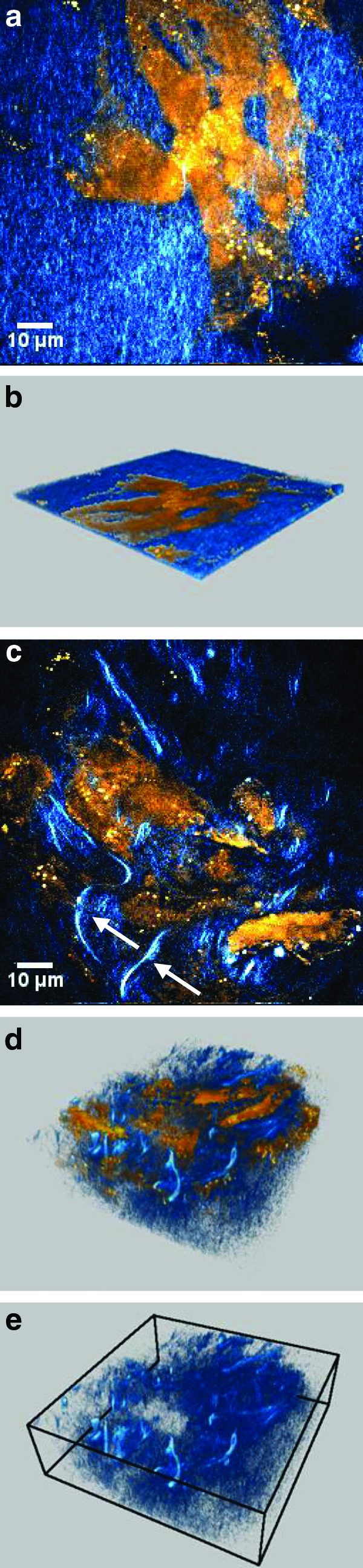
Early cell proliferation and collagen production. SHG/CARS overlay images and rendered volume views of MC3T3-E1 cells grown on cellulose scaffold using standard growth medium for one day.
This indicates an initiated collagen production at this early stage, also observed for the samples of category I (Fig. 1a, b). Additionally, the detection of individual fiber segments highlights the feasibility of SHG microscopy for detailed studies of collagen production. Collagen synthesis is characteristic for osteoprogenitor cells in their mature stage and, in particular, for cells differentiated into osteoblasts. 34 The generation of an extracellular collagen matrix has been identified as a prerequisite for osteoblast differentiation of MC3T3-E1 cells, and initial collagen production during the first two days of growth was followed by increasing expression of the osteoblast phenotype and further collagen synthesis. 35 In addition, ECM collagen production after one day of growth has previously been noted for MC3T3-E1 cells grown on collagen scaffolds. 36
Gradually, during growth, denser multi-cell regions developed and could be observed in the cellulose micropores (Fig. 3) in agreement with previous observations 19 and indicating proliferation characteristic for the osteoprogenitor phenotype. 34 Simultaneous SHG and CARS measurements in these dense regions clearly show developed networks of collagen fibers within the cellular matrix (Fig. 4a–c) at depths ranging from 10 to 80 μm below the upper cell layer. Thus, SHG/CARS microscopy allows for noninvasive imaging under native conditions below the surface of the tissue construct without need of the sectioning carried out for samples of category I (Fig. 1). Based on excitation using near-infrared radiation, nonlinear techniques have a distinct advantage compared with microscopy using visible light, which generally shows higher extinction during tissue penetration. The maximum attainable imaging depth depends on experimental parameters such as excitation intensity, focusing optics, and extinction of laszr beam and propagating signal in the sample. In the presented measurements, scanning depths down to 100 μm was adequate to cover the cell regions at the probed positions. Under appropriate experimental conditions, nonlinear microscopy techniques allow for imaging deep inside tissue, demonstrated for collagen down to a depth of 550 μm using femtosecond laser pulses for excitation, and an objective with long working distance for focusing. 21
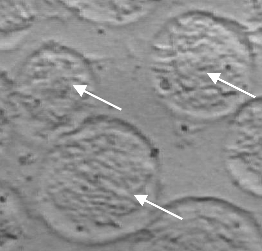
Bright field image of microporous cellulose scaffold after six days of growth with dense clusters of MC3T3-E1 cells located in the pores (arrows).
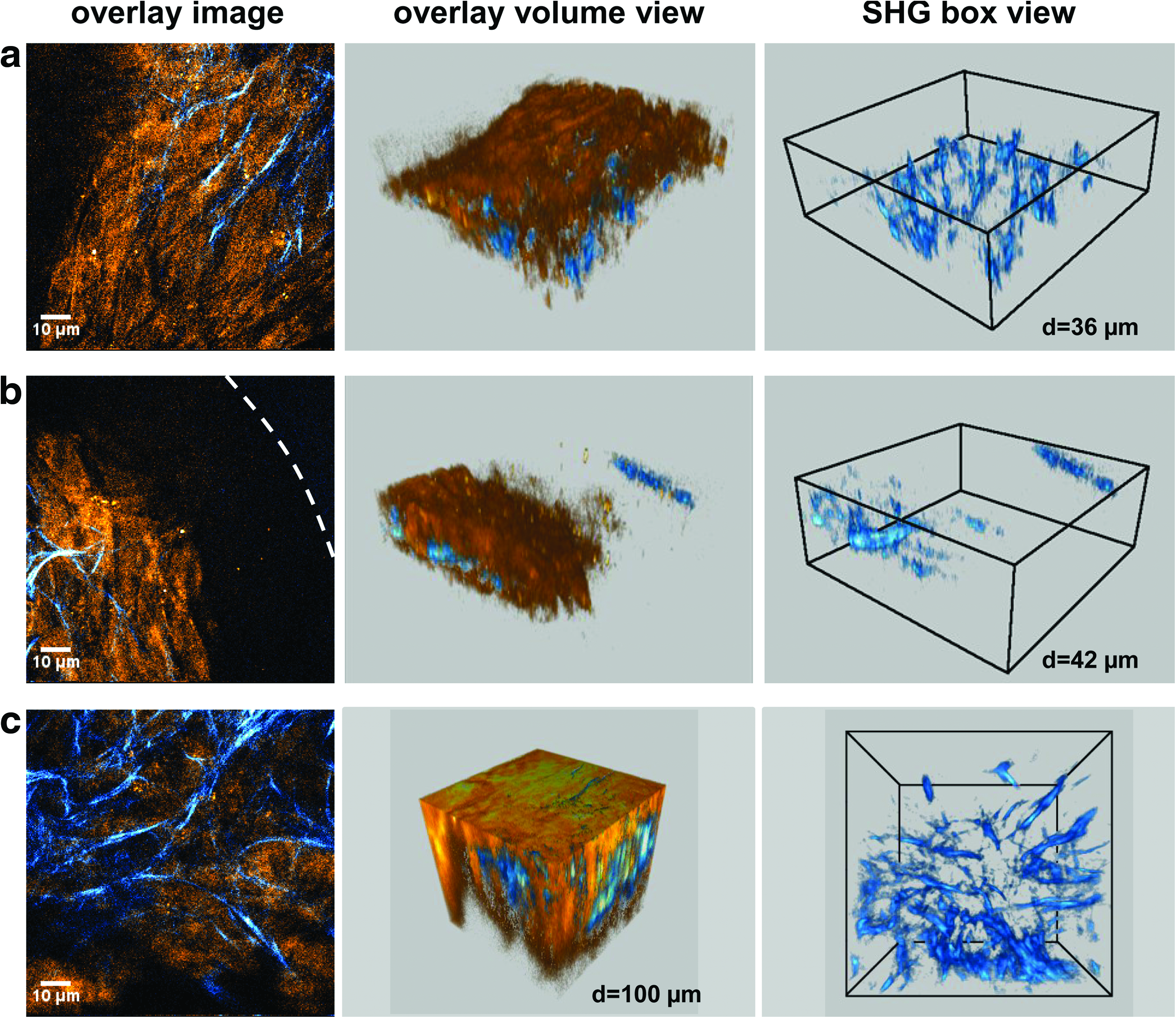
Collagen production in cellulose micropores. Synthesized collagen fibers (SHG, blue color) can be distinguished embedded in compact cells clusters (CARS, orange color) established in the scaffold pores. Measurements on six-
Evaluated collagen fiber diameters were typically 1 μm, although some fiber bundles a few microns wide could also be observed. Since high collagen expression is a hallmark of the osteoblast phenotype and differentiation of MC3T3-E1 cells grown on cell cultivation dishes have been observed after four days of growth, 35 it is possible that category II samples of the later time points also contain cells differentiated into osteoblasts.
An alignment of ECM collagen fibers along the long axis of MC3T3-E1 cells has previously been observed for cells grown on synthesized collagen scaffolds, and a similar arrangement could be seen in some of the measured SHG/CARS images, as exemplified in Figure 4a. Moreover, other studies of MC3T3-E1 cells have reported an alignment of ECM collagen fibers in the main migration direction of the cells.36,37 However, the dense structures of randomly oriented cells found in many micropores make such fiber alignment less apparent (Fig. 4c, box view). Furthermore, networks of ECM collagen fibers can be found close to pore boundaries (Fig. 4b) as well as in centers (Fig. 4c), showing collagen build-up within the entire volume of a cellulose micropore and confirming supply of nutrition and oxygen.
For the images of Figure 4a and b, measured in a 6-day-old sample, collagen fibers are located in regions around 25 μm thick, whereas the image in Figure 4c, taken in an 8-day-old sample, shows an entangled collagen network covering a depth of around 70 μm. The development in these samples indicates a first step toward matrix mineralization and confirms the functionality of BC as a scaffold for osseous tissue.
Production of ECM collagen by osteoprogenitor cells has been reported to depend on multiple parameters such as scaffold material structure, 38 cell density, 39 mechanical cues, 40 and oxygen availability.28,29 A dependence on local cell density could be observed in our data when evaluating the cell matrix collagen content from SHG images. Figure 5 shows the percentage of imaged cell area covered by collagen fibers versus the thickness of the cell region. Highest values are obtained for SHG images measured in thick regions of multiple cell layers, that is, of high local cell density.
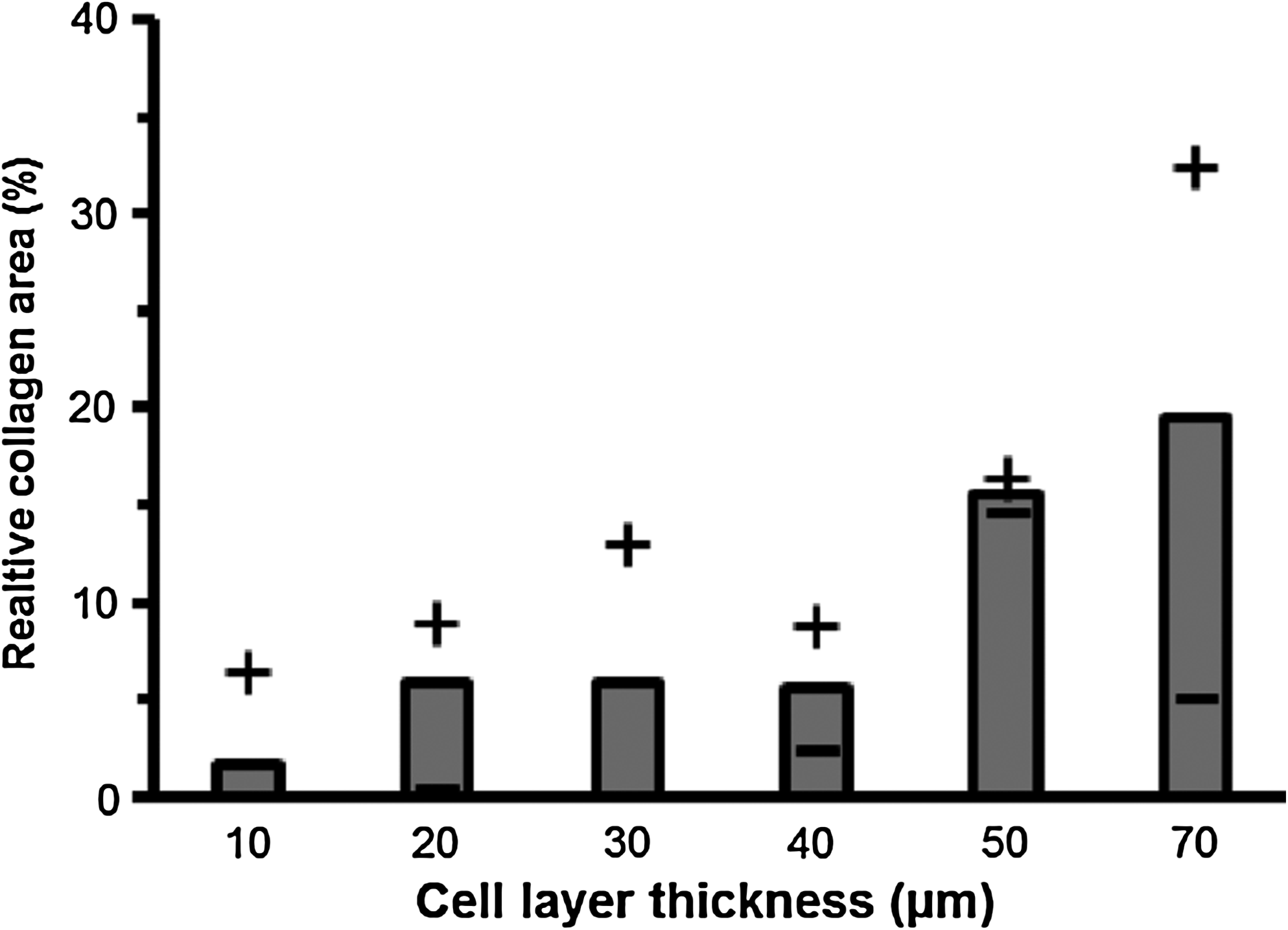
Percentage collagen fiber area relative to cell region area, evaluated from SHG/CARS images, plotted versus cell region thickness. Bars represent averages, whereas (+) and (−) indicate maximum and minimum values, respectively. Highest amounts of collagen were obtained in images from thick cell regions, indicating a coupling to local cell density.
In general, there is potential for control of the ECM collagen synthesis by modification of scaffold structure and composition as well as via cell cultivation conditions. Recent technical developments allowing for improved control of cellulose scaffold architecture, cell seeding, and growth conditions facilitate the possibility to obtain an optimal cellulose-based bone graft.
Conclusion
Nonlinear optical microscopy for label-free specific imaging provides powerful tools for use in biology, chemistry, and material science. For tissue-engineering constructs, in many cases consisting of soft materials seeded with cells, methods for noninvasive imaging of intact samples are able to provide highly relevant information not accessible by conventional techniques. This is highlighted here in an application of BC as a scaffold material for osseous tissue. Using SHG microscopy, we have shown that the material permits a rather immediate collagen synthesis by seeded osteoprogenitor MC3T3-E1 cells during their first days of growth on the scaffold, thus initiating osteogenesis. Simultaneous SHG and CARS microscopy on intact scaffolds allowed detailed imaging of three-dimensional collagen fiber arrangement inside dense cell matrices, mainly localized to the cellulose micropores, which is beneficial for bone formation. Altogether, this illustrates the potential of BC as a scaffold for bone tissue engineering and clearly manifests the applicability of nonlinear microscopy techniques as powerful tools within tissue engineering.
Footnotes
Acknowledgments
The authors gratefully acknowledge the histology preparation and staining carried out by HistoCenter AB, Göteborg, Sweden. The financial support provided by the Swedish Research Council is gratefully acknowledged.
Disclosure Statement
All authors declare that no competing financial interests exist.