
Abstract
For bone regeneration applications, scaffolds made from a composite of a biodegradable polymer and ceramic have advantages over scaffolds made from only one component (biodegradable polymer or ceramic alone). In this study, a simple and rapid method was developed to induce hydroxyapatite (HA) nanoparticle adsorption on polyglycolic acid (PGA) scaffold surfaces. PGA meshes were coated with HA nanoparticles by immersing the scaffolds in a buffer solution containing 3,4-dihydroxyphenylalanine (DOPA), a critical, functional element in mussel adhesive protein known to strongly bind to various materials. Substantial HA coating on PGA scaffolds was achieved within 24 hours of immersion, as determined according to selective staining of ceramic particles, scanning electron microscopy, X-ray photoelectron spectroscopy, and energy-dispersive spectroscopy. To evaluate the osteoconduction efficacy of the scaffolds in vivo, PGA scaffolds, DOPA-coated PGA scaffolds, PGA scaffolds immersed in HA solution, and HA- and DOPA-coated PGA (HA-DOPA-PGA) scaffolds were implanted in critical-sized defects in mouse skulls for 8 weeks. Micro-computed tomography and histological analyses showed that bone regeneration in vivo was far more extensive on HA-DOPA-PGA scaffolds than on the other scaffolds. DOPA offers an efficient and simple method of HA coating on polymer scaffolds. HA–polymer composite scaffolds fabricated using this method could be useful as bone graft.
Introduction
There has been increasing interest in the development of biodegradable polymer–ceramic composite materials as a solution to these problems.5,10–21 Biodegradable polymers such as poly(lactic-coglycolic acid) (PLGA) combined with a calcium phosphate ceramic has been shown to provide better manipulation and control of the scaffold structure to conform to bone defects. 11 Biodegradable polymers in the composite materials can reduce the brittleness of the ceramics by acting as binders. 20 Calcium phosphate ceramics in the composite materials can offer environments favorable for cell seeding, survival, migration, and growth and differentiation of osteogenic cells. 1 In addition, ceramics can create a local alkaline environment that may reduce the inflammatory reaction of acidic degradation byproducts of polyester by neutralization. 22
Many methods have been studied to develop biodegradable polymer––ceramic composite scaffolds. These methods include ceramic coating by simulated body fluid (SBF),12,23 solvent casting and particulate leaching (SC/PL), 17 phase separation, 16 and gas forming and particulate leaching (GF/PL),10,11 but fabrication of composite scaffolds using GF/PL and SBF is time consuming, requiring at least 4 to 5 days.10,23 Cytotoxic organic solvents are used in the SC/PL and phase-separation methods.16–19 Moreover, the use of polymer solutions may hinder ceramic exposure on the scaffold surface, decreasing the contact between osteogenic cells and bioactive ceramics in the composite scaffolds. 10 This study offers a simple and rapid method of inducing HA nanoparticle adsorption on polymer scaffold surfaces using 3,4-dihydroxyphenylalanine (DOPA) coating. DOPA is a critical functional element that is responsible for the adhesive properties of mussel adhesive proteins; 24 in this substance, the catechol moiety appears to bind strongly to various synthetic materials, 25 suggesting that HA nanoparticle coating on polymer scaffolds may occur by binding of DOPA to the HA nanoparticles and the polymer scaffolds. The DOPA method would have advantages over previous methods, because it is rapid (<24 hours) and solvent free. HA nanoparticle coating on polymer scaffolds was achieved by immersing the polymer scaffolds in a buffer solution containing DOPA and HA nanoparticles. HA nanoparticle coating on polymer scaffolds was confirmed using various analyses. The osteoconduction efficacy of the HA-coated polymer scaffolds was evaluated by examining bone formation on scaffolds transplanted in critical-sized defects in mice skulls.
Materials and Methods
HA nanoparticle adsorption on polymer scaffolds
Polyglycolic acid (PGA) meshes (2-mm thick; bulk density, 59 mg/m:; Albany International, Albany, NY) were coated with HA nanoparticles (100- to 200-nm diameter; Berkeley Advanced Biomaterials, Berkeley, CA) by immersing the scaffolds in a Tris buffer solution (10mM, pH 8.5; Sigma, St. Louis, MO) containing DOPA (2 mg/mL; Sigma) only (16-hour immersion), HA nanoparticles (20 mg/mL) only (16-hour immersion), or DOPA/HA mixture (8, 16, and 24 hours). The DOPA concentration and the choice of the buffer solution were determined according to the DOPA coating method described previously. 26
Characterization of scaffolds
The extent of HA coating on the scaffolds was visualized using silver nitrate staining. Scaffolds were immersed in 5% (w/v) silver nitrate (Sigma) solution and placed directly in front of a bright lamp for 30 minutes, after which the scaffold coloration was examined to determine the extent of staining. The size of HA nanoparticles was examined using transmission electron microscopy (JEM1010, JEOL, Tokyo, Japan) at 120 kV. The tensile mechanical properties of the scaffolds were measured using an Instron Universal testing machine (Instron UTM, Model 5543, Instron Ltd., Buckinghamshire, United Kingdom) with a 50-N load cell at a cross-head speed of 3 mm/min. The HA nanoparticle coating on the scaffolds, the pore size, and the porosity of the scaffolds were examined using scanning electron microscopy (SEM; JSM-6330F, JEOL, Tokyo, Japan). Samples were dried and mounted on an aluminum stub using double-sided carbon tape. The specimens were coated with platinum using a sputter coater (Cressington 108, Cressington Scientific Instruments, Cranberry, PA) and examined using SEM at an acceleration voltage of 10 kV. Energy-dispersive spectroscopy (EDS; INCA Energy, Oxford Instruments Analytical Ltd., Bucks, United Kingdom) was used to identify and semiquantitatively characterize chemical elements on scaffold surfaces. To determine the chemical composition of the scaffold surface, X-ray photoelectron spectroscopy (XPS; PHI 5000 VersaProbe, ULVAC-PHI, Chigasaki, Japan) analyses were performed using a microfocused (100 μm, 25 W) Al X-ray photon source; the C 1s, O 1s, Ca 2p, and P 2p peaks were evaluated. The residual pressure in the spectrometer was 1×10⁻⁷ Pa. The constant pass energy was 57.8 eV. All XPS data were acquired at a nominal photoelectron takeoff angle of 45°. The integrated areas beneath the XPS peaks were determined after background subtraction, and the atomic percentages were determined by normalizing the peak areas of each element to the total area of all elements.
In vivo implantation
Six-week-old, female imprinting control region mice (Koatech, Pyeongtaek, Korea) were anesthetized with xylazine (10 mg/kg) and ketamine (100 mg/kg) intraperitoneally. After shaving the scalp hair, a longitudinal incision was made in the midline of the cranium from the nasal bone to the posterior nuchal line, and the periosteum was elevated to expose the surface of the parietal bones. Using a surgical trephine bur (Ace Surgical Supply Co., Brockton, MA) and a low-speed micromotor, two transosseous defects (4-mm diameter, circular) were produced in the skull. The defect size corresponded to the critical defect size for the mouse calvarial defect model. 27 The drilling site was irrigated with saline, and bleeding points were electrocauterized. The periosteum was removed and never restored. The defect was filled with PGA, DOPA-coated PGA (DOPA-PGA), PGA immersed in HA solution (HA-PGA), or DOPA- and HA-coated PGA (HA-DOPA-PGA) (n=8 implants per group). The periosteum and skin were then closed in layers using resorbable 6-0 Vicryl sutures (Ethicon, Edinburgh, United Kingdom). The mice were housed singly after surgery. The Institutional Animal Care and Use Committee of Seoul National University approved the study (SNU-100203-5). The implants were retrieved for analysis 8 weeks after the surgery.
Micro-computed tomography analyses
The animals were killed using carbon dioxide asphyxiation 8 weeks after implantation, and the craniums, including the implanted scaffolds, were retrieved. Samples were fixed in 10% (v/v) neutral buffered formalin solution. To analyze bone formation, specimens were evaluated using micro-computed tomography (CT) (n=8 per group; SkyScan-1172; Skyscan, Kontich, Belgium).
Histological analyses
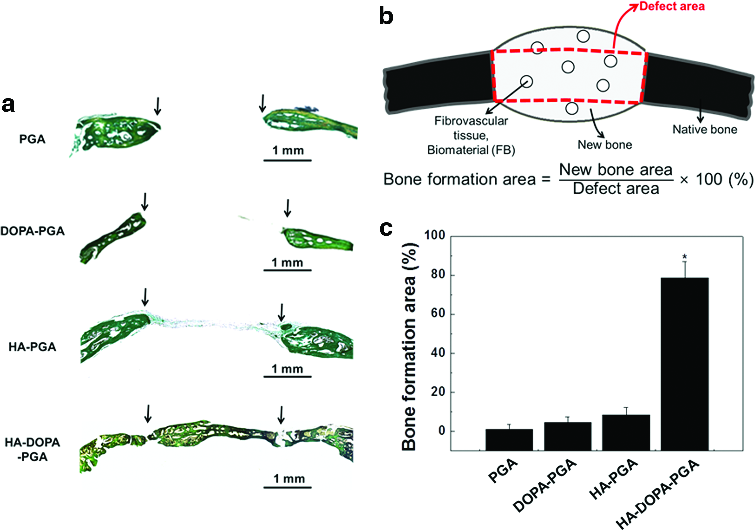
After collection of X-ray and CT scans, all samples were decalcified in ethylenediaminetetraacetic acid (pH 6.0) for 7 days and embedded in paraffin. The tissue blocks were sectioned at 5-μm thickness and stained with Goldner's trichrome. The Goldner's trichrome–stained sections were examined with a microscope to determine histomorphometry. The percentage of bone-occupying space within the construct was measured using an image analysis system (KS400, Zeiss, Munich, Germany) coupled to a light microscope. The bone formation area was expressed as a percentage relative to the defect area (bone area/defect area ×100%).
Statistical analyses
Quantitative data were expressed as means±standard deviations. Statistical analyses were performed using analysis of variance (ANOVA). A p-value less than 0.05 was considered statistically significant.
Results
Characterization of HA-DOPA-PGA scaffolds
To determine whether DOPA coating induced HA nanoparticle adhesion to the scaffold surface, the calcium ions on the scaffold surfaces were stained with silver nitrate (Fig. 1a). HA exhibited abundant staining in the HA-DOPA-PGA scaffold. By contrast, PGA, HA-PGA, and DOPA-PGA scaffolds showed no staining. Transmission electron microscopy showed that the size of the particles was approximately 20 to 50 nm wide and 100 to 200 nm long (Fig. 1b).

Instron UTM analyses of PGA, DOPA-PGA, HA-PGA, and HA-DOPA-PGA scaffolds showed that DOPA, HA, or DOPA/HA coating did not affect the mechanical properties of the scaffolds (Table 1). No significant differences in tensile strength, Young's modulus, or strain at break were observed between any two groups.
All Scaffolds were Immersed in Either DOPA, HA, or HA/DOPA Solution for 16 Hours. No Significant Difference was Observed Between any Two Groups.
PGA, polyglycolic acid; DOPA, 3,4-dihydroxyphenylalanine; HA, hydroxyapatite.
SEM of PGA, DOPA-PGA, HA-PGA, and HA-DOPA-PGA scaffolds at a lower magnification (Fig. 2) showed that DOPA, HA, and DOPA/HA coating did not affect the pore size and the porosity of the scaffolds. SEM of PGA, DOPA-PGA, and HA-PGA scaffolds at a higher magnification (Fig. 3) showed no mineral crystal precipitations. On the other hand, PGA immersed in a buffer solution containing DOPA and HA nanoparticles exhibited mineral crystal precipitations on the smooth surface of PGA scaffolds within 8 hours. The mineral coating was observed to grow progressively throughout the immersion period.
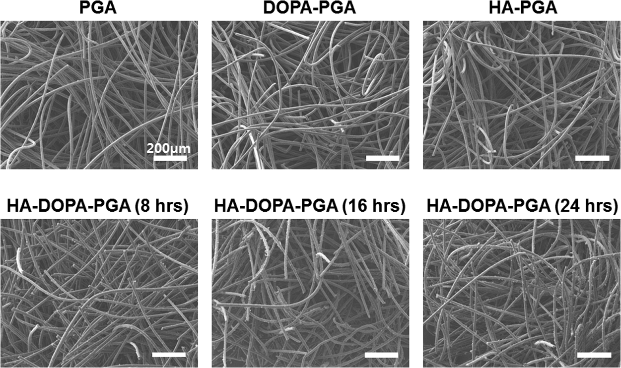
Scanning electron micrographs of PGA, DOPA-PGA, HA-PGA, and HA-DOPA-PGA scaffolds (8, 16, and 24 hour immersion). HA, DOPA, and HA/DOPA coating did not affect the pore size and the porosity of the scaffolds. The scale bars indicate 200 μm.

Scanning electron micrographs of PGA, DOPA-PGA, HA-PGA, and HA-DOPA-PGA scaffolds (8, 16, and 24 hour immersion). HA nanoparticles adsorbed on polymer fibers are observed on HA-DOPA-PGA scaffolds. The scale bars indicate 5 μm.
EDS analysis of HA-DOPA-PGA scaffolds showed an increasing tendency for HA nanoparticles to adhere to the scaffold surface during the immersion time period. Visualization of phosphate showed increasing phosphate adhesion on HA-DOPA-PGA scaffolds during the immersion time period (Fig. 4a). EDS spectra revealed that the main elements on the scaffold surfaces were carbon, oxygen, calcium, copper, zinc, and platinum (Fig. 4b). It was hypothesized that atoms other than calcium were components of the PGA scaffolds. Quantitative analysis showed that calcium, a component of HA, increased progressively on the HA-DOPA-PGA scaffolds (Fig. 4c).

EDS analyses of various elements on the surfaces of PGA, DOPA-PGA, HA-PGA, and HA-DOPA-PGA (8, 16, and 24 hours) scaffolds.
The surface composition of the HA-DOPA-PGA composite scaffolds was analyzed using XPS (Fig. 5a, Table 2). The amount of calcium and phosphate was significantly higher on the surface of HA-DOPA-PGA scaffolds than on PGA, HA-PGA, or DOPA-PGA scaffolds (Fig. 5b). Atomic ratios of calcium and phosphate on the HA-DOPA-PGA scaffolds increased during the immersion time period (Table 1). No calcium or phosphate peaks were observed on PGA, HA-PGA, or DOPA-PGA scaffolds.
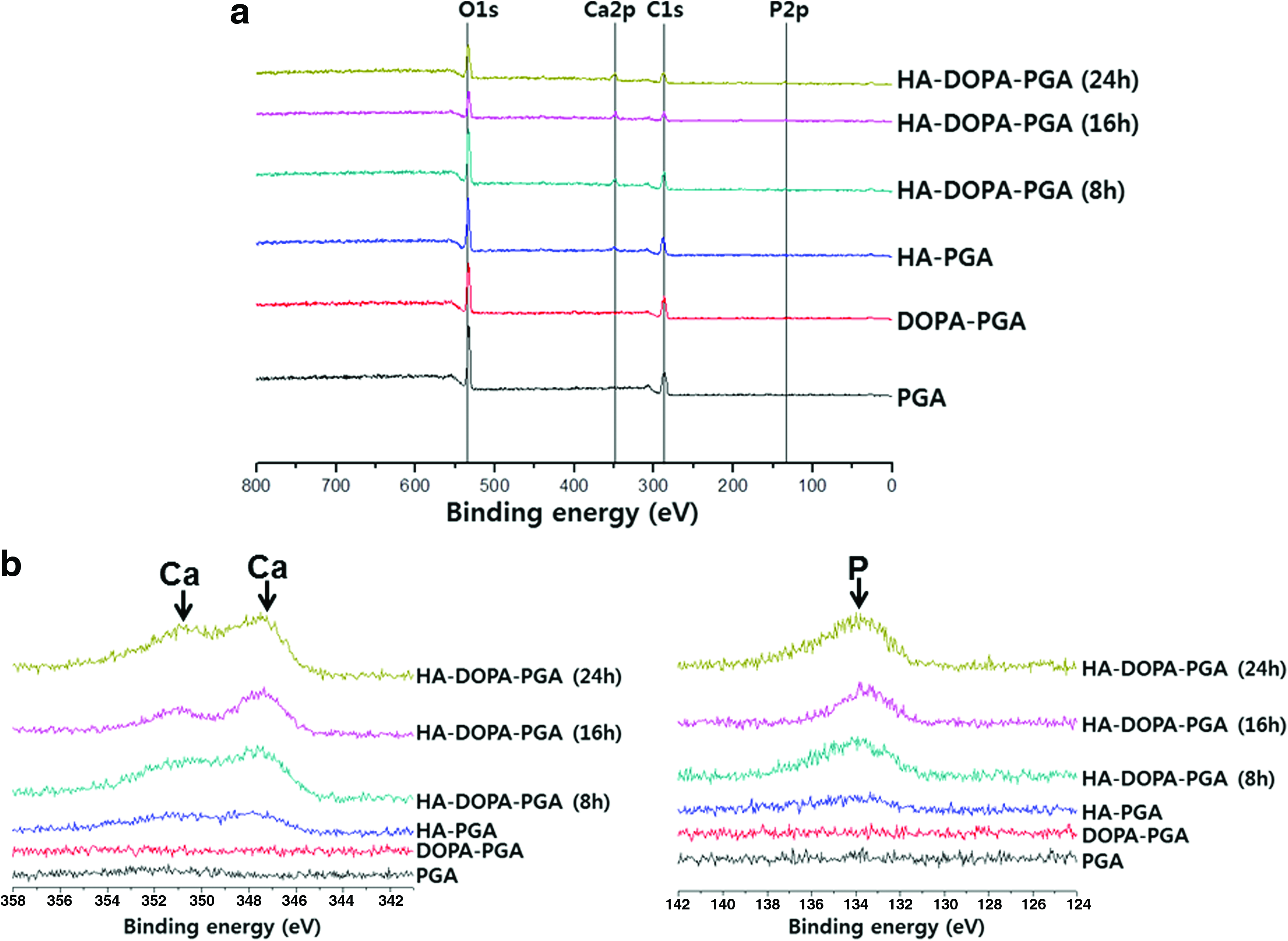
XPS of PGA, DOPA-PGA, HA-PGA, and HA-DOPA-PGA (8, 16, 24 hour immersion) scaffolds.
Osteoconductivity of HA-DOPA-PGA scaffolds
Eight weeks after the surgery, HA-DOPA-PGA composite scaffolds implanted into critical-sized defects in mouse skulls exhibited greater bone formation in vivo than PGA, HA-PGA, and DOPA-PGA scaffolds. Micro-CT evaluations (Fig. 6) allowed the mineralized tissue to be distinguished from the remaining soft tissue present inside the defects. The images showed significantly greater new bone formation in the HA-DOPA-PGA scaffolds than in the other scaffolds. Goldner's trichrome staining of the histological sections (Fig. 7a) and histomorphometric analysis (Fig. 7b) of PGA, HA-PGA, and DOPA-PGA scaffolds showed little bone formation in vivo. By contrast, significant mineralization of the bone was observed on the defects treated with HA-DOPA-PGA scaffolds.
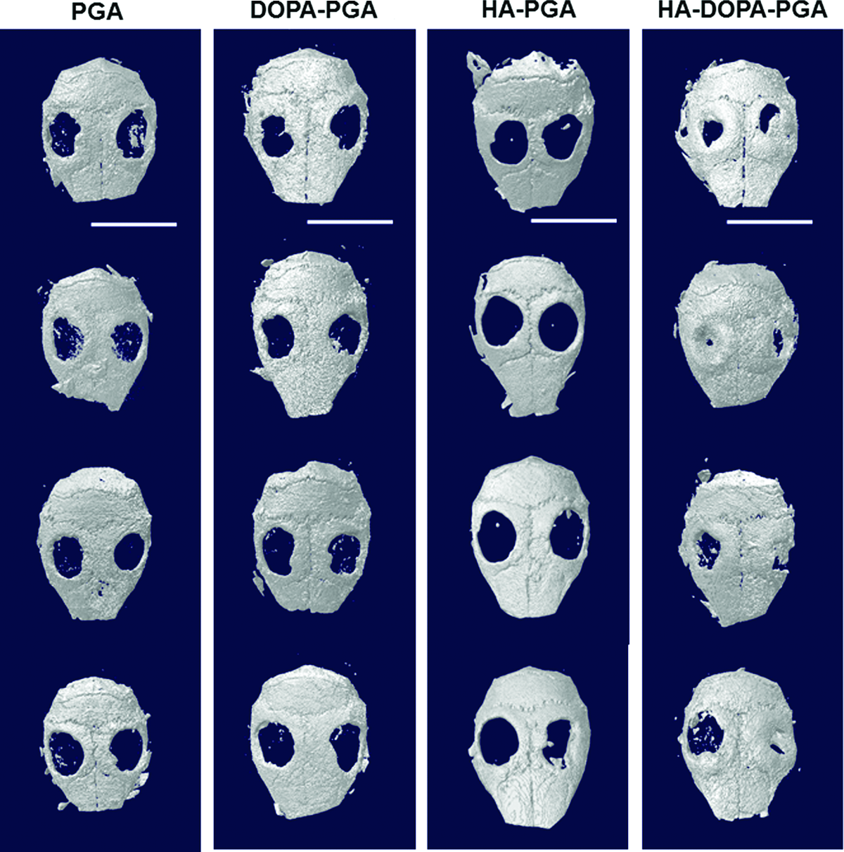
Osteoconductive properties of implants evaluated by micro-CT analyses of PGA, DOPA-PGA, HA-PGA, and HA-DOPA-PGA scaffolds implanted into calvarial defects on mice for 8 weeks. Scale bars indicate 10mm. Color images available online at
Osteoconductive properties of implants.
Discussion
A novel method to fabricate biodegradable polymer–bioceramic composite scaffolds for efficient bone regeneration was developed. The method of DOPA-induced bioceramic composite coating on a biodegradable polymer could be advantageous over conventional methods for fabricating biodegradable polymer–bioceramic scaffolds (e.g., immersion in SBF, SC/PL, and GF/PL). This method avoids the use of organic solvents, which can remain residually in the scaffolds and may cause harm to surrounding tissues and newly formed tissues. 28 HA-DOPA-PGA scaffolds may be easily and rapidly fabricated. Substantial HA coating on PGA scaffolds was achieved within 24 hours of incubation, which is less than half the time required with the GF/PL and SBF methods.14,29 Moreover, HA-DOPA-PGA can be fabricated through a simple immersion of biodegradable polymer in a solution of ceramic nanoparticles and DOPA.
The nanosized HA particles used to fabricate the composite scaffolds in this study have several advantages over microsized HA particles. A long period is needed for degradation of crystalline HA in vivo, and residual HA may hinder or retard complete bone healing. 30 The use of nanosized particles rather than microsized HA particles can achieve greater distribution of HA on scaffold surfaces with the same total amount of HA. Furthermore, nanosized HA particles are expected to yield better osteoactivity in composite scaffolds. 31
The use of DOPA leads to efficient coating of HA on the surface of the composite scaffold. Instron UTM analysis and SEM demonstrated that scaffolds can be coated with DOPA, HA, or DOPA/HA without affecting the mechanical properties, pore size, or porosity of the scaffolds. Silver nitrate staining, SEM morphology, EDS, and XPS analyses showed that HA was adsorbed abundantly on scaffold surfaces when DOPA was used for HA coating. Negligible adsorption of HA on scaffold surfaces was observed when the scaffolds were immersed in HA solution without DOPA. This indirectly demonstrates that DOPA was coated on scaffold surfaces and was necessary for HA adsorption on scaffold surfaces. The SC/PL, SBF, and phase-separation methods use polymer solutions that may cause imbedding of bioceramics in the polymer layer and poor exposure of bioceramics on polymer surfaces. Therefore, by directly inducing HA adsorption on the scaffolds surfaces, HA-DOPA-PGA scaffolds can enhance osteoconductivity by increasing the probability of osteogenic cells making contact with HA on scaffold surfaces.4,5,32
Finally, HA-DOPA-PGA scaffolds resulted in excellent bone regeneration efficacy in vivo. The use of HA-DOPA-PGA scaffolds resulted in greater bone formation than the use of PGA, HA-PGA, and DOPA-PGA scaffolds (Fig. 7). The results showed that HA coating or DOPA coating alone on a PGA scaffold surface does not have a significant effect on bone regeneration (Figs. 6 and 7). Therefore, osteoconductivity was enhanced by HA adsorption on the scaffold surfaces, stimulating the migration of osteogenic cells from surrounding tissues and osteogenic differentiation of the recruited cells. The bone formation area was found to be less than 5% in a previous study using the GF/PL method to fabricate a composite scaffold of PLGA and HA, 33 whereas composite scaffolds fabricated using DOPA showed a bone formation area of nearly 80% (Fig. 7c).
Conclusion
HA nanoparticle coating on polymer scaffolds was achieved within 24 hours using DOPA. The HA coating on polymer scaffolds created with this method dramatically enhanced osteoconductivity. The HA nanoparticle coating process is simple, rapid, and solvent free. The polymer–ceramic composite scaffold fabricated with this method may be useful as a bone graft.
Footnotes
Acknowledgments
This study was supported by grants from the National Research Foundation of Korea (2009-0080769) and the Korea Health 21 R&D Project, Ministry of Health and Welfare, Republic of Korea (A101539).
Disclosure Statement
No competing financial interests exist.