
Abstract
In this article, we describe the design and manipulation of charged nanomatrices and their application as efficient platforms for modulating cell behaviors. Using electrospraying technology and well designed biomaterials, poly(ɛ-caprolactone; PCL) and polyethylenimine, the negatively charged PCL nanomatrix (nPCL nanomatrix) and the positively charged PCL nanomatrix (pPCL nanomatrix) were fabricated. It was demonstrated that cell adhesion, affinity, and shape were sensitively modulated in negatively and positively charged nanomatrices. Our results showed that the pPCL nanomatrix promoted adhesion of NIH 3T3 fibroblast cells as compared to the nPCL nanomatrix. When fluid shear stress was applied, cell affinity on the pPCL nanomatrix increased even more. NIH 3T3 fibroblast cells adopted a relatively spherical shape on the pPCL nanomatrix while adopting an aligned, narrow shape on the nPCL nanomatrix. It was also found that charged nanomatrices influenced the cross-sectional cell shape. The cross-sectional cell shape on the pPCL nanomatrix was extremely flattened, whereas the cross-sectional cell shape was relatively round on the nPCL nanomatrix and some of the adhered cells floated. We also showed that the surfaces of the nPCL and pPCL nanomatrices adsorbed the different serum proteins. These results collectively demonstrated a combination of environmental factors including nanoscale structure, electrostatic forces, and absorption of biomolecules on charged substrates affected cell response in terms of cellular adhesion and shape.
Introduction
The in vivo extracellular microenvironment consists of charged materials such as ECM and biomolecules. The cell membrane has a net negative charge, therefore, electrostatic interactions between the cell and extracellular microenvironment (or between cells) may play critical roles in modulating cell functions.8–11 A number of studies have focused on how cell adhesion and shape are affected by chemically charged substrates with corresponding functional chemical groups such as -COOH (negatively charged at pH 7.4) and -NH2 (positively charged at pH 7.4).9,12–15 Few studies have investigated the effects of electric charge 12 or charged nanoparticles13,14 on cell adhesion and shape. It is widely accepted that negative and positive charges influence cell behavior, however, it remains unclear whether cell adhesion and shape are affected by charge alone8,11–18 because it is difficult to separate charge effects on cell adhesion and shape. Functional groups that generate charges also influence the surface wettability of substrates. For example, plasma treatment is the useful method for charge surface of substrates, which generate functional group such as hydroxyl and amino groups on the surface of substrates. However, this method also influences hydrophilic-hydrophobic property of the surface. 19
Here, we propose that the combination of charge cues with nanoscale structure is essential for the design and manipulation of engineered ECM. We hypothesized that negative or positive charges on a nanoscale substrate would provide unique cues to control cell adhesion, affinity, and shape, providing a simple but powerful strategy for cell and tissue engineering applications and for understanding cell biology. More specifically, we designed and fabricated charged nanopatterned matrices as an appropriate model system using well designed biomaterials and electrospraying technology and investigated whether cell responses were precisely modulated by the charged nanomatrices.
Materials and Methods
Design and fabrication of charged nanomatrices
We fabricated a negatively charged nanomatrix using poly(ɛ-caprolactone) (PCL, Mw: 80,000; Sigma-Aldrich) and electrospraying technology. After dissolving PCL in chloroform (CL; Sigma-Aldrich)/dimethylformamide (DMF; Sigma-Aldrich) (3:1 v/v) at a concentration of 20 wt.%, the PCL solution was passed through a custom-made metal nozzle using a syringe pump at a slow, well-controlled flow rate. A controllable high voltage (10–15 kV) source was connected to the metal nozzle with a size of 50 mm while the PCL solution was pumped at various flow rates (0.4–1.2 mL/h) to control the fine spray of the PCL solution. By maintaining a uniform jet size, we were able to create a nanomatrix with a controlled nanofiber diameter. The positively charged nanomatrix was fabricated by electrospraying technology using PCL and polyethylenimine (PEI). After dissolving the PCL (20 wt.%) and PEI (1 wt.%) in CL/DMF (3:1 v/v), the solution was electrosprayed under controlled conditions
Imaging the structures of the charged nanomatrices using field-emission scanning electron microscope
The fabricated nanomatrices were coated with gold and imaged by a field-emission scanning electron microscope (FESEM; JEOL, JSM-5410LV) at an accelerating voltage of 2 kV. Five images of each nanomatrix were used to quantify the diameters of the nanofibers using Adobe Photoshop (Adobe). Briefly, 10–20 nanofibers of the nanomatrix shown in each FESEM image were considered to determine the mean diameters of the nanofibers.
Wide-angle X-ray scattering patterns of the charged nanomatrices
Wide-angle X-ray scattering (WAXS) patterns were acquired over a diffraction angle of 2θ=5°–45° at room temperature using an angle X-ray diffractometer with Cu Kα radiation, operated at 40 kV and 20 mA.
Contact angle measurements of the charged nanomatrices
Water contact angles on the fabricated nanomatrices were measured using a video contact angle analyzer (VCA; Phoenix 600). Ten microliters of water was dropped onto the surface of each nanomatrix. The water contact angle was measured as the tangent to the interface of the droplet on the nanomatrices. Measurements were repeated at least 10 times for each sample and averaged. All experiments were performed at room temperature.
Zeta potential measurements of charged nanomatrices
The zeta potentials of the charged nanomatrices were measured by an electrophoretic light scattering spectrophotometer (ELS-8000; OTSUKA Electronics Co. Ltd.) using a cylindrical glass cell. An electrolyte solution (10 mM NaCl) was placed onto the charged nanomatrix. Each cell was then mounted between a pair of gold-coated electrodes to assess the electrical potential difference between the plug ends. The zeta potential was determined using the Smoluchowski equation. 20
Culturing of cells seeded on charged nanomatrices
NIH 3T3 fibroblast cells (4×10⁴ cells/matrix) were seeded onto charged nanomatrices and cultured for up to 14 h in Dulbecco's modified Eagle's medium (Sigma-Aldrich) with 10% fetal bovine serum (FBS; Sigma-Aldrich) and 1% antibiotics (Sigma-Aldrich) at 37°C in a humidified atmosphere containing 5% CO2.
Immunofluorescence staining and quantification of cell adhesion on charged nanomatrices
NIH 3T3 fibroblast cells (4×10⁴ cells/matrix) were seeded onto charged nanomatrices and allowed to spread for 14 h. Adhered cells on charged nanomatrices were fixed with a 4% paraformaldehyde solution (Sigma-Aldrich) for 20 min, permeabilized with 0.2% Triton X-100 (Sigma-Aldrich) for 15 min, and stained with TRIT conjugated phalloidin (Millipore) and 4, 6-diamidino-2-phrnykinodole (DAPI; Millipore) for 1 h. Images of the stained cells were taken using a fluorescence microscope (Zeiss). Before DAPI staining, unattached cells on the charged nanomatrices were washed twice using phosphate-buffered saline (PBS; Sigma-Aldrich). After DAPI staining, 10 fluorescent images with 50–100 cells per image were used to quantify cell adhesion on the charged nanomatrices. The quantitative analysis of the cell adhesion on charged nanomatrices was also performed using WST-1 assay (EZ-Cytox Cell Viability Assay Kit; Daeillab Service Co., Ltd.).
Imaging cell shapes on charged nanomatrices using FESEM
NIH 3T3 fibroblast cells (4×10⁴ cells/matrix) were seeded onto the charged nanomatrices and allowed to spread for 14 h. Adhered cells on charged nanomatrices were fixed with modified Karnovsky's fixative consisting of 2% paraformaldehyde and 2% glutaraldehyde (Sigma-Aldrich) in a 0.05 M sodium cacodylate buffer (Sigma-Aldrich) for 4 h. The samples were washed with 0.05 M sodium cacodylate buffer three times for 10 min and fixed with 1% osmium tetroxide (Sigma-Aldrich). The samples were washed with distilled water and dehydrated with graded concentrations (50, 70, 80, 90, and 100% v/v) of ethanol. The samples were then treated with hexamethyldisilazane (Sigma-Aldrich) for 15 min. Finally, the samples were coated with gold prior to cell shape observation by the FESEM.
FIB preparation and FESEM observation of cross-sectional cell shapes on charged nanomatrices
The samples used for FESEM imaging were also used for focused ion beam (FIB) milling and imaging of cross-sectional cell shape on the charged nanomatrices. The method used for the FIB preparation was previously reported in detail. 21 Briefly, after observation of samples by the FESEM, cells of interest on the charged nanomatrix were selected as target samples. Each target sample was then deposited with platinum using metal-organic chemical vapor deposition, and ablated with gallium ions accelerated at 30 kV with an ion current of 2 nA. After coarse milling, a lower beam current of 240 pA was applied at an acceleration voltage of 30 kV to polish the cross section of the target sample. The target sample was imaged at an acceleration voltage of 2 kV by the FESEM.
Quantification of cell shape on charged nanomatrices
For the quantitative analysis of cell shape, images of the fibroblast cells on charged nanomatrices obtained by the FESEM or the fluorescence microscopy were analyzed using a custom-written MATLAB script. Cell shape parameters such as width, length, perimeters, and area were determined. These values were then used to calculate the cell shape index (CSI) according to the following equation:
Here, A is the cell area, and P is the cell perimeter. The CSI value ranges from 0 to 1 and is used to indicate whether the cell shape is linear or circular. A larger CSI corresponds to a more circular cell shape.
Cell affinity assay
Fluid shear stress was used to evaluate cell affinity to the charged nanomatrices. NIH 3T3 fibroblast cells (4×10⁴ cells/matrix) were seeded onto charged nanomatrices and allowed to spread for 14 h. Fluid shear stress was applied through the rocking system (Rocker; Vision), with respect to different periods (15, 30, and 60 min).
22
Twenty-four well culture dishes with 500 μm of culture medium were fixed to a platform in the vertical plane to prevent slipping, and a fluid shear stress was generated by rocking the platform up and down±20° at 2 cycles per second. The wall shear stress calculated using the mathematical model described by Zhou et al.
22
was ∼1 dyn/cm3. We removed the cells that had detached from the charged nanomatrices, and then quantified the adherent cells using WST-1 assay. The affinity of cells was calculated by the following equations:
Here, [A]sample and [A]100% denote the absorbance of the adherent cell viability on the charged nanomatrices (after applying fluid shear stress) and the total cell viability on the charged nanomatrices (before applying fluid shear stress), respectively. All experiments were repeated three times.
Biomolecule adsorption on charged nanomatrices
The charged nanomatrices were incubated in a 10% FBS solution at a room temperature for 2 h before being carefully washed three times with a PBS solution. The adsorbed biomolecules (e.g., proteins) were analyzed by X-ray photoelectron spectroscopy (XPS, ESCA2000; Thermo VG Scientific). XPS analysis was conducted using an Mg Kα line as the X-ray source under ultrahigh vacuum (10−9 torr). Spectra were taken at a pass energy of 0.1 eV.
Statistical analysis
All quantitative results are presented as mean±standard deviation and unpaired student's t-tests were used for statistical analysis. p-Values<0.05 were considered statistically significant.
Results
Design and fabrication of charged nanomatrices
Positively- or negatively charged substrates are most commonly fabricated using biomaterials with chemical functional groups. However, it is difficult to maintain wettability of the charged substrates using this method. Therefore, we avoided using biomaterials with different functional groups such as -COOH or -NH2 that influence wettability. The surfaces of PCL capsules or fibers are negatively charged due to the orientations of carbonyl groups, 20 whereas PEI has many amine functional groups that provide positive charges due to the ionization of amine groups in the physiological environment or in vitro cell culture media. 11 However, since PEI substrates have hydrophilic surfaces compared to PCL substrates, the pure PEI substrates cannot be used as a model system to investigate the effects of positive charge on cell adhesion and shape. In a previous study, we determined that PEI can be dissolved in an organic solvent (e.g., DMF) and blended with PCL, and that substrates fabricated using PCL and small amounts of PEI did not affect the surface wettability of substrates. 23 Using this phenomenon,, we successfully fabricated positively charged nanostructured matrices without altering wettability, in contrast to nanostructured matrices fabricated with only PCL that have negatively charged surfaces (Fig. 1A, B, respectively).

Characteristics of the charged nanomatrices used in this study. Schematic illustrations of the
There are several recent technologies available for bio-nanopatterning surfaces such as dip-pen nanolithography, micro- and nanocontact printing, electron beam lithography, and electrospraying. 2 Electrospraying was used in this study because it allows more freedom to use materials such as PCL and PEI for bio-nanopatterning than other technologies. However, precise manipulation is required to fabricate controlled nanoscale structures by electrospraying. PCL solution (20 wt.%) was electrosprayed to fabricate the negatively charged PCL nanomatrix (nPCL nanomatrix). To fabricate the positively PCL charged nanomatrix (pPCL nanomatrix), a small amount of PEI (1 wt.%) was added into a PCL solution (20 wt.%) and electrosprayed. To fabricate precisely controlled nPCL and pPCL nanomatrices, we carefully controlled working conditions such as flow rate and applied voltage. We controlled the flow rate of the polymer solution and the voltage, allowing us to maintain a suitable uniform jet size and tailor the nanofiber diameter. Therefore, we were able to fabricate nPCL and pPCL nanomatrices with similar structures (Fig. 1C, D).
Characteristics of the charged nanomatrices
We investigated the characteristics of the charged nanomatrices (Fig. 1). The morphologies of the nPCL and pPCL nanomatrices are shown in Figure 1C and D, respectively. FESEM images demonstrate that the nanomatrices contained nanoscale fibrous networks similar to ECM structure. The average nanofiber diameters of the nPCL and pPCL nanomatrices were 480.8±136.0 nm and 459.4±182.6 nm, respectively, and the nPCL and pPCL nanomatrices had similar nanofiber diameter distributions (Fig. 1C, D). The hydrophilicities of the nPCL and pPCL nanomatrices were evaluated by water contact angle testing, and detected no significant differences although the pPCL nanomatrix had amine functional groups, as confirmed by Fourier transform infrared analyses (Figs. 1C and Supplementary Fig. S1; Supplementary Data are available online at
We then investigated the zeta potentials of the fabricated nPCL and pPCL nanomatrices to confirm their surface charges (Fig. 1F). As expected, the nPCL nanomatrix had a negatively charged surface due to the orientations of surface carbonyl groups (Fig. 1A). In contrast, the surface of the pPCL nanomatrix fabricated with a small amount of PEI had a positive charge due to the ionization of cationic amine groups (Fig. 1B). Together, these results demonstrate that negatively and positively charged nanomatrices were successfully fabricated and could be used to investigate the modulation of cell behaviors in nanoscale structure.
Analysis of cell adhesion on the charged nanomatrices
To investigate whether cell adhesion was influenced by the charged nanomatrices, we cultured NIH 3T3 fibroblast cells on the nPCL and pPCL nanomatrices for 14 h. We observed that cell adhesion was sensitive to the charged nanomatrices, suggesting that electrostatic interactions between cells and charged nanomatrix play important roles in cell adhesion. Interestingly, cell adhesion was greater by around 20% on the pPCL nanomatrix than on the nPCL nanomatrix (p<0.05; Fig. 2). This indicates that cell membranes may adhere more closely to positively charged nanomatrix than to negatively charged nanomatrix, and that attractive electrostatic forces between cells and positively charged nanomatrix promote cell adhesion. In addition, biomolecules in the cell culture medium that adsorb on the pPCL nanomatrix may lead to increased cell adhesion compared with the nPCL nanomatrix (Fig. 5). This is consistent with results obtained by Keselowsky et al., 24 who reported that the adsorption of fibronectin, which is one of the specific binding sites for adhesion-mediating proteins of the surface receptors of cells, followed the surface trend of NH3+>CH3>COO−>OH. Therefore, positively or negatively charged surfaces may lead to different biomolecule adsorption behaviors, and modulate cell adhesion differently.24,25 Our results also agree with previous findings that cell adhesion depends on the surface charges of substrates with chemical functional groups, and that positive charges promote cell adhesion although they fail to maintain the wettability of substrates.17,18
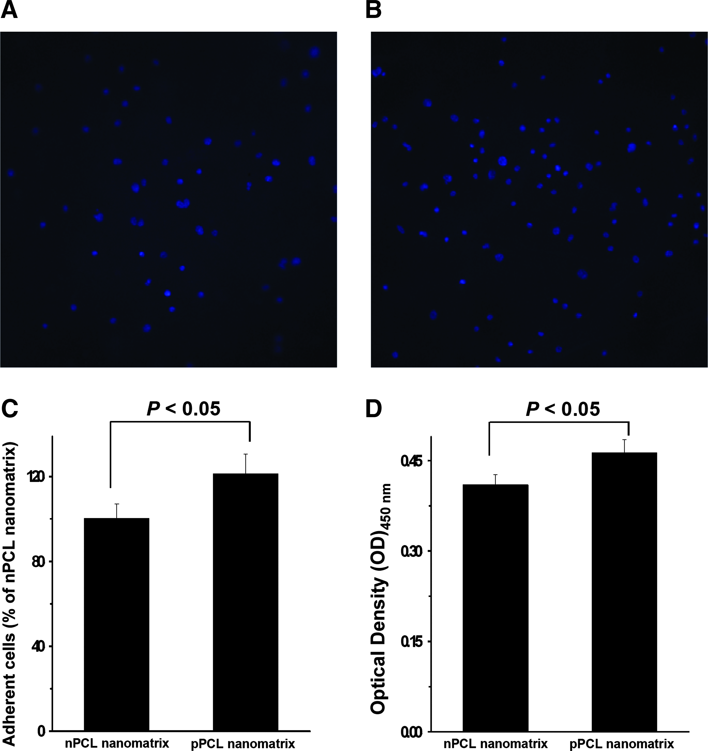
Representative immunofluorescent images (4, 6-diamidino-2-phrnykinodole [DAPI]) of the adhered cells on the
Analysis of cell shape on the charged nanomatrices
A cell shape on the charged nanomatrices was investigated. As shown in Figure 3A–D, both nanomatrices greatly influenced cell shape. FESEM images demonstrate that the NIH 3T3 fibroblast cells adhered to and spread well on both nanomatrices. Interestingly, FESEM images also revealed that the NIH 3T3 fibroblast cells showed a highly spread shape (relatively spherical shape) on the pPCL nanomatrices than those on the nPCL nanomatrices. The NIH 3T3 fibroblast cells also had a more narrow shape on the nPCL nanomatrices than on the pPCL nanomatrices. These results agree with immunofluorescence staining data observations (Fig. 3A, C) and suggest that different shape of cells on charged nanomatrices may be able to facilitate different cell functions such as migration, proliferation, and differentiation. 26
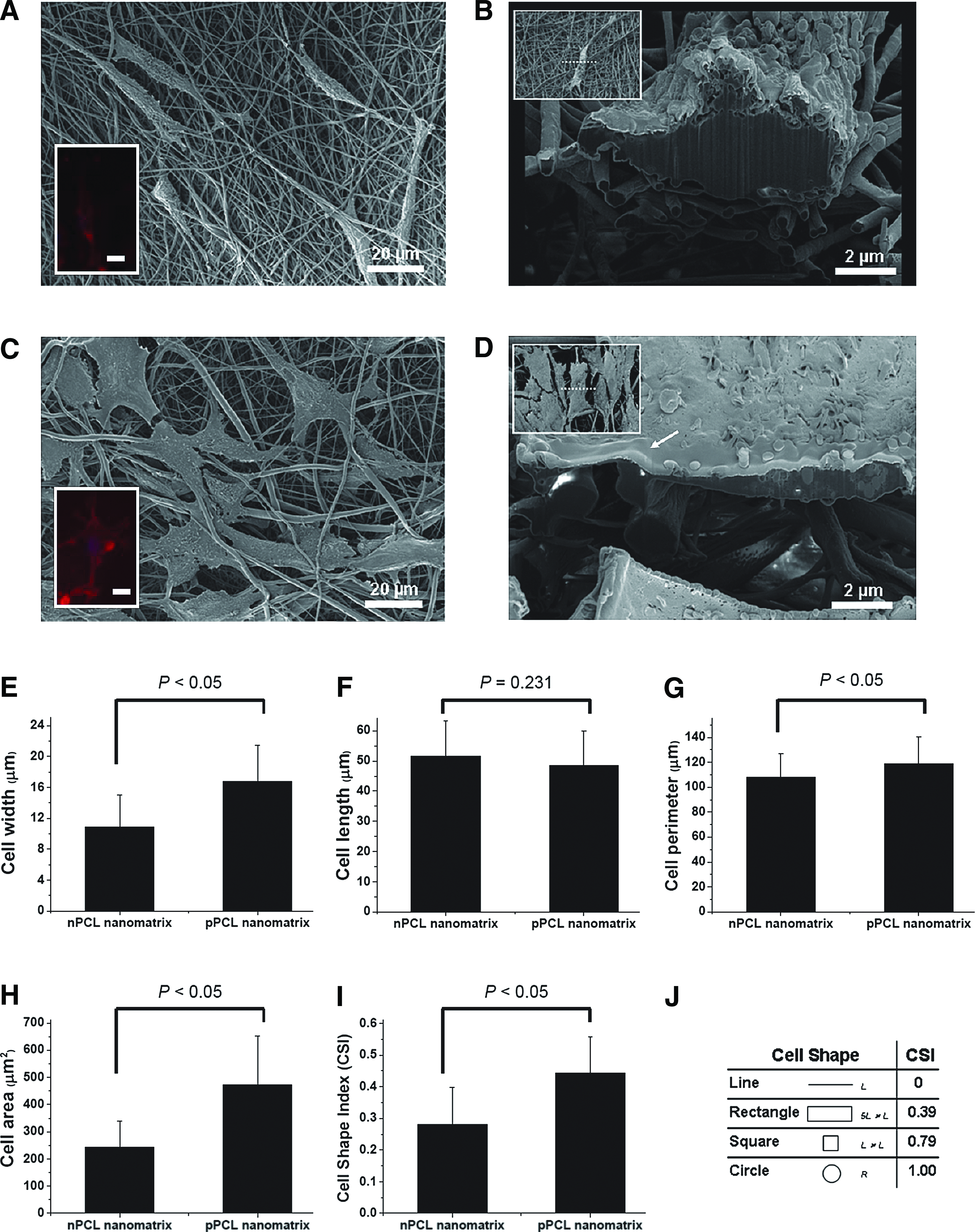
The modulated shape and adhesion of NIH 3T3 fibroblast cells on the charged nanomatrices.
To quantify cell shape observations, CSI=4πA/P2 values of cells on nPCL and pPCL nanomatrices were calculated. The cell widths were significantly larger on the pPCL nanomatrix than on the nPCL nanomatrix (p<0.05) (Fig. 3E), whereas the cells on the nPCL nanomatrix were slightly longer although there was no significant difference statistically (p=0.231) (Fig. 3F). We analyzed cell perimeters and areas on the charged nanomatrices. The perimeters and areas of cells on the pPCL nanomatrix were greater than on the nPCL nanomatrix (p<0.05; Fig. 3G, H). The NIH 3T3 fibroblast cells were highly spread on the pPCL nanomatrix than those on the nPCL nanomatrix. The CSIs of cells on the nPCL and pPCL nanomatrices were 0.280±0.116 and 0.443±0.114, respectively (Fig. 3I). The CSI of cells on the pPCL nanomatrix was significantly larger than on the nPCL nanomatrix (p<0.05), suggesting that cells on the positively charged nanomatrix were more circular in shape than those on the negatively charged nanomatrix, while the cells on the nPCL nanomatrix were more elongated. Together, these findings suggest that cell shape can be modulated by charged nanomatrices. In particular, the positively charged nanomatrix provides cues to cells resulting in highly spread and circular shapes compared with the negatively charged nanomatrix, on which cells demonstrated elongated shapes.
To test the hypothesis that charged nanomatrices influence cross-sectional cell shape due to electrostatic forces between cells and charged nanomatrix, we investigated cross-sectional cell shapes on the nPCL and pPCL nanomatrices (Fig. 3B, D) using FESEM and FIB milling technology. The cells adhered well to both charged nanomatrices with direct contact between the cell cytoplasm and the charged nanomatrices, although some portions of the cell membranes were not directly connected due to the limited cell length and nanopatterned substrate structure. 26 Interestingly, the cross-sectional cell shape on the pPCL nanomatrix was extremely flattened because electrostatic forces lead to attraction between the negatively charged cell membrane and the positively charged nanomatrix. In contrast, the cross-sectional cell shape was relatively round on the nPCL nanomatrix and some of the adhered cells floated, suggesting that electrostatic repulsive forces between the negatively charged cell membrane and negatively charged nanomatrix influence cell adhesion and shape.
To show that charged nanomatrices influence other cell types as well, we cultured human umbilical vein endothelial cells (HUVECs) and human mesenchymal stem cells (hMSCs) separately on nPCL and pPCL nanomatrices for 14 h. As shown in Supplementary Fig. S2, the cell shape for the two additional cell lines was also greatly modulated by the charged nanomatrices. FESEM images revealed that the HUVECs and hMSCs showed highly spread shapes on the pPCL nanomatrices, whereas more narrow shapes were shown on the nPCL nanomatrices.
Other properties of nPCL and pPCL nanomatrices influenced cell shape even though we fabricated the charged nanomatrices in the present study with similar morphologies. Chen et al. 27 reported that the shapes of NIH 3T3 fibroblast cells did not obviously differ when exposed to PCL fiber networks of various nanofiber diameters (117–1,647 nm). Bashur et al. 28 and Kim et al. 29 reported that the shapes of NIH 3T3 fibroblast cells were modulated by the sizes of nanopatterned poly(D,L-lactic-co-glycolic acid) or poly(urethane acrylate) substrates, becoming narrow in shape and smaller in area when nanopatterned substrates of smaller sizes were used as cell culture substrates. Therefore, we investigated the shapes of NIH 3T3 fibroblast cells on a pPCL nanomatrix with small nanofiber diameters (Supplementary Fig. S3). The resulting cell shapes were not significantly different than those on the pPCL nanomatrix with relatively large nanofiber diameters. This indicated that the slightly different structures of nPCL and pPCL nanomatrices didn't modulate cell shape, but their charges did.
Cell affinity assay
Fluid shear stress was used to investigate the effects of charged nanomatrices on cell affinity in this study. Figure 4B shows the affinity of cells on the nPCL and pPCL nanomatrices with the application of fluid shear stress. After culturing of NIH 3T3 fibroblast cells on the nPCL and pPCL nanomatrices for 14 h, we applied fluid shear stress using the rocking culture system with respect to the different number of minutes (15, 30, and 60 min). In the presence of shear stress, the pPCL nanomatrices showed better cell affinity than the nPCL nanomatrix. In particular, when the shear stress was applied for 60 min, 15.9%±4.3% of cells were adhered on the pPCL nanomatrices while 5.1%±2.3% of cells were adhered on the nPCL nanomatrices (p<0.05). This result indicates that positively charged nanomatrices may provide the stronger environments for cell affinity under fluid shear stress, compared with negatively charged nanomatrices. Our observations suggest that the cell-substrate affinity greatly depends on the charge of a substrate surface. This may provide us with critical information for the design and manipulation of engineered ECM.
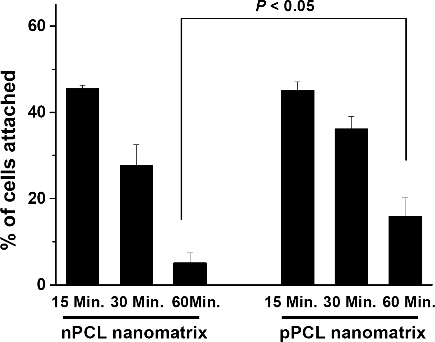
Affinity of cells on the nPCL nanomatrices and pPCL nanomatrices after applying fluid shear stress (∼1 dyn/cm2) with respect to different periods (15, 30, and 60 min). The affinities of cells were significantly different according to the charged nanomatrices. The error bars represent the SD about the mean (n=3 for each group).
Discussion
The nanoscale structure of the ECM provides instructive signals and a physical framework to regulate cell behaviors.1,2 In this study, charged nanomatrices were described as an effective strategy for design and manipulation of engineered ECM. Simply, we suggest using suitable charged materials (by adding a small negative or positive charge for fabricating nanoscale engineered ECM) that allow the fabrication of unique engineered ECMs that can be used in cell and tissue engineering applications and for a better understanding of cell biology.
General processes of cell behaviors follow these steps: cell adhesion on substrate→spreading→cytoskeleton development (cell shape)→functions. Therefore, the differently modulated cell adhesion and shape according to the charged nanomatrices would reflect different cell functions.30–32 For instance, Li et al., reported that highly elongated fibroblasts cells (the cell shape adopted on the nPCL nanomatrix) showed 40% higher collagen type I expression compared with fibroblasts cells with circular shape (the cell shape adopted on the pPCL nanomatrix). 31 In this case, we may be able to use the charged nanomatrices as an analysis platform for cell functions according to the modulated cell shapes.
In a human body, living cells experience a wide variety of mechanical stimuli.33,34 In particular, the cells of blood vessels are continuously exposed to shear stress from blood flow. 35 Therefore, mechanical stimuli may play a critical role in regulation cell signaling and function, especially within the context of cell-shape modulation via charged nanomatrices. To address this issue, we attempted to calculate the shear stress of the surface surrounding a modulated single cell on the nPCL and pPCL nanomatrices using a simple computational model and an analysis method. Although our predicted shear stresses were calculated with numerous simplifying assumptions about the system (the model blood-like fluid was flowed with a steady velocity (0.053 ms−1) around the single cell on the simple model substrates of charged nanomatrices in a parallel-plate chamber) our simulation results indicate that the shear stress around the surface of cells on the nPCL and pPCL nanomatrix are significantly different due to the modulated cell shape such as width and height (cross-sectional cell thickness); the maximum shear stress around a single cell on the nPCL nanomatrix was more than two times higher than that of a cell on the pPCL nanomatrix (Supplementary Fig. S4). This is consistent with the report of Salvi et al., stating that cell shape greatly influences shear stress of cell surface 36 and is therefore one of the possible reasons why pPCL nanomatrices showed higher cell affinity under fluid flow conditions than the nPCL nanomatrices in this study (Fig. 4).
The mechanisms underlying the effects of charged nanomatrices on differential cell responses such as adhesion and shape remain unknown. We hypothesize that a combination of factors including nanoscale structure, electrostatic forces, and absorption of biomolecules on charged substrates affect cell response (Fig. 6). In our nPCL and pPCL nanomatrices, cells exposed to nanoscale structural cues were not directly connected to nanomatrices, and were generally aligned with the orientations of the nanomatrices (Figs. 3A–D). It is widely accepted that the materials implanted into our body are immediately coated with proteins from blood and interstitial fluids and that cells initially respond to the implanted materials based on these adsorbed proteins; 8 namely, the composition of the adsorbed proteins is one of the key mediators for cell behavior, including adhesion and shape. Our study showed that negatively or positively charged nanomatrices absorbed different biomolecules from media in vitro (Fig. 5). XPS indicated that the surface chemical compositions and biomolecule adsorption differed on oppositely charged nanomatrices. After treatment with 10% FBS, the intensity of the C1s peak increased on the nPCL nanomatrix (Fig. 5B) and decreased on the pPCL nanomatrix (Fig. 5E), possibly due to the differences in electrostatic interactions between charged nanomatrices and biomolecules in the biomolecule adsorption process. Positively charged proteins with amines would be electrically bound to negatively charged nPCL nanomatrix due to the orientations of carbonyl groups, suggesting that the presence of exposed proteins with carboxyls may increase the intensity of the C1s peak. Likewise, negatively charged proteins with carboxyls would be electrically bound to amine groups on the surface of pPCL nanomatrix, suggesting that the presence of exposed proteins with amines may decrease the intensity of the C1s peak. We confirmed similar results for the amide carbonyl peak (-NH-C=O-), which appeared at ∼288.0 eV in both the nPCL and pPCL nanomatrices (data not shown). This agreement was also confirmed by the presence of a higher N1s peak for the pPCL nanomatrix than for the nPCL nanomatrix (Fig. 5C, F), suggesting that negatively charged proteins might be bound to the positively charged nanomatrix, and that the exposed proteins (amines) might increase the N1s peak on the pPCL nanomatrix. As cell membranes have net negative charges, cells are contacted differently on charged nanomatrices by opposing (attractive or repulsive) electrostatic forces (Fig. 6). As a result, cell adhesion and shape were modulated by the charged nanomatrices. Further, we may expect that the nPCL and pPCL nanomatrices may be able to adsorb different proteins of ECMs such as immunoglobulins, vitronectin, fibrinogen, and fibronectin or focal adhesion proteins, which would influence important cell functions. For example, pPCL nanomatrices would strongly modulate α5β1 integrin than nPCL nanomatrices since it has been known that α5β1 integrin binding affinity is higher at the positively charged surface compared with the negatively charged surfaces. 24
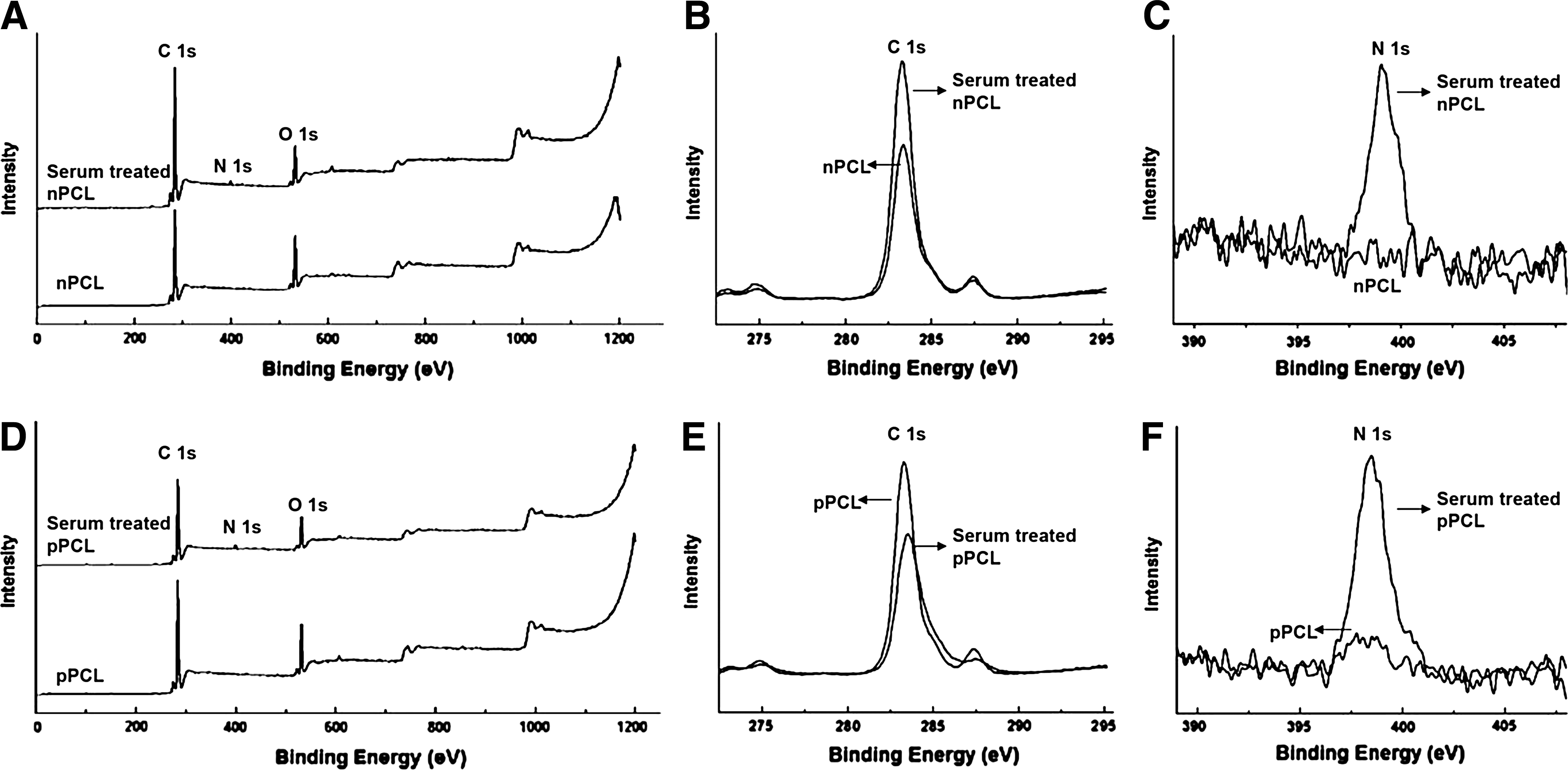
Determination of the surface chemical compositions on the nPCL and pPCL nanomatrices. X-ray photoelectron spectroscopy (XPS) measurements of the nPCL nanomarix before and after the 10% fetal bovine serum (FBS) treatment:
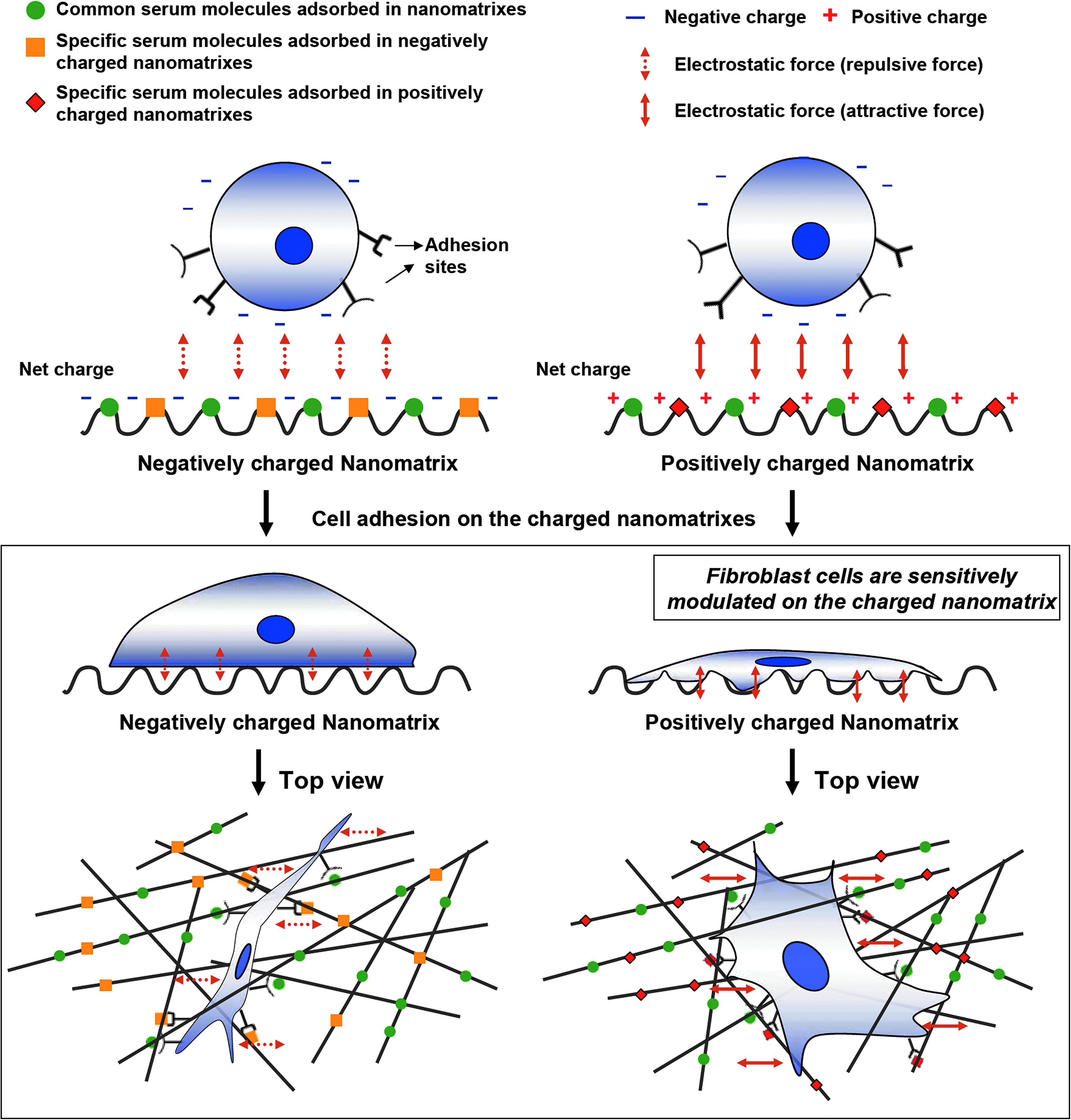
The proposed mechanism modulating cell adhesion and shape on negatively- and positively-charged nanomatrices. Color images available online at
Conclusions
Our study supports the notion that charged nanomatrices can be used to modulate cell adhesion and shape. Using PCL, PEI, and electrospraying technology, the nPCL and pPCL nanomatrix were developed as model systems for investigating modulation of cell responses with charges. We showed that cells can be sensitively discriminating in their responses of adhesion, shape, and affinity to the charged nanomatrices. We also confirmed that negatively- or positively charged nanomatrices absorbed different biomolecules from media in vitro. We propose that a combination of factors including nanoscale structure, electrostatic forces, and absorption of biomolecules on charged substrates can affect cell responses. We conclude that charged nanomatrices are efficient platforms to design and manipulate engineered ECMs for biological, cell, and tissue engineering applications.
Footnotes
Acknowledgment
This study was supported by Mid-career Research Program through National Research Foundation of Korea (NRF) grant funded by the Ministry of Education, Science and Technology (MEST; 2011-0028922). We thank Prof. Chong-Su Cho and In-Yong Kim for giving advice on this article. We also thank Saenggeul Baek who is a research scientist at the National Instrumentation Center for Environmental Management (NICEM) in Seoul National University for supporting FIB preparation and FESEM observation. D.H. Kim thanks the Department of Bioengineering at the University of Washington for the new faculty startup fund. D.H. Kim is also supported by a Perkins Coie Award for Discovery.
Disclosure Statement
No competing financial interests exist.
References
Supplementary Material
Please find the following supplemental material available below.
For Open Access articles published under a Creative Commons License, all supplemental material carries the same license as the article it is associated with.
For non-Open Access articles published, all supplemental material carries a non-exclusive license, and permission requests for re-use of supplemental material or any part of supplemental material shall be sent directly to the copyright owner as specified in the copyright notice associated with the article.