
Abstract
Natural polymers share recognition sequences that promote cell adhesion, rendering them attractive candidates for scaffolding in tissue engineering applications. However, challenges remain with regard to the fabrication of robust and porous structures of such raw materials for the design of extracellular matrix (ECM) mimics of living tissues. In this study, we present a fibrous scaffold that solely consists of albumin, the most abundant protein in mammalian blood plasma. The scaffold was fabricated using the electrospinning method, and resulted in microscale fibers that demonstrated mechanical properties which were similar to those of elastin fibers, a common component of connective tissue ECM. Albumin scaffolds proved nontoxic and supported adhesion and the spreading of fibroblasts, muscle cells, and endothelial cells (ECs) in vitro. In vivo studies demonstrated ∼50% biodegradation of the albumin scaffolds within 3 weeks of implantation. In addition, it was found that the fibers were encapsulated by dense fibrosis and evoked a weak inflammatory response, similar to that triggered by poly(L-lactide)/poly(lactic-co-glycolic acid) scaffolds. Albumin tubular structures fabricated to mimic blood vessels successfully guided the formation of blood vessel-like bi-layer structures made of fibroblasts and ECs. Thus, albumin scaffolds featuring biologically relevant characteristics pose a readily applicable alternative to synthetic scaffolding materials.
Introduction
A number of approaches have been taken toward designing adequate scaffolds, relying on the use of materials ranging from purely synthetic to purely biological, each possessing advantages and drawbacks. Scaffolds generated from synthetic materials, such as biodegradable polymers, feature adequate mechanical properties, degradation rates, porosity, and architecture, but are less supportive of cell attachment and spreading. In contrast, although natural polymer-based scaffolds engineered from proteins,12–14 or polysaccharides, 15 bear recognition sequences that promote cell adhesion, their mechanical properties are typically inferior to those of naturally occurring matrix equivalents. 13 Combinations of synthetic polymers that are grafted or surface treated with natural sequences have been shown to provide the most advanced support for cell adhesion.16–21
Electrospinning is a well-established procedure that is used for creating 3D scaffolds which are destined to mimic in vivo tissue topographies.4,11,22–24 However, the resulting product often lacks relevant biochemical cues. 25 The method draws solid nanofibers from a polymer solution by applying a strong electrostatic field. 26 Dror et al. 27 successfully applied the electrospinning technique to fabricate ultra-thin fibers of the globular bovine serum albumin (BSA) protein, which demonstrated high strength in comparison to other synthetic and natural fibers. The BSA spinning process was established by unfolding the protein chain conformation, an act that could potentially affect its natural role in the transportation of free fatty acids and drugs in the blood stream. 28 However, we hypothesize that BSA-cells interactions and subsequent biodegradation will not be impacted, as in the body, similar denaturized forms of the protein bind vascular endothelial cells (ECs) via receptor-mediated interactions, 29 which then stimulate further protein degradation through the endosome-lysosome pathway. 28
In this work, we explored the mechanical and biological features of electrospun scaffolds that solely consist of albumin fibers, and compared them with those of scaffolds made of polycaprolactone (PCL) and poly(L-lactide)/poly(lactic-co-glycolic acid) (PLLA/PLGA), which are fabricated by electrospinning and a salt-leached technique. These widely used synthetic polymers represent different morphologies and mechanical properties (at room temperature), as well as different surface energy properties that may affect cell-matrix interactions. Mechanical properties were determined under physiological conditions, and biological behavior was assessed both in vitro and in vivo. In addition, several cell types, including ECs, were seeded on the albumin scaffolds, and cell adherence and organization were evaluated.
Materials and Methods
Electrospinning of fibrous scaffolds
BSA (Fraction V; MP Biomedicals) was dissolved in a 9:1 (w/w) mixture of 2,2,2-trifluoroethanol (ReagentPlus®; Sigma-Aldrich) and distilled water, to form a 10% BSA solution (w/w). Beta-mercaptoethanol (β-ME, molecular biology grade; Merck) was added (0.2 g β-ME per 1 g of BSA) to denaturate the protein, 30 thus forming a spinnable solution.
Electrospinning of the BSA solution was conducted under room conditions (22°C±1°C and 40%±2% relative humidity), using a syringe pump (Harvard Apparatus), a 23-gauge needle (inner diameter ∼0.37 mm), and a custom-built high-voltage (30 kV max) direct current (DC) supply. Solution flow rate was 2 mL/h under a voltage supply of 12 kV, and a tip-to-collector distance of 14 cm. Random mats were collected on an horizontal metal plate, and tubes were collected using a rotating aluminum wire with a diameter of 2 mm at 350 rpm. Samples were dried from residual solvents in a vacuum at a pressure of ∼10−3 atm.
A 10% solution of PCL (Mw 80,000 Da; Sigma-Aldrich) in a 80:20 (w/w) mixture of Chloroform (Frutarom) and dimethylformamide (Frutarom) was electrospun using the following parameters: an applied voltage of 12 kV, a flow rate of 2 mL/h, and a tip-collector distance of 17 cm.
Preparation of porous polymer scaffold
PLLA/PLGA scaffolds were made of 50% PLLA (Polysciences) and 50% PLGA (Boehringer) via a salt-leaching process. PLLA/PLGA (1:1) were dissolved in chloroform to yield a 5% (w/w) polymer solution, 0.24 mL of which was then cast into cylindrical molds containing 0.4 g of 200–600 μm sodium chloride particles. After solvent evaporation, the disc-like sponges were detached from their molds and immersed in distilled water for 6–8 h (water was changed every hour). Once the salt dissolved, the porous structure was obtained.
Structure and morphology
Samples from the fabricated scaffolds were coated with gold and characterized using a Phenom desktop scanning electron microscope (SEM) (5 kV accelerating voltage; FEI Company) and a Gemini high resolution-SEM (1 kV accelerating voltage; Carl Zeiss). Fiber size and mean pore size were measured using ImageJ software. The porosity, P, of scaffolds was calculated according to P=1−ρs/ρa, where ρs is the measured scaffold density, determined from sample weight and volume, and ρa is the standard albumin density (1.4 g/cm3).
Mechanical properties
Tensile tests (strain ramp) were carried out in a custom-built uniaxial tensile machine, under a strain rate of ∼33%/min. Small rectangles, ∼10 mm long, were cut from fiber mats and mounted using rubber clamps. For tests performed under wet conditions, samples were immersed in phosphate-buffered saline (PBS) for 15 min before the test. All tests were conducted at room temperature (∼25°C). The engineering stress was calculated over the cross-sectional area A=W · δ, where the initial width W and thickness δ of samples were measured with a caliper and micrometer, respectively. Mat stiffness (Young's modulus) was calculated over a strain range of 0.1–0.2 for BSA mats, and 0–0.03 for PCL mats. Extensibility was defined as the strain until breakage, and toughness (energy to breakage) was calculated as the area under the stress-strain curve.
In vitro biocompatibility test
The Griess assay (Promega) was performed, as per the manufacturer's instructions, on albumin scaffolds. PLLA/PLGA salt-leached sponges and electrospun PCL scaffolds served as control materials, due to their well-established biocompatibility.31,32 RAW and J774 macrophage cell lines (TIB-71; American Type Culture Collection [ATCC]) were plated separately, 1 day before the assay (2×10⁵ cells per well in 6-well plate) and then incubated in 2 mL/well high-glucose (4.5 g/L) Dulbecco's modified Eagle's medium (DMEM) (Gibco), supplemented with 10% fetal bovine serum (FBS) (Hyclone) and 1% pen-strep antibiotics (Biological Industries). A 10 mg scaffold sample was cut into the smallest possible pieces to allow for maximal interaction with the cells and the reagents, and incubated with the cells for 24 h. Control samples of cells cultured without scaffolds were treated with 500 ng/mL lipopolysacharide (LPS) (Pseudomonas aeruginosa, 1 mg/mL; Sigma), to obtain positive controls, or left untreated, as negative controls. A fresh reference curve was prepared in parallel to each assay, using the nitrite standard diluted in the aforementioned culture medium. Absorbance at 520 nm was recorded over 24 h.
In vivo biodegradability and biocompatibility test
All the described surgical processes were performed following the protocols approved by the Institutional Animal Care and Use Committee. Three groups of 6-week-old, ICR male mice (n=4 per group; Harlan Laboratories) were anesthetized using a ketamine: xylazine cocktail at a dose of 35 μL/20 g, delivered with a 25-gauge needle. Disc-like samples (∼5 mm in diameter) of electrospun albumin mats were subcutaneously implanted to the dorsal of the anesthetized mice. Electrospun PCL mats or salt-leached PLLA/PLGA sponges were also subcutaneously implanted as controls. Tissue samples of the graft area were extracted for histological analysis at 1, 2, and 3 weeks postimplantation.
Cell culture
Mouse skeletal myoblast cells (C2C12) were cultured in 10 mL DMEM supplemented with 10% FBS, 2.5% HEPES 1M, and 1% pen-strep antibiotics. Human foreskin fibroblasts (HFF) were cultured in 10 mL DMEM supplemented with 10% FBS, 1% minimum essential medium enriched with nonessential amino acids×100, 0.2% β-ME, and 1% pen-strep solution. Umbilical artery smooth muscle cells (SMCs) were cultured in 10 mL DMEM supplemented with 10% FBS, 2.5% HEPES 1 M, and 1% pen-strep. Human umbilical vein endothelial cells (HUVEC) were cultured in 10 mL EGM-2 supplemented with Single Quote (Lonza). Albumin scaffold samples (∼5×3 mm2) were sterilized by exposure to 365 nm UV for 15 min and then seeded with ∼106 cells per scaffold. Cell-seeded scaffolds were incubated for 30 min at 37°C, to allow for cell adhesion. Culture media were replaced thrice during the 1-week culture period.
Histology and immunofluorescence staining
Constructs were fixed in 10% neutral buffered formalin (Sigma Aldrich), and embedded in paraffin after undergoing standard fixation. Transverse, 5 μm-thick sections were placed on silanized slides for immunofluorescence and hematoxylin and eosin staining, or for Masson's Trichrome staining. Epitope recovery was achieved by heating samples (90°C, 20 min) in ReVeal buffer (Biocare Medical). Tissues were then blocked with 10% FBS (Hyclone) blocking solution for 10 min. The sections were then simultaneously incubated with mouse anti-human CD31 antibodies (1:20 dilution) and rabbit anti-human vWF antibodies (1:200) (both from Epitomics), at room temperature, for 30 min, before being extensively washed with PBS. Sections were then labeled with Cy2-conjugated anti-rabbit IgG and Cy3-conjugated anti-mouse IgG2b (1:100; Jackson Immunoresearch Laboratory), in addition to 4′6-diamidino-2-phenylindole (DAPI), a nuclear counterstain (Sigma Chemical Co.; 1:1000). Constructs were characterized using a fluorescence microscope (Axiovert 200M; Zeiss) and AxioVision imaging software (version 3.1; Carl Zeiss).
Results and Discussion
Fiber morphology and mechanical behavior
Images of electrospun scaffolds and salt-leached sponges are shown in Figure 1. Electrospun albumin fibers were smooth with a ribbon-like shape (see the inset in Fig. 1A), a morphology that is likely due to fiber collapse which is consequential of entrapped solvents. 33 Fiber width ranged between 1 and 6 μm, and thickness at the center of the ribbon was below 0.5 μm. Electrospun PCL fibers were uniform with a mean diameter of 1.1±0.4 μm. Both BSA and PCL nonwoven fiber mats were 100–200 μm thick, with a porosity of 85%–90%, and a pore size range of 10–20 μm. Salt-leached PLLA/PLGA sponges were 700–800 μm thick, and bore pores sizes ranged between 120 and 450 μm (Fig 1C).
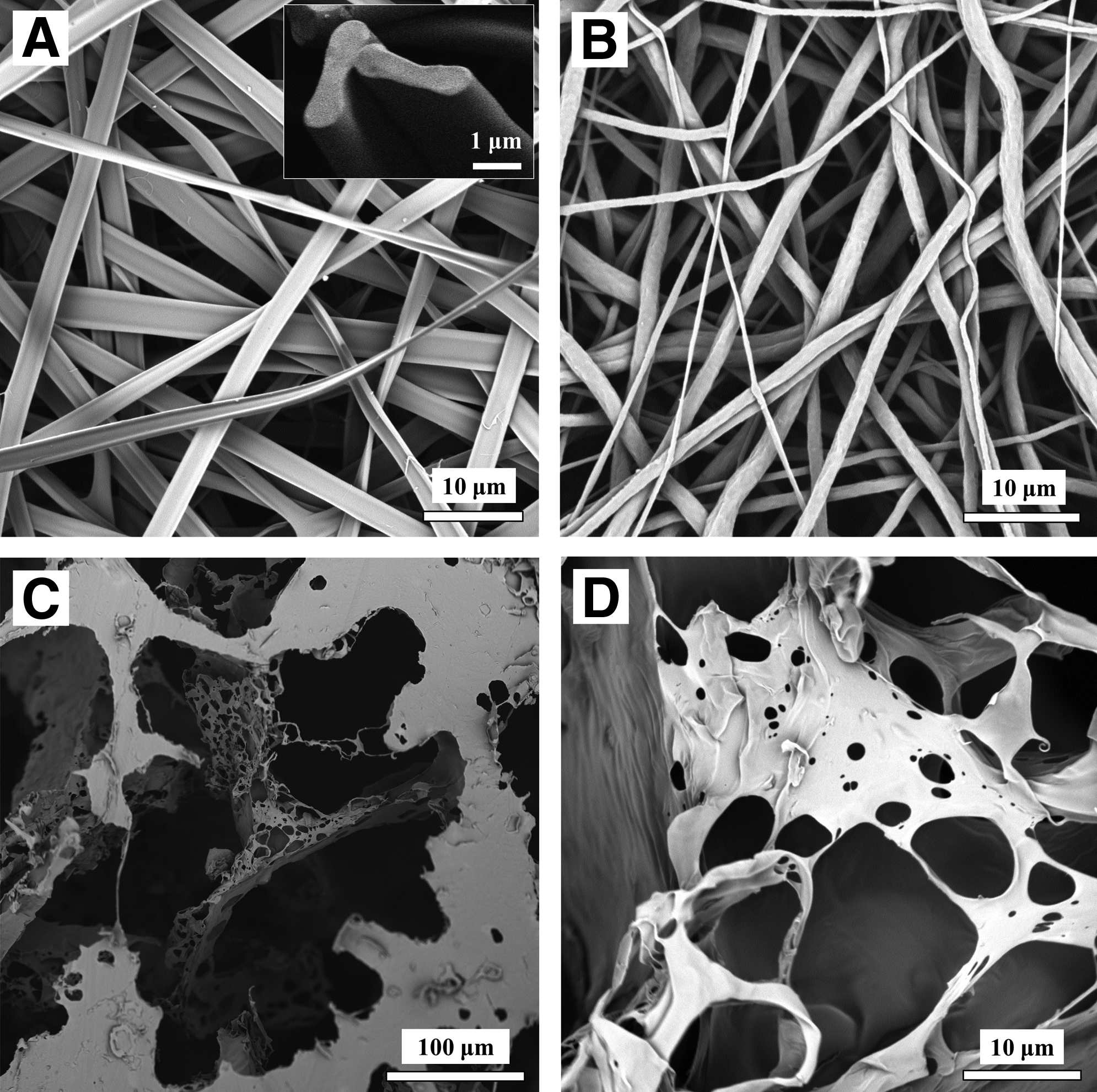
Scanning electron microscope images of the morphology of tested scaffolds.
Quasi-static tensile tests of electrospun albumin scaffolds immersed in physiological medium (PBS) yielded a stress-strain curve that was similar to the responses of natural tissues, with some nonlinearity 34 (Fig. 2). Mat strength (ultimate tensile stress) was 1.52±0.30 MPa (n=8), stiffness (Young's modulus) was 0.35±0.04 MPa, extensibility was 1.63±0.10, and toughness was 0.83±0.16 MJ/m3. In general, biological fibers can be classified as either stiff or soft. Stiff fibers, such as collagen and actin, feature stiffness in the order of a few GPa, with low extensibility (below 20%); while soft fibers, such as elastin and resilin, have stiffness in the order of a few MPa and are highly extensible (above 100%).35,36 On comparison of the mechanical properties of the albumin-based fibers to other biological fibers (Table 1), the albumin fibers showed mechanical behavior that was typical of soft fibers. Tensile tests performed on PCL mats immersed in PBS yielded a stiffness of 15 MPa (n=3), a strength of 30 MPa, and an extensibility of more than 6 (see Table 1).
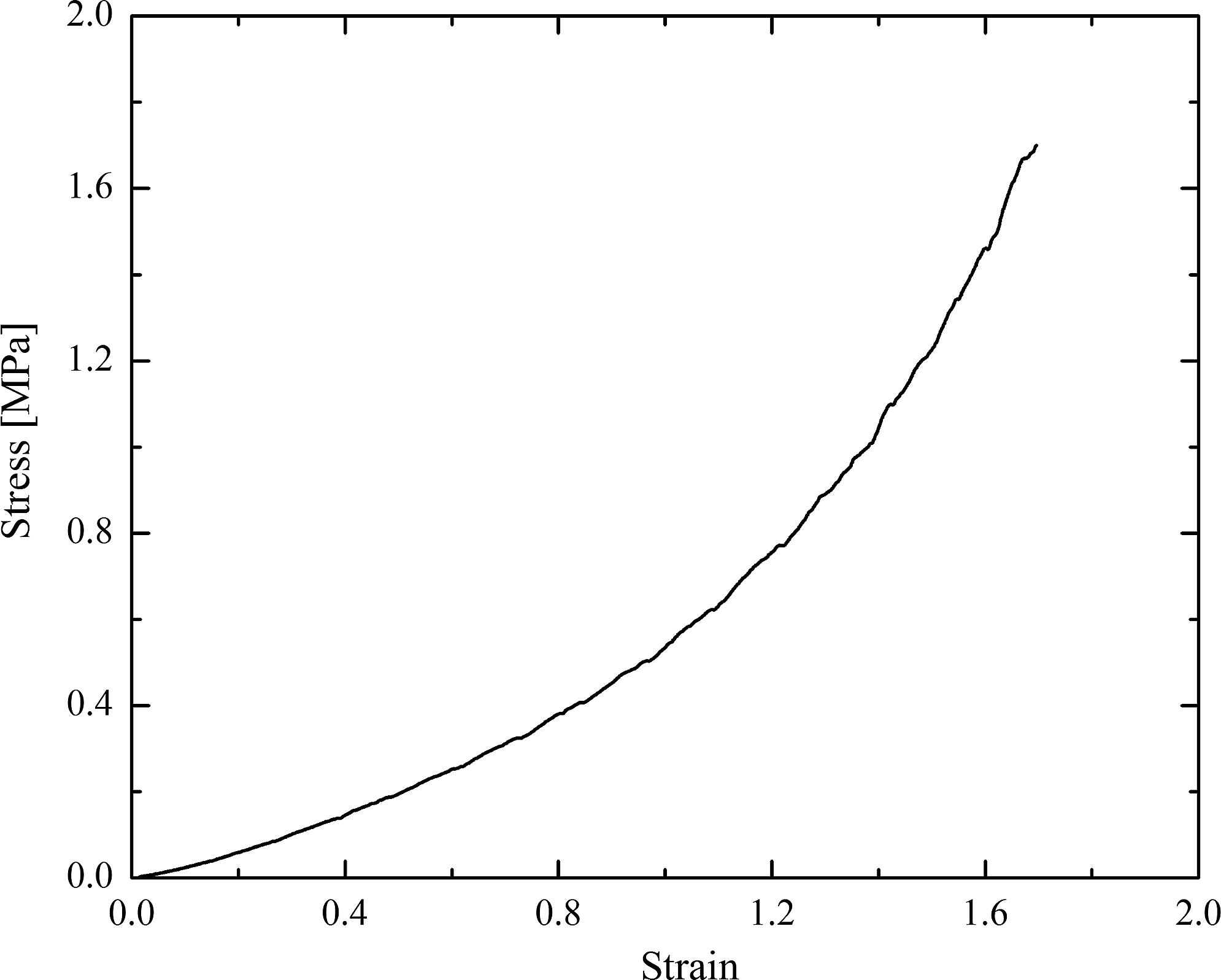
Stress-strain curve of the tested albumin fibers. Graph presents representative data recorded until maximal tensile stress was reached. Sample was immersed in phosphate-buffered saline (PBS) for 15 min before the test. Strain rate was ∼33%/min.
Wet samples were immersed in phosphate-buffered saline/water while testing their mechanical properties.
Cross-linked with HMDI.
Electrospun random mat.
End of diastole.
BSA, bovine serum albumin; PCL, polycaprolactone; PLLA/PLGA, poly(L-lactide)/poly(lactic-co-glycolic acid).
In vitro biocompatibility test
Tissue engineered grafts should prove to be safe, noninflammatory, and nontoxic before use in vivo. In vitro biocompatibility tests, 45 such as the Griess assay, can offer an early indication of expected scaffold biocompatibility in vivo. Nitric oxide (NO), secreted from macrophages, acts as a toxic agent toward infectious organisms and regulates host immune cell functioning.46,47 One of the most common methods for measuring NO production involves spectrophotometric monitoring of nitrite, a spontaneous oxidation product of NO. This Griess reaction 48 occurs during immunological responses, on macrophage stimulation, where the level of secreted NO correlates with the degree of stimulation. Thus, the Griess assay was performed immediately after the addition of scaffold samples to macrophage cultures, as well as at 4, 16, 20, and 24 h thereafter (Fig. 3). Increasing amounts of nitrite were detected for each sample type (n=9 for each scaffold type), in linear correlation with the time it was incubated with macrophages. Responses were similar to those previously reported for the same cell lines cultured under parallel conditions. 45 As expected, nitrite concentrations measured in the PLLA/PLGA and PCL samples were lower than those detected in the LPS-treated positive control samples, and were similar to those measured in the scaffold-free negative controls. Nitrite levels detected in cells incubated with electrospun albumin samples were similar to those measured in cultures exposed to PLLA/PLGA and PCL samples, indicating that this new scaffold material is inert and does not stimulate immune responses in RAW and J774 cells. These data serve as an indicator of the expected immune responses and cell tolerability toward electrospun albumin scaffolds in vivo.
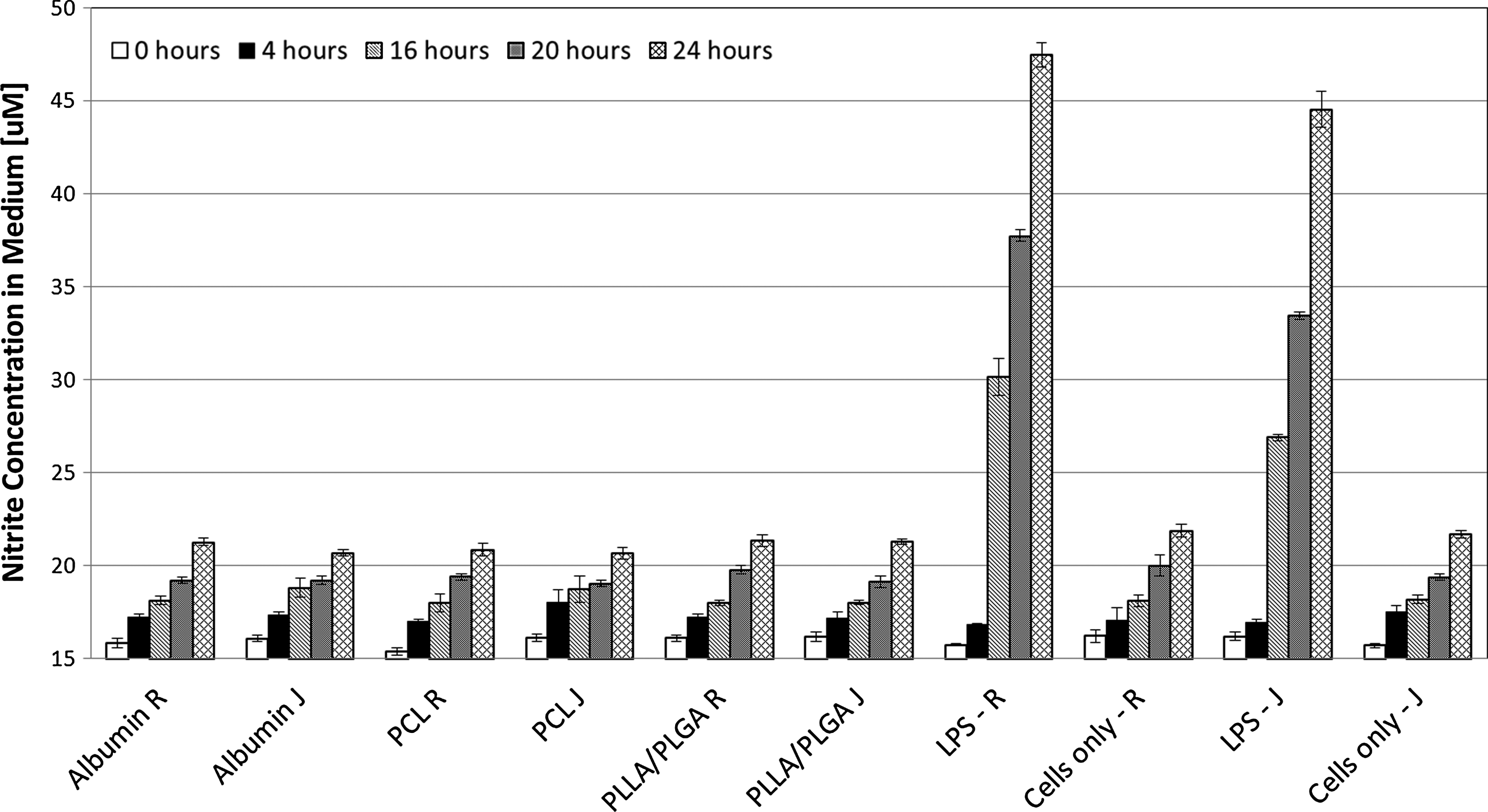
Macrophage-based inflammatory response to the presence of scaffolds. Macrophages (R indicates RAW cells and J indicates J774 cells) were incubated with bovine serum albumin (BSA), PLLA/PLGA, or PCL scaffolds. Nitrite concentrations in 50 μL medium samples were measured after 0, 4, 16, 20, and 24 h of incubation, using the Griess assay. Lipopolysacharide (LPS)-treated cells served as a positive control, whereas untreated cells served as a negative control. Mean responses (n=9) are presented, along with standard deviations.
In vivo biodegradability and biocompatibility analyses
The complexity of a living physiological system can never be matched by an in vitro culture assay, as a multitude of in vivo factors can integratively influence the biological response to implanted scaffolds. Albumin scaffolds are made of a naturally occurring polymer and are not expected to elicit immunologic reactions in vivo, whereas the implanted scaffolds may be recognized as foreign materials and will be isolated from the surrounding tissue. 49 For this purpose, the biodegradability and inflammatory response of subcutaneously sutured albumin fiber mats were evaluated in ICR mice and compared with those of implanted PLLA/PLGA and PCL disc-like scaffolds. The PLLA/PLGA and PCL scaffolds were chosen as controls due to their known lengthy degradation times (∼6 months for PLLA/PLGA 50 and several years for electrospun PCL scaffolds 31 ). In addition, the PLLA/PLGA scaffold is widely utilized in biomedical applications, with well-characterized biocompatibility characteristics. 49
No behavioral changes indicating systemic toxicity were noted during the 3-week postoperative period. Histological analyses demonstrated that within 1 week of implantation, BSA scaffolds were encapsulated by a thick fibrous layer, seemingly composed of collagen fibrils, macrophages, and giant cells (Fig. 4A, D). High-magnification images revealed a number of inflammatory cells that had succeeded in entering and integrating with the albumin scaffold, especially at its margins. On examination of the implanted PCL samples (Fig. 4B, E), discontinuous scaffold fractions were revealed, seemingly due to inappropriate integration of the scaffold with the host tissue, which led to slicing difficulties on extraction. At this same time point postimplantation, PLLA/PLGA scaffolds were encapsulated by a thin fibrous tissue (Fig. 4C, F), and inflammatory cells were mainly observed in the periphery of the scaffold; a few cells infiltrated the implant area. All tested scaffolds evoked a minimal inflammatory response in the surrounding bulk tissue, when compared with the response at the implant-tissue interface.
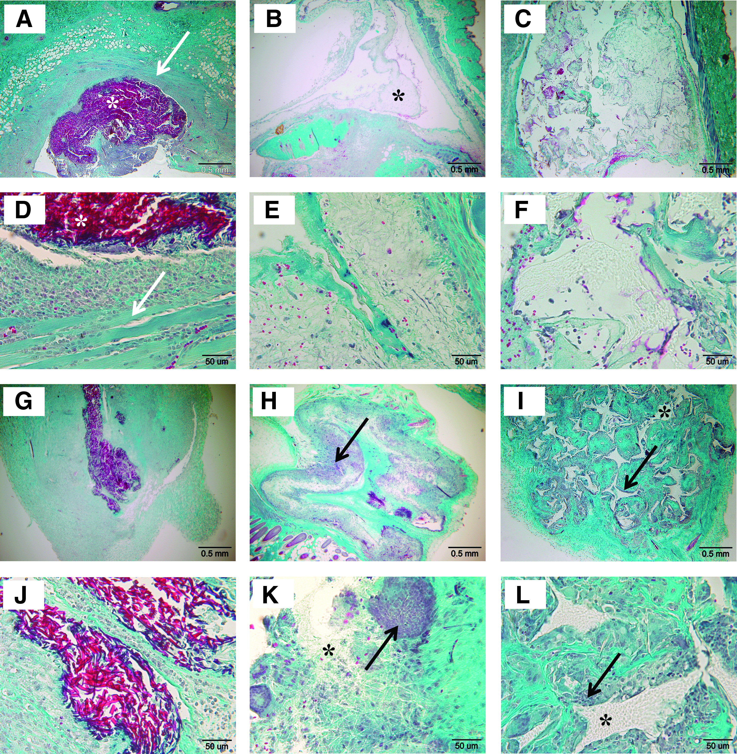
Inflammatory response to subcutaneously implanted BSA
At 3 weeks postimplantation, BSA implants were encapsulated by dense fibrosis, and an inflammatory response was evoked, which was indicated by the infiltration of a few macrophages (Fig. 4G, J). In sharp contrast, PCL constructs that were extracted at this same time point postimplantation were characterized by dense inflammatory cells detected throughout the surrounding tissue and the scaffold site (Fig. 4H, K). Clusters of giant cells were abundant (Fig. 4K). PLLA/PLGA scaffolds had been infiltrated by giant cells and tissue ECM, owing to the relatively large pores (few hundreds of micrometers) of this scaffold material (Fig. 4I, L). The overall tissue response to PLLA/PLGA scaffold resembled the mild inflammatory response previously reported for this kind of biomaterial, whose biocompatibility is considered satisfactory. 49
Qualitative visual and microscopic inspection at 3 weeks postimplantation showed that albumin scaffolds were at least 50% of the size of the originally implanted discs. No degradation was observed in vitro for identical samples immersed in culture mediums for several weeks. It is, therefore, concluded that albumin fibers are enzymatically degraded in vivo. PLLA/PLGA and PCL scaffolds showed almost no reduction in size during the 3-week implantation period.
In vitro cell culture
In order to examine the potential of albumin scaffolds as a clinically relevant 3D scaffold, their capacity to support cell growth was determined. Various cell types were seeded (monoculture) on either disc-like or tubular electrospun albumin scaffolds. Parallel and perpendicular construct sections were collected after 1 week in culture, and demonstrated that scaffolds supported cell growth (Fig. 5). Cell-scaffold interactions promoting cell adherence to the albumin scaffold surface are assumed to be albumin binding protein (ABP)-mediated. 29 Alternatively, adherence can be mediated by the adsorption of serum proteins, such as fibrinogen and fibronectin, to the negatively charged albumin fibers, which are highly supportive of strong cell-matrix interactions. Only a small number of cells successfully penetrated the fibers and settled deep within the scaffold, which may be a result of the scaffold's small pore size.
Cell adhesion to electrospun albumin scaffolds. Hematoxylin and eosin (H&E)-stained sections of
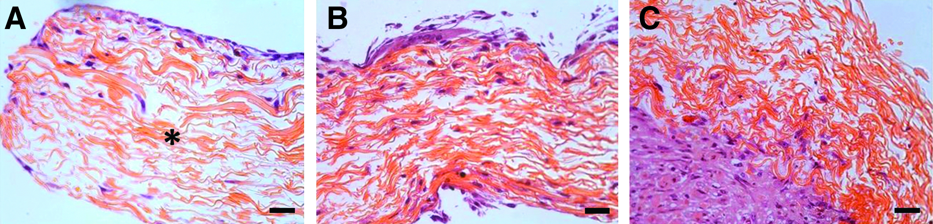
Albumin scaffolds were then further tailored to match a vessel-like morphology. Such applied structures can be effectively used to produce in vitro vascular grafts.20,51,52 Blood vessels are hierarchical structures, where the inner (lumen) layer of ECs is coated with SMCs and fibroblasts (connective tissue cells). In order to develop a small tissue-engineered blood vessel, we fabricated tubular scaffolds made of electrospun albumin fibers and conducted a two-step in vitro experiment, in which fibroblasts were first cultured for 1 week within the tubes, and then supplemented with HUVEC for a second week in culture. Such cocultivation drives the differentiation potential of foreskin fibroblasts toward the formation of smooth muscle-like cells. 53 The initial stages of fibroblast growth within the tubes were examined via histological images of cell-embedded constructs (Fig. 6). HFF adhered to the lumen of the albumin tube and formed a vessel-like structure. Immunofluorescence sections of HFF-HUVEC embedded tubes collected after 2 weeks in culture demonstrated the creation of a bi-layer structure of fibroblasts lined with ECs on the lumen of the tubes. In addition, ECs were able to penetrate deep within the tube walls and to organize into small lumen-like structures (Fig. 7).

HFF adhesion to electrospun albumin tubes. Albumin tubes were incubated for 1 week with ∼3×10⁶ HFFs. Sections were then fixed and stained. H&E-stained perpendicular sections of HFF-seeded tubes, fixed after 1 week in culture. Asterisk indicates the scaffold region. Magnification is

Cell interactions with albumin tubes after 2 weeks of incubation in vitro.
Conclusions
In the present work, we fabricated and characterized electrospun albumin-based scaffolds. The albumin scaffold proved biodegradable and provoked what we believe is a milder inflammatory response, when compared with implanted PLLA/PLGA and PCL scaffolds. Electrospun albumin fibers feature mechanical properties that are similar to those of elastin fibers and are, therefore, expected to provide a highly flexible matrix. Various cell types successfully grew on planer- and tubular-shaped albumin scaffolds, indicating scaffold support of cell adhesion and proliferation. Penetration of cells into the scaffold was limited; however, the pore size can be further increased, for example, by spinning thicker fibers from a higher concentration polymer solution54,55 or by following the technique set forth by Tzezana et al. 10 In conclusion, this work highlights the attractiveness of electrospun albumin scaffolds for application in tissue engineering protocols. Further work will investigate the nature of cell-albumin scaffold interactions, as well as the differentiation of fibroblasts into SMCs on the fabricated tubular scaffolds under flow conditions.
Footnotes
Acknowledgments
The authors gratefully acknowledge the financial support of the Russell Berrie Nanotechnology Institute (RBNI) at the Technion, and the Singapore National Research Foundation under the CREATE Program: The Regenerative Medicine Initiative in Cardiac Restoration Therapy (NRF-Technion). The technical assistance of Aviva Kabala is appreciated.
Disclosure Statement
No competing financial interests exist.