
Abstract
Supercritical fluids are used in various industrial fields, such as the food and medical industries, because they have beneficial physical and chemical properties and are also nonflammable and inexpensive. In particular, supercritical carbon dioxide (ScCO2) is attractive due to its mild critical temperature, pressure values, and nontoxicity. Poly(L-lactide-co-ɛ-caprolactone) (PLCL), which is a biocompatible, biodegradable, and very elastic polymer, has been used in cartilage tissue engineering. However, organic solvents, such as chloroform or dichloromethane, are usually used for the fabrication of a PLCL scaffold through conventional methods. This leads to a cytotoxic effect and long processing time for removing solvents. To alleviate these problems, supercritical fluid processing is introduced here. In this study, we fabricated a mechano-active PLCL scaffold by supercritical fluid processing for cartilage tissue engineering, and we compared it with a scaffold made by a conventional solvent-casting method in terms of physical and biological performance. Also, to examine the optimum condition for preparing scaffolds with ScCO2, we investigated the effects of pressure, temperature, and the depressurization rate on PLCL foaming. The PLCL scaffolds produced by supercritical fluid processing had a homogeneously interconnected porous structure, and they exhibited a narrow pore size distribution. Also, there was no cytotoxicity of the scaffolds made with ScCO2 compared to the scaffolds made by the solvent-pressing method. The scaffolds were seeded with chondrocytes, and they were subcutaneously implanted into nude mice for up to 4 weeks. In vivo accumulation of extracellular matrix of cell–scaffold constructs demonstrated that the PLCL scaffold made with ScCO2 formed a mature and well-developed cartilaginous tissue compared to the PLCL scaffold formed by solvent pressing. Consequently, these results indicated that the PLCL scaffolds made by supercritical fluid processing offer well-interconnected and nontoxic substrates for cell growth, avoiding problems associated with a solvent residue. This suggests that these elastic PLCL scaffolds formed by supercritical fluid processing could be used for cartilage tissue engineering.
Introduction
In our previous studies, we developed an elastic biodegradable poly(L-lactide-co-ɛ-caprolactone) (PLCL) scaffold that could effectively deliver the mechanical signals associated with the surrounding biological environment, and we showed that these properties might enhance and facilitate the cartilage regeneration process. 9 Also, the mechanical property of the PLCL scaffold was similar to that of native cartilage. 10 In terms of mechano-active tissue engineering, it is important to consider the mechanical properties of materials to provide cells that are grown in an environment similar to that of a real organ.8,11,12
Although many studies have been carried out using PLCL scaffolds, conventional methods, such as solvent casting, have usually been used for the fabrication of PLCL scaffolds; these methods start with dissolving the polymer in organic solvents, such as chloroform or dichloromethane. This leads to a cytotoxic effect and long processing time for removing solvents.8,11,12 To alleviate these problems, a supercritical fluid processing could be introduced.6,13–16
Supercritical fluids are used in various industrial fields, such as foods, pharmaceutics, medicines, and chemical engineering. The unique physical properties of supercritical fluids exhibit liquid-like density, gas-like viscosity, and no surface tension. These properties render supercritical fluids advantageous for use as a medium for various processes. Among supercritical fluids, the use of supercritical carbon dioxide (ScCO2) is particularly attractive for medical applications, because it is nontoxic as well as inexpensive and nonflammable. Moreover, its supercritical temperature (Tc) and pressure values (Pc) are relatively mild (Tc=31.5°C and Pc=75.8 bar).17–19 In the ScCO2, some polymers are highly swollen, allowing processing, such as porous structure formation. Furthermore, ScCO2 can be used to impregnate biomolecules, such as growth factors or genes, into a polymer. These properties can be used to develop porous constructs with encapsulated biomolecules for controlled, sustained release of drugs.17,20–22
In this study, we fabricated mechano-active PLCL scaffolds by supercritical fluid processing for cartilage tissue engineering, and we compared it with scaffolds made by a conventional solvent-pressing method in terms of physical and biological performance. To examine the optimum condition for preparing scaffolds with ScCO2, we investigated the effects of pressure, temperature, and depressurization rate on PLCL foaming. Also, to evaluate the possibility of PLCL scaffolds produced by supercritical fluid technology for mechano-active cartilage tissue engineering, in vitro and in vivo experiments were performed with chondrocytes.
Materials and Methods
Fabrication of PLCL scaffolds by supercritical fluid foaming technique
PLCL was synthesized, as described previously. 23 Briefly, l-lactide (100 mmol; Purac) and ɛ-caprolactone (100 mmol; Sigma) were polymerized at 150°C for 24 h in the presence of stannous octoate (1 mmol; Sigma) as a catalyst. Then, we weighed 70 mg of PLCL (Mn=152,000) and put it into each well of a Teflon mold. After the mold was fed into a reactor, the reactor was heated to a predefined temperature. Afterward, the reactor was purged three times with nitrogen (N2) and filled with CO2 until a certain pressure level was reached and the PLCL/CO2 mixture maintained at a constant pressure for soaking time. Lastly, the reactor was then depressurized back to the ambient pressure over a period of time (venting time [VT]). While this supercritical fluid process for producing scaffolds was occurring, the process was controlled by a mass flow controller (MFC, TSC-100, MKP nano high-tech). 6 Figure 1 shows the overall experimental scheme. For the control experiments, PLCL scaffolds were also fabricated using a solvent-casting method. PLCL was dissolved in chloroform (5% w/v), and then mixed with NaCl particles (100–300 μm) of 70 wt% of PLCL weight (0.12 g/mL). The chloroform was evaporated in air to form PLCL gels, which were then pressed in a mold. The residual chloroform was evaporated for 24 h at room temperature, and subsequently removed under vacuum for 72 h. The salts were leached out and the resulting scaffolds were freeze-dried. 8

Apparatus for supercritical fluid processing. MFC, mass flow controller.
Characterization of scaffolds
The morphology of the scaffolds was examined by scanning electron microscopy (SEM; Hitachi) operating at 15 kV. The samples were coated with gold using a sputter-coater (Eiko IB3). A mercury intrusion porosimeter (Autopore IV; Micrometrics) was used to examine the surface and determine the scaffold pore size distributions. Additionally, the pore interconnectivity within the scaffolds was measured by the conventional method. 24 Briefly, a 1% aqueous solution of rhodamine B was mixed with anhydrous ethyl alcohol, and 60 μL of solution was dropped on each scaffold. The solution that remained on the scaffolds after 1 h was removed and dried. The stained area was measured by an image analysis program, ImageJ (Image J 1.43; National Institute of Mental Health, Image Proversion 4.5; Media Cybernetics). Additionally, the mechanical properties of the PLCL scaffold were examined by performing compression tests using a universal testing machine (TMS-Pro, Food Technology Corp.) with a 1 kN-maximum load cell at a cross-head speed of 3 mm·min−1.
Cytotoxicity tests
The PLCL scaffolds were tested for cytotoxicity following the ISO 10993-5 guidelines. A total of 0.2 g of the scaffolds were placed in conical tubes containing 1 mL of culture medium (DMEM/F12 [Welgene] supplemented with 10% fetal bovine serum [Welgene] and 1% penicillin–streptomycin [Welgene]) and incubated at 37°C in a shaking water bath for 72 h. In addition, L929 mouse fibroblasts (1.8×10⁴ cells/cm2) were cultured in 48-well plates for 1 day; the extracts were added to the culture medium at a 1:1 ratio. Cell viability was evaluated by a water soluble tetrazolium salts (WST) assay (Cell counting kit-8; Dojindo). 25 The culture medium was used as a negative control, and the extracts of a latex film (6 cm2/mL) were used as a positive control.
In vivo implantation studies
To investigate their biocompatibility and cartilaginous tissue formation, in vivo implantation studies were performed. The chondrocytes were isolated by enzymatic digestion of the knee joint articular cartilage from 3-week-old rabbits, as previously described. 7 Afterward, the cultured chondrocytes (passage number=2) were collected by trypsin (Gibco BRL) treatment and resuspended in the culture medium (5×10⁷ cells/mL). Seventy μL of this cell suspension was inoculated into scaffolds (9-mm diameter, 3-mm thickness, disk type) and allowed to stabilize for 2 h, after which a culture medium was added. Then, the cell–polymer constructs were implanted into the subcutaneous dorsum of 7-week-old male athymic mice (SLC). Implants were harvested for analysis after 4 weeks. 2 For histological analysis, the cell–polymer construct explants were fixed in 10% (v/v) buffered formalin, dehydrated in a graded ethanol series, and embedded in paraffin. The specimens were cut into 5-μm thick sections and stained with hematoxylin and eosin (H&E). Collagen and sulfated glycosaminoglycans in the retrieved constructs were stained using Masson's trichrome (M-T) for the former and Alcian blue and Safranin O for the latter. 26
Statistical analysis
All the samples were assayed in triplicates, and the results obtained were expressed in standard deviations (SD) above and below the mean±SD. All statistical analyses were carried out using the t-test. A result was considered to be statistically significant when the p-value was less than 0.05.
Results
Effect of processing conditions on the morphology of PLCL scaffolds
To study the effect of processing conditions on the morphology of PLCL scaffolds, scaffolds were fabricated at various temperatures. Figure 2 shows the gross views of scaffolds processed at 35°C and 45°C, respectively. We found that polymer foaming did not occur below the soaking temperature of 45°C. Figures 3 and 4 show the results from the mercury intrusion porosimeter. From these data, it was confirmed that the scaffolds had a higher porosity and smaller pores at the higher pressure. Also, we could observe that there were many small pores in the scaffolds at the processing conditions of high temperature or long VT (Table 1). These data correlated with the morphologies of scaffolds obtained by SEM (Figs. 5 and 6).

Gross-views of the poly(L-lactide-co-ɛ-caprolactone) (PLCL) polymers processed at
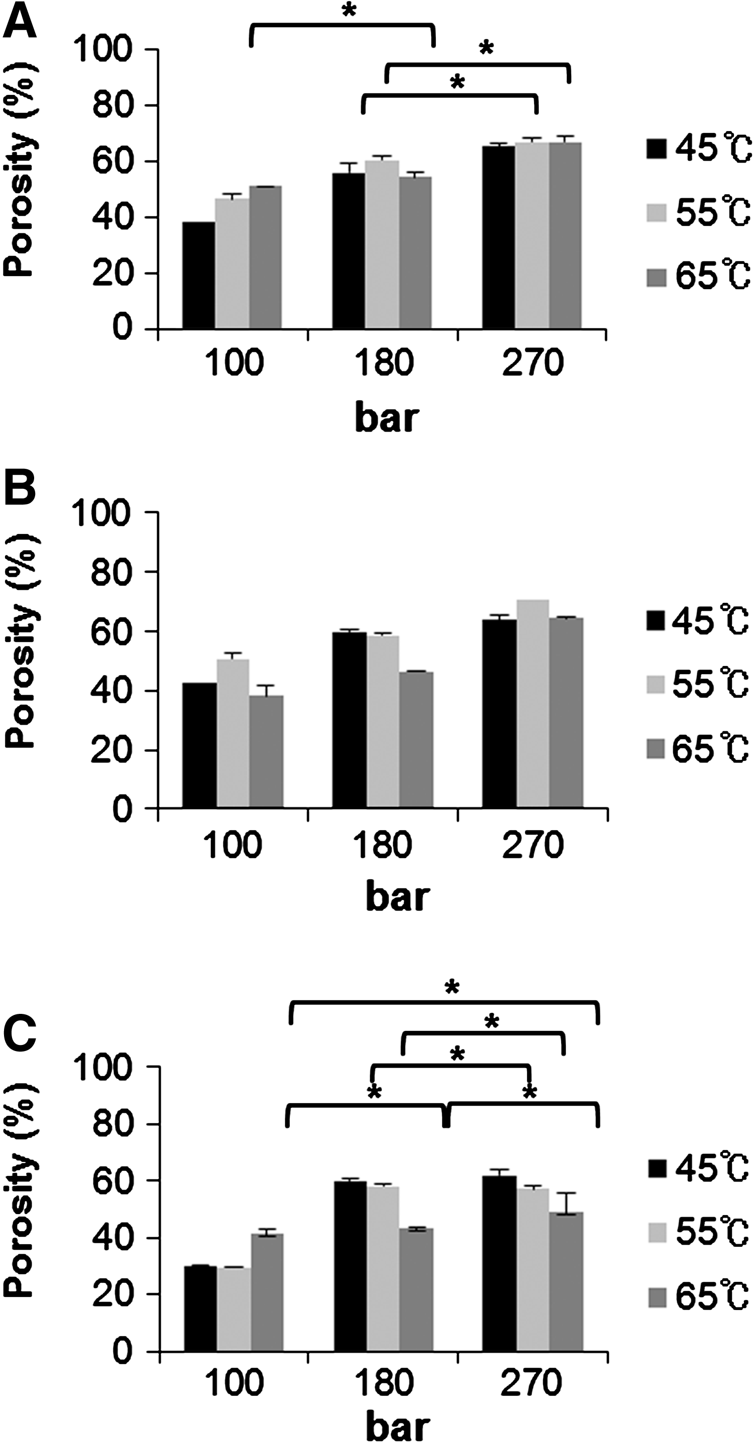
Porosity of scaffolds fabricated by supercritical fluid processing.

Pore size distribution of scaffolds fabricated by supercritical fluid processing.

The effects of pressure and temperature on the morphology of PLCL scaffolds: scanning electron microscopy (SEM) images. Processing conditions: VT=50 min and soaking time (ST)=120 min.

The effect of VT on the morphology of PLCL scaffolds: SEM images. Processing conditions: P=270 bar and ST=120 min.
P, pressure; T, temperature; ST, soaking time; VT, venting time.
Interconnectivity and compressive modulus of PLCL scaffolds
To examine the interconnectivity between pores in the scaffold, we stained the scaffold with an aqueous solution of rhodamine B and estimated the amount of rhodamine B solution that was penetrated through the scaffold pores. Figure 7A and B show that the stained area increased with an increase in the porosity of the scaffold by supercritical fluid foaming, except in the case of the scaffold of processing temperature (T)=45°C, VT=200 min and T=55°C, VT=50 min. That is why they had very small pores, and therefore, the solution could not penetrate these samples. Also, we compared the pore interconnectivity of the scaffolds fabricated by solvent casting with that fabricated by supercritical fluid processing. Although the scaffold (porosity=57.8%±0.1%, and mean pore size=211.6±91.3 μm) of T=55°C, VT=200 min had similar porosity and mean pore size as the scaffold (porosity=60.5%±3.4%, and mean pore size=223.8±51.8 μm) by solvent casting, the scaffold fabricated by supercritical fluid processing had a larger stained area than that fabricated by solvent casting. Also, these data correlated with the morphologies of scaffolds obtained from SEM (Fig. 7C, D).

Interconnectivity of PLCL scaffolds.
We performed the compression tests for the PLCL scaffolds that had similar porosity and mean pore size produced by supercritical fluid processing or solvent casting. The scaffold fabricated by the solvent-casting method had a higher compressive modulus (175.9±11.6 kPa) compared with the scaffold fabricated by supercritical fluid processing (104.9±8.2 kPa). That is thought to be why the frame of pore structures of the PLCL scaffold fabricated by the solvent-casting method was more solid and contained more closed pores.
Cytotoxicity tests
The cell metabolic activity was measured to evaluate cytotoxicity of the PLCL scaffolds. The viability of cells incubated with scaffold extracts was measured by a WST-1 assay (Fig. 8). We found that viability of the L929 cells exposed to the extract of the scaffolds fabricated by solvent casting was estimated to be 35.1%±7.8%, but scaffolds fabricated by supercritical fluid foaming were noncytotoxic, and there was statistically negligible significant difference in cell viability when compared to a negative control, namely the culture medium.
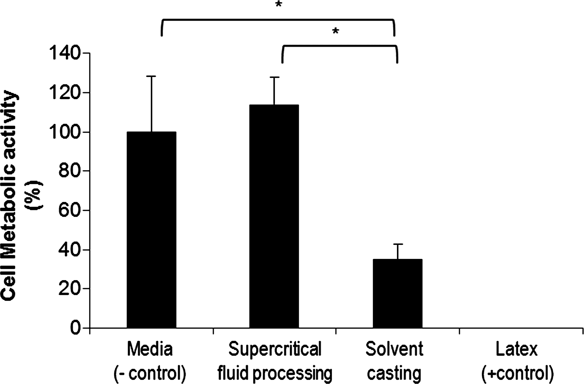
Cell viability (water soluble tetrazolium salt [WST-1]) of L929 fibroblasts (cytotoxicity tests). Media was used as a negative control, and extracts of latexes were used as a positive control. (*p<0.05).
In vivo implantation studies
To investigate whether the cells seeded onto the scaffolds were able to form cartilaginous tissue structures in vivo, chondrocyte/PLCL constructs were subsequently implanted subcutaneously into athymic mice. In histological analyses, H&E staining showed that the PLCL implants formed mature and well-developed cartilaginous tissues that were similar to the native cartilage, as evidenced by chondrocytes within lacunae (Fig. 9). The results of M-T staining indicated the presence of abundant collagens, and Alcian blue and Safranin O staining provided evidence of the accumulation of sulfated glycosaminoglycans, the extracellular matrix material produced by differentiated chondrocytes in newly-formed tissues. However, we confirmed that many chondrocytes into the PLCL scaffold fabricated by supercritical fluid processing were penetrated well, and cartilaginous tissues were distributed widely compared to the group of solvent casting.

Histological studies of implants at 4 weeks. The sections were stained with hematoxylin and eosin
Discussion
In this study, using supercritical fluid processing, we fabricated highly elastic and biodegradable PLCL scaffolds, which are useful in mechano-active tissue engineering. Usually, polymeric scaffolds, such as PLGA and poly-D, L-lactic acid, which were widely used as a scaffold in tissue engineering, can be fabricated at 35°C by supercritical fluid foaming, because these polymers have enough free volume for CO2 to penetrate into the polymer chains. 6 On the other hand, the linear structure of PLCL, whose hard domain consists of caprolactone chains, does not contain any substituents in the -[(CH2)5]n- part of the caprolactone chain, which are fixed more strongly in position by the intermolecular interactions between them. It is therefore difficult for CO2 to penetrate into the polymer chains, because the free volume between the chains is very small. Only at temperatures higher than 45°C could CO2 penetrate into the polymer chains of PLCL. However, PLCL foams cannot be processed for a long period of time at above 70°C, as they are sensitive to heat. Under these conditions, scaffolds were produced at 45°C, 55°C, and 65°C, and it was found that pores were larger and more open at a higher temperature. At a constant pressure, the solubility of CO2 in the polymers decreases with increasing temperatures, because the density of CO2 decreases as well. However, the diffusion rate of CO2 in the polymers is higher at a higher temperature. In addition, CO2 can more easily penetrate into the polymer chains due to the increased mobility of the polymer chains. It is therefore expected that pores will grow larger and interconnect effectively at a higher temperature. Also, pressure is a critical factor for PLCL scaffold morphology, such as the porosity or pore size. At a constant temperature, the density of CO2 in the polymer matrix is higher at a higher pressure. The scaffold has a high porosity at high pressure, because the pores were produced from the CO2 incorporated with the polymer. Thus, the porosity of a scaffold can be tailored by careful control of the pressure conditions. The concentration and diffusion coefficient of CO2 in the polymer matrix increases with increasing pressure, and the relationship between them is approximately linear. In addition, a higher pressure has been found to increase dissolution of CO2 and, as a consequence, the glass transition temperature (Tg) decreases. Therefore, at a higher pressure, the amount of CO2 incorporated into the polymers is greater, and hence, the substrate is more highly supersaturated upon release of the pressure. These higher supersaturation pressures lead to higher nucleation densities; hence, many smaller pores were observed in the scaffolds. Additionally, CO2 has sufficient surface energy, so CO2 can create a new surface if the driving force is sufficient. A high depressurization rate would thus lead to a higher nucleation rate and a larger number of nucleation sites that would ultimately become small-sized pores. If there is enough time for growth, CO2 molecules tend to get together. Furthermore, bigger CO2 drops with the same amount of CO2 as smaller drops have lower surface energy and are more stable according to thermodynamics. Thus, a decrease in the depressurization time would result in scaffolds with larger pores. 6
By measuring the interconnectivity and the compressive modulus of PLCL, we found that the PLCL scaffolds fabricated by supercritical fluid processing had good interconnectivity and lower compressive modulus compared to the PLCL scaffolds fabricated by the solvent-casting method. On the basis of these results, it is considered that scaffolds fabricated by supercritical fluid processing ensure a relatively high mass transfer rate if they are used in tissue engineering. Furthermore, in mechano-active cartilage tissue engineering, the mechanical properties, especially the compressive modulus of the scaffold, are very important factors, because the scaffold should deliver compressive signals to adherent cells onto the scaffold in dynamic physiological systems. Thus, the softness of the PLCL scaffolds produced by supercritical fluid processing would be helpful in mechano-active cartilage tissue engineering.7–10,12
By the cytotoxicity study, we confirmed that PLCL scaffolds fabricated by supercritical fluid processing had good biocompatibility. Thereafter, we investigated the possibility of these PLCL scaffolds as a substrate for cartilage tissue engineering by in vivo tests. Here, we could observe in vivo accumulation of the cartilaginous extracellular matrix into the implants of the PLCL scaffolds made with ScCO2 mature and well-developed cartilaginous tissue. We thought that this result was considered to be from good pore interconnectivity, which encourages cells to penetrate into the scaffold, and biocompatibility due to cytotoxicity.
Conclusion
In cartilage regeneration, it is important that the scaffolds both maintain their mechanical integrity as well as deliver mechanical signals to adherent cells onto the scaffolds in the body. With this point in mind, the nontoxic and highly porous PLCL scaffold could by useful for cartilage tissue engineering. Here, using supercritical fluid processing, we could tailor scaffolds by controlling processing conditions, such as pressure, temperature, and VT. The PLCL scaffolds produced by supercritical fluid processing offer well-interconnected and nontoxic substrates that have mechano-active properties for cartilage tissue engineering, avoiding problems associated with solvent residue, as compared to the scaffolds fabricated in this article by solvent casting. Also, from the in vivo studies, we confirmed that these PLCL scaffolds more enhanced chondral extracellular matrix deposition. Consequently, these results suggest that these elastic PLCL scaffolds formed by supercritical fluid processing could be used for cartilage tissue engineering.
Footnotes
Acknowledgment
This study was supported by grants from the Korea Healthcare Technology R&D Project, Ministry of Health & Welfare (MOHW), Republic of Korea (A110962 and A110328).
Disclosure Statement
No competing financial interests exist.