
Abstract
Immobilization of biomolecules onto implant surfaces is highly relevant in many areas of biomaterial research. Recently, a 2-step immobilization procedure was developed for the facile conjugation of biomolecules onto various surfaces using self-polymerization of dopamine into polydopamine. In the current study, a 1-step polydopamine-based approach was applied for alkaline phosphatase (ALP) and bone morphogenetic protein-2 (BMP-2) immobilization, and compared to the conventional 2-step polydopamine-based immobilization and plain adsorption. To this end, ALP and BMP-2 were immobilized onto titanium and polytetrafluoroethylene (PTFE) substrates. The absolute quantity and biological activity of immobilized ALP were assessed quantitatively to compare the three types of immobilization. Plain adsorption of both ALP and BMP-2 was inferior to both polydopamine-based immobilization approaches. ALP was successfully immobilized onto titanium and PTFE surfaces via the 1-step approach, and the immobilized ALP retained its enzymatic activity. Using the 1-step approach, the amount of immobilized ALP was increased twofold to threefold compared to the conventional 2-step immobilization process. In contrast, more BMP-2 was immobilized using the conventional 2-step immobilization approach. Retention of ALP and BMP-2 was measured over a period of 4 weeks and was found to be similar for the 1-step and 2-step methods and far superior to the retention of adsorbed biomolecules due to the formation of covalent linkages between catechol moieties and immobilized proteins. The biological behavior of ALP and BMP-2 coatings immobilized using polydopamine (1- and 2-step) as well as adsorption was assessed by culturing rat bone marrow cells, which revealed that the cell responses to the various experimental groups were not statistically different. In conclusion, the 1-step polydopamine-based immobilization method was shown to be more efficient for immobilization of ALP, whereas the conventional 2-step method was shown to be more efficient for attachment of BMP-2 onto implant surfaces.
Introduction
Inspired by the composition of mussel adhesive proteins (which are rich in
So far, 1-step immobilization of biomolecules using polydopamine has not been compared with 2-step polydopamine-based immobilization or simple adsorption. Therefore, we have for the first time compared the efficacy of the novel polydopamine-assisted 1-step immobilization with conventional polydopamine-assisted 2-step immobilization as well as simple adsorption, assuming that a 1-step polydopamine-based immobilization procedure not only simplifies immobilization of biomolecules even further, but also increases the total amount of immobilized biomolecules at implant surfaces.
To test the above-mentioned hypothesis, the metalloenzyme alkaline phosphatase (ALP) was selected as a model protein. This enzyme catalyses the hydrolysis of organic phosphate monoesters (such as β-glycerophosphate) into inorganic phosphates. As such, ALP plays a crucial role in mineralization of bone tissue. 21 ALP has recently been applied successfully as a thin film onto bone implant surfaces.22,23 ALP coatings were shown to induce surface mineralization of bone implant surfaces and accelerate bone apposition onto ALP-coated titanium implants in vivo.22,23 The mechanism behind this was expected to be the conversion of phosphate monoesters by the coating, resulting in a local increase of phosphate ions. De Jonge et al. immobilized ALP via an electrospray deposition method, resulting in a noncovalently attached coating.22,23 Retention of the electrosprayed ALP was recently found to be poor (<10%, data not shown) due to the lack of covalent linkages. The use of ALP as a model protein allows for straightforward measurement of enzymatic activity by means of commercially available biochemical assays. Further, ALP is relatively cheap and can be labeled easily with radioactive iodine to allow for accurate determination of absolute quantities of immobilized biomolecules using gamma-counting. These advantages make ALP an interesting candidate to compare the new 1-step method to the conventional 2-step. The applicability of the 1-step method was further explored by immobilizing BMP-2.24,25 BMP-2 is recognized as one of the most potent osteogenic growth factors, but its high cost limits applicability of BMP-2 as surface coating onto implant surfaces. Irreversible immobilization of BMP-2 onto implant surfaces using an industrially upscalable 1-step procedure would open up new opportunities for improved control over the retention and therapeutic efficacy of BMP-2. 26 Two types of substrate materials with completely different physicochemical properties were tested, that is, titanium and polytetrafluoroethylene (PTFE). Titanium was selected because of its bioinertness and widespread usage in the field of orthopedics and dentistry. 27 PTFE is also widely used in biomedical applications and was mainly selected, since this highly hydrophobic polymer represents an ultimate challenge for immobilization of biomolecules.
Materials and Methods
Commercially obtained pure titanium discs (12-mm diameter, 1.5-mm thickness) or sheets of PTFE (DS groep, product No.: 6900.010 cut to 10 mm×10 mm×1 mm) were used as substrates for the immobilization of ALP (Sigma; P7640, ALP from bovine intestinal mucosa) or BMP-2 (R&D Systems; 355BM/CF, carrier-free recombinant human BMP-2). Before use, the titanium discs and PTFE sheets were cleaned ultrasonically in acetone (10 min) and isopropanol (10 min). Dopamine*HCl (dopamine) was purchased from Sigma (H8502). Tris(hydroxymethyl)aminomethane (Tris) was purchased from Gibco BRL (15506-017).
Solutions
Unless stated otherwise, all solutions were aqueous solutions buffered at pH=8.5, using 10 mM Tris. ALP solution was prepared by dissolving ALP at a concentration of 1 mg/mL. 22 The dopamine solution was prepared by dissolving dopamine at a concentration of 2 mg/mL. 7 The ALP–dopamine solution consisted of 1 mg ALP and 2 mg dopamine per mL. BMP-2 solution contained 4 μg/mL of BMP-2 per mL and BMP-2–dopamine solution 4 μg of BMP-2 and 2 mg of dopamine per mL. The BMP-2 concentration was selected based on a study by Liu et al., who observed that coprecipitation of BMP-2 with calcium phosphate was effective in concentrations of 1–10 μg/mL to increase the ALP activity of rat bone marrow stromal cells. 28
Coating procedures
Biomolecules were immobilized onto substrates using (1) simple adsorption (ads), (2) conventional sequential immobilization of dopamine and the biomolecule of interest (2-step), or (3) simultaneous immobilization of dopamine and the biomolecule (1-step).
For immobilizing ALP, these three approaches were performed as follows: for ads, substrates were soaked in 1 mL ALP solution for 16 h. In the case of 2-step, substrates were first soaked in 1 mL dopamine solution for 16 h, followed by flushing with the Tris buffer and immersion in 1 mL ALP solution for 16 h. 11 One-step coating, on the other hand, was performed by immersing the substrates in 1 mL ALP–dopamine solution for 16 h.
BMP-2 coatings were prepared in a similar way from BMP-2 solution and BMP-2–dopamine solution, but instead of immersion in 1 mL, 75-μL aliquots were added on top of the substrates. After the immobilization procedure, all experimental substrates were washed four times for 2 min in a 24-well plate containing 2 mL Tris buffer before being dried in air.
Contact-angle measurements
Static contact-angle measurements were performed for 1-step, 2-step, ads, and uncoated materials, as well as substrates modified with only dopamine (via immersion in dopamine solution for 16 h) after washing with H2O. To this end, a dataphysics OCA 30 instrument using deionized water as the probe liquid was used. A sessile drop technique was applied to add the liquid (4 μL) to the surface. Contact-angle measurements were performed in triplicate.
Quantification of absolute amount of immobilized ALP and BMP-2
The absolute amount of ALP and BMP-2 immobilized was quantified using radiolabeling. Briefly, for ALP, 10 μL of 0.5 M phosphate buffer (pH 7.4), 80 μL ALP solution (1 mg/mL in 50 mM phosphate-buffered saline [PBS]), and 8 μL 125I (800 μCi) were added to an Eppendorf vial coated with 100 μg iodogen. For BMP-2, 10 μL of 0.5 M phosphate buffer (pH=7.4), 15 μL BMP-2 solution (0.33 μg/mL in 50 mM PBS), and 10 μL 125I (1000 μCi) were added to an Eppendorf vial coated with 100 μg iodogen. The mixture was incubated for 10 min at room temperature (RT) and subsequently purified on a PD-10 column eluted with PBS. Radiochemical purity of 125I-ALP and 125I-BMP-2 exceeded 95% as measured by instant thin-layer chromatography. For ALP, 315 μCi (∼30 μg) labeled ALP (hot) was mixed with 50 mg unlabeled (cold) ALP and volumized to 50 mL in Tris buffer (yielding a final ALP concentration of 1 mg/mL with 6 μCi/mL radiation). For BMP-2, 56.9 μCi (∼1.72 μg) labeled BMP-2 was mixed with 10 μg unlabeled and volumized to 976 μL for a final concentration 12 μg/mL (which was further diluted with the Tris buffer to obtain 4 μg/mL with 19.4 μCi/mL radiation). These solutions were used to immobilize the biomolecules as described above, followed by radiation measurements using a shielded well-type gamma-counter (Wizard, Pharmacia-LKB) in glass vials (n=4). Standards (n=3) containing 5 μL (for ALP) or 25 μL (for BMP-2) of the solutions in similar glass vials were measured simultaneously with the coated substrates to correct for radioactive decay. Further, to measure the retention of the immobilized biomolecules, coated substrates (n=4) were soaked in 3 mL PBS (pH 7.4 at 37°C). At selected time points, the PBS was replaced, and the radioactivity of the experimental substrates was measured.
Quantification of biochemical activity of immobilized ALP
The biochemical activity of ALP that was immobilized onto titanium using the three different coating procedures was monitored colorimetrically by measuring the conversion of para-nitrophenylphosphate to 4-nitrophenol. Briefly, ALP-coated substrates were placed into a 24-well plate containing 200 μL ultrapure water, 50 μL of 0.5 mM 2-amino-2-methyl-1-propanol (AMP) buffer (pH 10.3), and 250 μL of 5 mM paranitrophenylphosphate. A standard curve with 4-nitrophenol was prepared by dilution from a stock solution 250 of μM (range: 0–62.5 nmol). After incubation at 37°C for 60 min, the ALP-coated substrates were removed, and 500 μL of 0.3 M sodium hydroxide (NaOH) was added to denature the enzyme. Subsequently, the plate was read at 405 nm. These values were converted to activity units, where 1 unit is the amount of enzyme that can convert 1 μmol para-nitrophenylphosphate/min at 37°C and pH 10.3.
Rat bone marrow cell culture
Rat bone marrow cells (RBMCs) were applied to evaluate the in vitro behavior of different coatings. RBMCs were isolated and cultured using a method adapted from Maniatopoulos et al. 29 Briefly, the femora of male Wistar rats were extracted, and the epiphyses were cut off. RBMCs were flushed out of the remaining diaphyses using the primary culture medium, consisting of alpha-minimal essential medium (Gibco), supplemented with 10% fetal bovine serum (Gibco) and 100 U/mL penicillin and 100 μg/mL streptomycin (Gibco). The flush-out of the two femora was cultured for one day in a humidified incubator (37°C, 5% CO2), after which the medium was refreshed to remove nonadherent cells. After primary culture for 3 additional days, the cells were detached using trypsin/ethylenediaminetetraacetic acid (EDTA) (0.25% w/v trypsin and 0.02% w/v EDTA). Subsequently, the cells were expanded for another 5 days before they were detached, counted, and seeded onto the experimental substrates with a density of 104 cells/cm2. After cell seeding, an osteogenic medium was used for cell culture that contained 50 μg/mL ascorbic acid (Sigma), 10−8 M dexamethasone (Sigma), and 10 mM sodium β-glycerophosphate (Sigma) on the basis of the primary culture medium. The medium was refreshed after 1 day and thereafter three times per week. The DNA amount, ALP activity, and calcium content (mineralization) of the substrates (n=3) were measured as markers for cell proliferation, early osteogenic differentiation, and late differentiation, respectively. Therefore, after 4, 8, 16, and 24 days of culturing, the medium was removed, and substrates were washed twice with PBS. One milliliter of milliQ water was added to each substrate, after which two freeze–thaw cycles were performed. Subsequently, the water was removed from the samples to analyze the DNA and ALP content. Afterward, substrates were incubated in 1000 μL of 0.5 M acetic acid solution on a shaking platform overnight, 1 mL 0.5 N, to extract the calcium.
DNA was quantified by the PicoGreen assay (Quant-iT PicoGreen dsDNA; Invitrogen). A DNA standard curve was made from the PicoGreen assay kit. About 100 μL sample or standard was added to 100 μL freshly made working solution into a 96-well plate. Finally, the fluorescent signal (λ excitation/emission: 485 nm/530 nm) was read using a microplate reader (Bio-Tek Instruments, Inc.). For ALP, 80 μL of sample or standard and 20 μL buffer solution (5 mM magnesium chloride and 0.5 M AMP) was pipetted into a 96-well plate, and 100 μL of substrate solution (5 mM paranitrophenylphosphate) was added per well. Subsequently, the plate was incubated for 1 h at 37°C, after which the reaction was stopped by adding 100 μL of 0.3 M NaOH. Serial dilutions of 4-nitrophenol (0–25 μM) were used for the standard curve. The plate was read in a spectrophotometric reader (Bio-Tek Instruments) at 405 nm. ALP activity results were normalized by the amount of DNA. The calcium content was quantified using the ortho-cresolphthalein complexone (OCPC; Sigma) method. To extract the calcium, substrates were incubated in 1000 μL of 0.5 M acetic acid solution on a shaking platform overnight. For analyses, 10 μL sample or standard solution was mixed with 300 μL of work reagent in a 96-well plate at RT for 10 min. The OCPC working solution was prepared by mixing 5 parts of a solution containing 18 mL ethanolamine, 2 mL milliQ, and 0.72 g boric acid; 5 parts of a solution containing 0.5 mL potassium hydroxide (1 M), 75 mL milliQ, 80 mg OCPC, and 0.5 mL acetic acid (0.5 M); and 2 parts of a solution containing 1000 mg 8-hydroxyquinoline in 20 mL 95% ethanol with 88 parts milliQ. The standards (0–100 μg/mL) were generated by serial dilutions of a calcium chloride stock solution (calcium concentration: 1 mg/mL). Subsequently, the plate was read in an enzyme-linked immunosorbent assay reader (Bio-Tek Instruments) at 570 nm.
Statistical analysis
Results were statistically evaluated via a one-way analysis of variance, followed by a Tukey–Kramer multiple comparison tests. Results were considered statistically significant at p-values below 0.05. Statistical analysis was performed using GraphPad InStat (GraphPad Software).
Results
Macroscopic observations and contact-angle measurements
For ads, no visual changes were observed for both the ALP solution and the coated substrates after soaking for 16 h. The dopamine solution used for 2-step turned gray after soaking, and aggregates were formed (see Fig. 1A). The ALP–dopamine solution used for 1-step, on the other hand, turned into dark brown without the presence of aggregates after 16 h. Although color changes were difficult to observe on top of the grayish titanium substrates, it was still possible to notice a darkening of the titanium substrates as a result of both 1-step and 2-step immobilization of ALP. In case of PTFE, the observed color changes toward dark brown were more explicit because of the white color of PTFE. Color changes for BMP-2 were similar to color changes as observed for ALP.
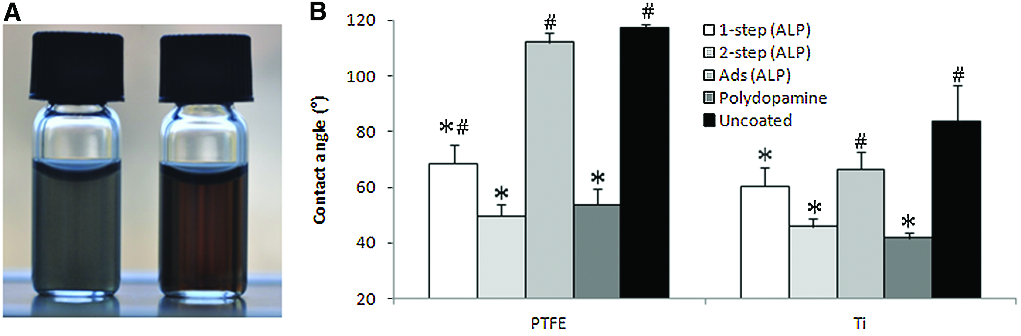
In Figure 1B, static contact angles are depicted. Substrates modified using 1-step (68±7° and 60±6° on PTFE and titanium, respectively), 2-step (50±4° and 46±3° for PTFE and titanium, respectively), and coated with polydopamine only (54±6° and 42±2° for PTFE and titanium, respectively) showed significantly (p<0.05) lower contact angles compared to substrates coated using adsorbed ALP (112±3° and 67±6° for PTFE and titanium, respectively) or uncoated substrates (117±1° and 84±13° for PTFE and titanium, respectively). For PTFE, the contact angles observed for the 1-step ALP immobilization procedure were significantly higher than substrates coated with polydopamine only or the 2-step ALP immobilization procedure.
Absolute quantity of immobilized ALP and BMP-2
The amount of immobilized ALP on titanium (Table 1) demonstrated significantly different values for all three groups (p<0.001), with amounts of 234±3, 534±40, and 1217±77 ng/cm2 for ads, 2-step, and 1-step, respectively. The sequential application of a double layer consisting of two consecutive 1-step immobilizations yielded a total amount of immobilized ALP of 2±0.2 μg/cm2. Absolute quantities of ALP immobilized on PTFE were significantly different (p<0.001) with 306±17, 943±60, and 1650±165 ng/cm2 for the ads, 2-step, and 1-step, respectively.
p<0.05 as compared to the ads group of the corresponding time interval and biomolecule/substrate combination; #p<0.05 as compared to the corresponding 2-step group. Data represent average±standard deviation (n=3 for BMP-2/PTFE; n=4 for the other biomolecule/substrate combinations).
ALP, alkaline phosphatase; PTFE, polytetrafluoroethylene; BMP-2, bone morphogenetic protein-2.
The quantity of BMP-2 adsorbed onto titanium was 17±3 ng/cm2, whereas the polydopamine-based approaches yielded a significantly (p<0.05) lower amount of immobilized BMP-2 (11±2 ng/cm2 and 5±1 ng/cm2, for 2-step and 1-step, respectively). On PTFE, the quantities of BMP-2 were 5.7±0.6, 5.9±0.5, and 0.9±0.1 ng/cm2 for ads, 2-step, and 1-step, respectively.
Retention of immobilized ALP and BMP-2
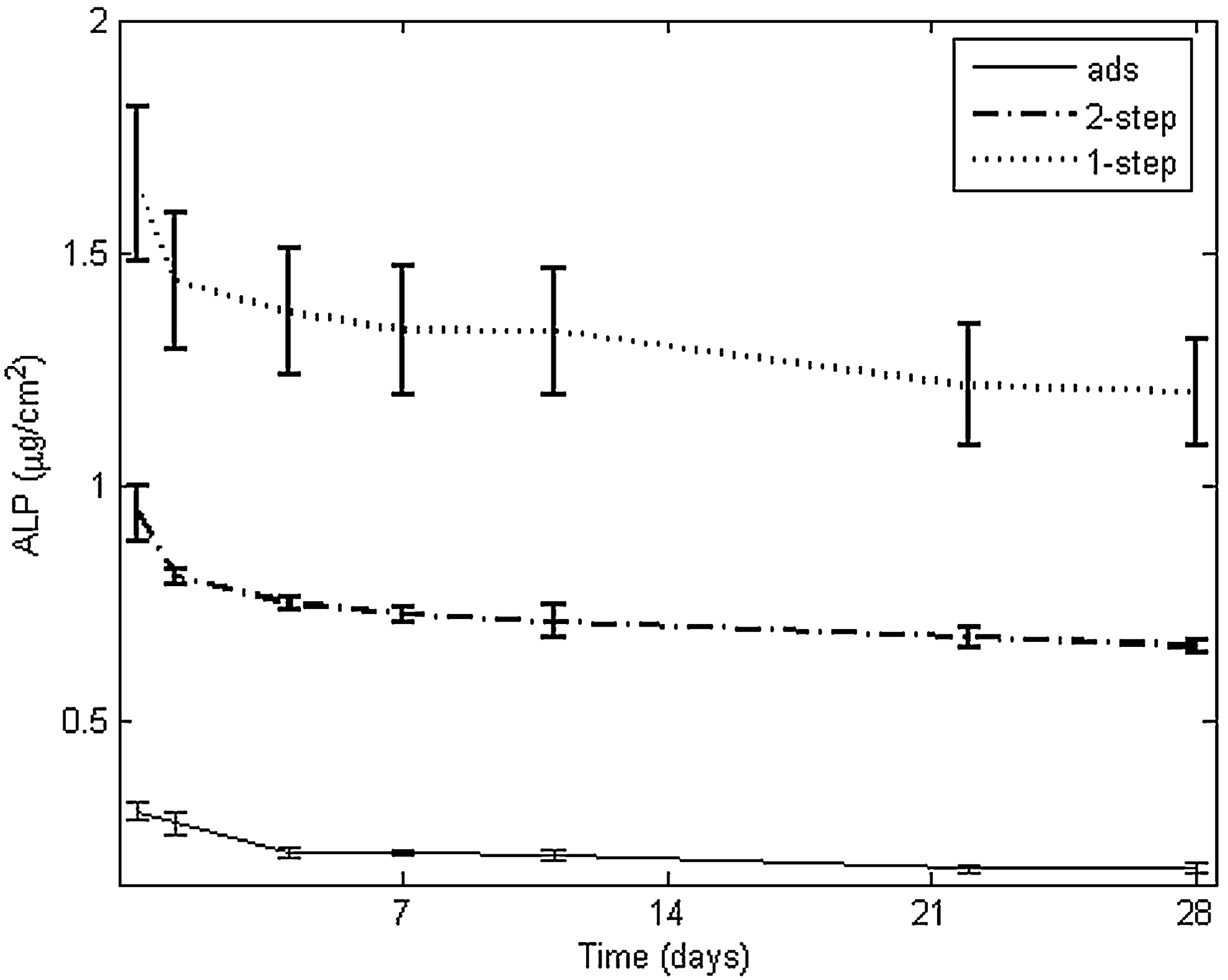
The relative retention and release of ALP and BMP-2 from all experimental substrates are presented in Table 1. The absolute quantities of ALP on titanium are depicted in Figure 2 as a function of immersion time in PBS. ALP immobilization onto titanium showed a burst release (within 1 day) followed by a low sustained release from day 1 to 28, irrespective of immobilization method. The sustained release for ads (1.2%±0.22%/day) was higher (p<0.01) than for 2-step and 1-step (0.7%±0.17% and 0.7%±0.04%/day, respectively). Relative to the initially immobilized amount at day 0, the final retention of ALP on day 28 was 61.2%±6.4% for ads, 67.2%±2.3% for 2-step, and 70.8%±1.0% for 1-step (p<0.05 for 1-step compared to ads).

Retention of ALP at the surface of titanium after soaking in phosphate-buffered saline for different time periods. Data represent average±standard deviation (n=4).
Retention of ALP on PTFE substrates followed a similar trend (Fig. 3) as compared to titanium substrates. After the initial burst release, a sustained release of 1.3%±0.15%, 0.7%±0.09%, and 0.6%±0.05%/day was observed from day 1 to 28 for ads, 2-step, and 1-step, respectively. This sustained release was significantly lower (p<0.001) for 2-step and 1-step than for ads. The final retention after 28 days was 59.0%±5.5% for ads, 70.1%±4.3% for 2-step, and 73.0%±1.3% for 1-step, compared to ads p<0.05 for both 1- and 2-steps.
Retention of ALP at the surface of PTFE after soaking in phosphate-buffered saline for different time periods. Data represent average±standard deviation (n=4).
In Figure 4, the absolute quantities of BMP-2 retained at the surface of titanium after various time points are shown. Compared to 1-step, 2-step and ads revealed a twofold and threefold increase on day 0, respectively. The burst release (29.6%±2.3%) of ads was significantly higher (p<0.001) compared to 2-step and 1-step (2.3%±0.8% and 5.5%±2.0%, respectively). A similar trend was observed for the sustained release (2.4%±0.28%, 0.4%±0.03%, and 0.4%±0.08%/day for ads, 2-step and 1-step, respectively), leading to a significantly (p<0.001) lower retention after 28 days for the ads group (21.8%±7% compared to 87.4%±1.0% and 83.9%±2.0% for 2- and 1-step, respectively). The absolute quantities of BMP-2 after 28 days of immersion were 10±2 and 4±1 ng/cm2 for 2-step and 1-step, respectively, whereas the amount of adsorbed BMP-2 decreased to a value similar to 1-step (4±1 ng/cm2).
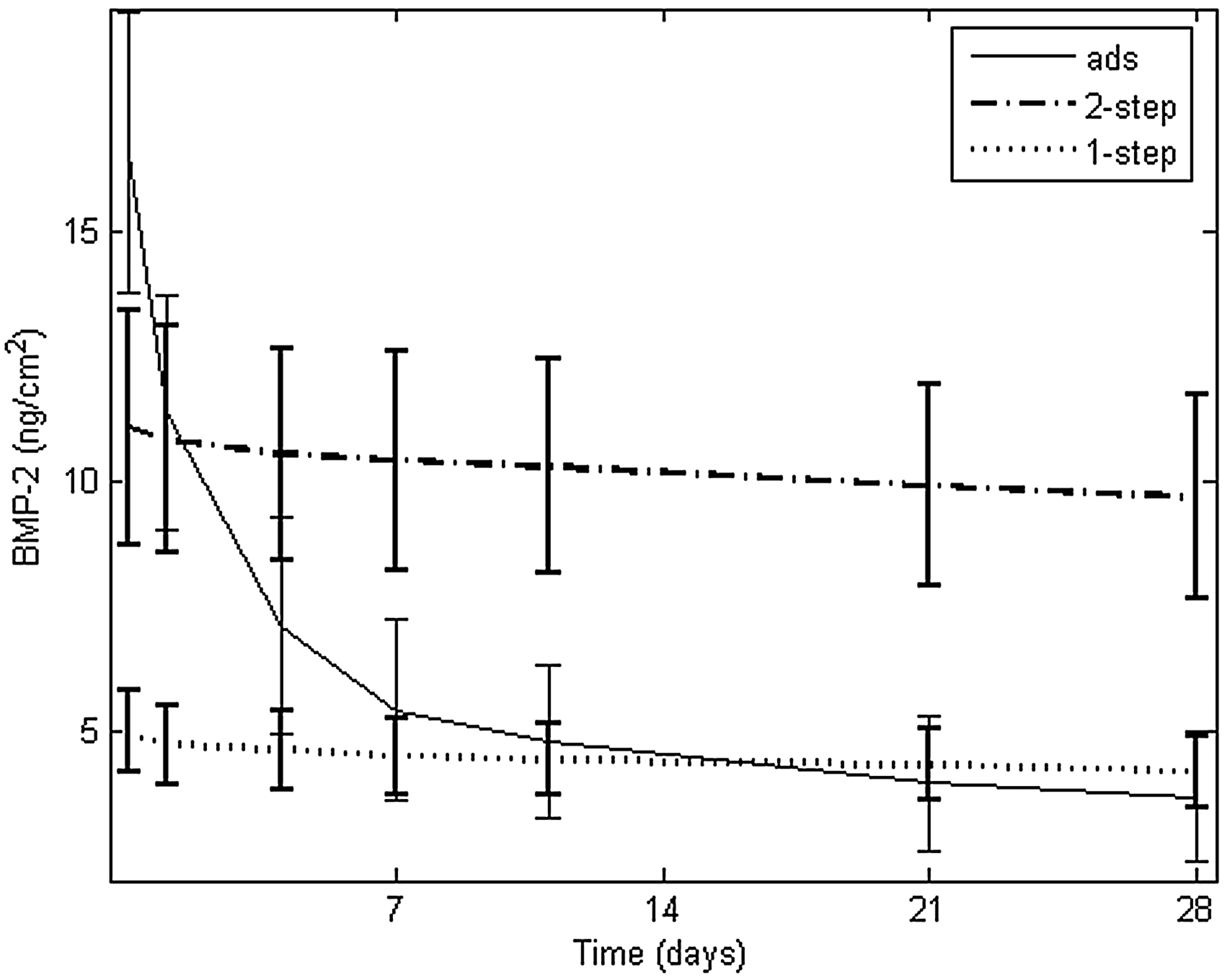
Retention of bone morphogenetic protein-2 (BMP-2) at the surface of titanium after soaking in phosphate-buffered saline for different time periods. Data represent average±standard deviation (n=4).
Biochemical activity of immobilized ALP
Titanium discs coated with ALP were shown to exhibit an enzymatic activity ranging from 17±11 μUnits/cm2 for the discs coated via ads (Fig. 5) to 46±12 and 128±24 μUnits/cm2 for 2-step and 1-step, respectively. The amount immobilized with 1-step was significantly higher (p<0.001) than ads.

RBMC culture
The proliferation of cells on the titanium surfaces modified with ALP or BMP-2 (as expressed by the amount of DNA) is depicted in Figure 6. For all the experimental groups, the DNA amount peaked at day 8, after which it gradually decreased until day 24. Figure 7 shows the ALP activity as a marker for early differentiation of the RBMCs. For all the experimental groups, the ALP activity increased continuously until day 24. On day 4 (1-step and 2-step, p<0.001) and day 8 (2-step, p<0.01), significantly lower ALP activities were found for immobilized BMP-2 compared to the ads group. The ALP activity of ALP-coated substrates without cells seeded on top was also measured after 4 days, and ALP activity could not be measured from this control. As a marker for late osteogenic differentiation, the calcium content is depicted in Figure 8. Calcium content was shown to increase until day 24 for all experimental surfaces. On day 12, the calcium content was significantly lower for 2-step as compared to ads and 1-step (p<0.05) for the ALP coatings. The BMP-2 coatings showed a significantly lower amount of calcium on day 8 (for 2-step compared to ads, p<0.05) and day 24 (for 1-step compared to ads and 2-step, p<0.01).
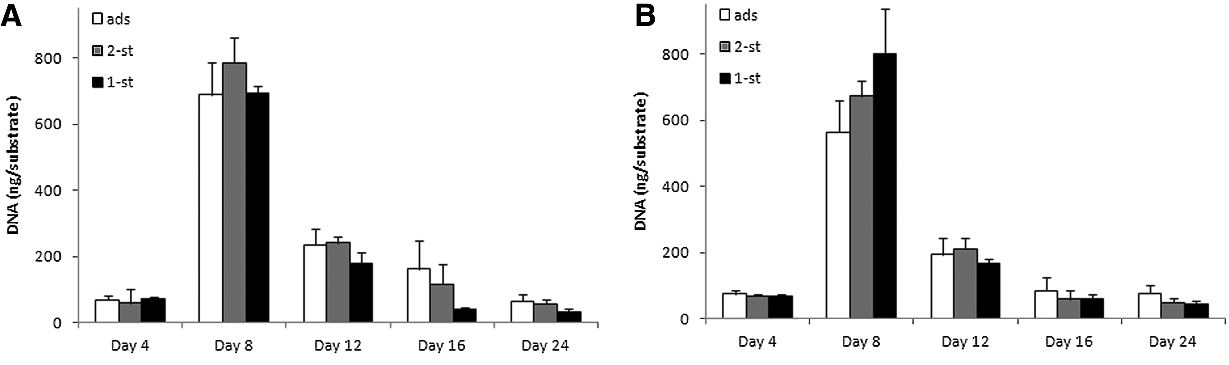
DNA content of rat bone marrow cells after culture on ALP-
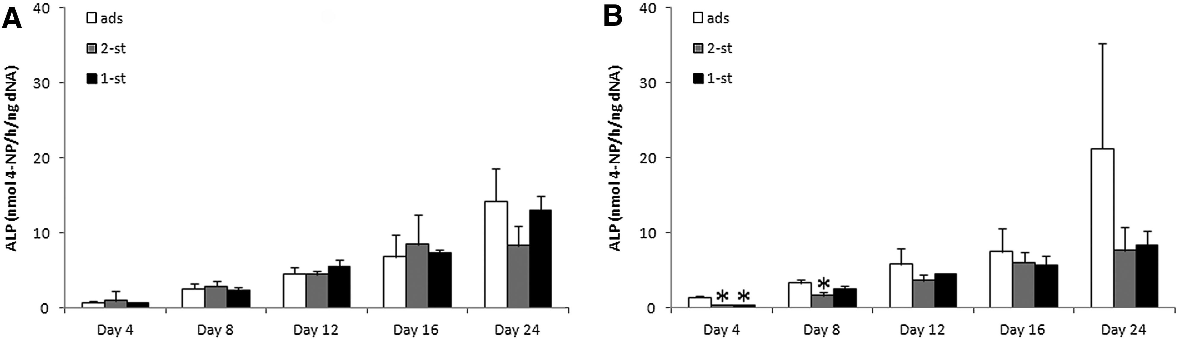
ALP activity of rat bone marrow cells after culture on ALP-
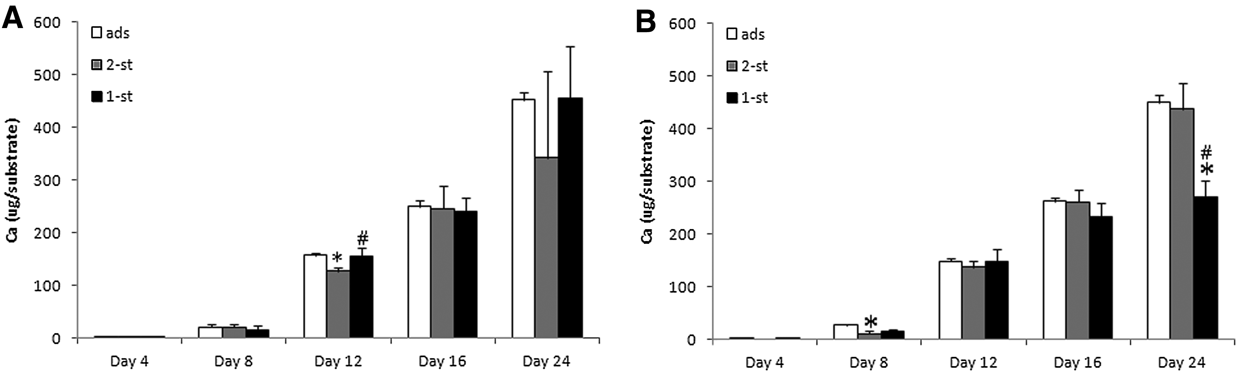
Calcium content of rat bone marrow cells after culture on ALP-
Discussion
The aim of the current study was to immobilize the osteogenic proteins ALP and BMP-2 onto titanium and PTFE surfaces using a 1-step procedure that involves self-polymerization of dopamine in the presence of biomolecules, thereby leading to homogeneous mixing of covalently linked biomolecules throughout a self-polymerized polydopamine layer. We hypothesized that this novel immobilization strategy would lead to a higher quantity of immobilized biomolecules compared to the conventional 2-step immobilization method involving self-polymerization of polydopamine before protein immobilization. The results confirmed that immobilization of ALP was more efficient using the 1-step procedure as reflected by higher absolute immobilized quantities and a higher enzymatic activity of these molecules. Immobilization of BMP-2, however, was more efficient using the conventional polydopamine-based 2-step immobilization, which was attributed to the much lower protein concentrations during immobilization.
Macroscopic observations and contact-angle measurements
Since no solid aggregates were formed when ALP was present, ALP appeared to exhibit an inhibitory effect the self-polymerization of polydopamine, which might be caused by incorporation of (soluble) ALP into the polydopamine aggregates. The low concentration of BMP-2 might explain why BMP-2 did not interfere with the self-polymerization of polydopamine. Further evidence for this statement is the measured contact angles. Wettability of materials is known to have an important influence on cell behavior, such as adhesion.30,31 Generally, contact angles for all polydopamine-treated experimental groups were considerably lower than uncoated surfaces. These data are in line with Lee et al.'s, who observed that regardless of the contact angle on the bare material, contact angles on polydopamine-coated surfaces were around 47°, stressing the universal applicability of the polydopamine coating. 7 Moreover, contact angles on PTFE surfaces coated with ALP using the 1-step process were significantly higher than the values obtained on substrates coated with polydopamine only. Although not statistically significant, a similar trend could be observed for the surface modifications on titanium substrates. This seems to suggest that the presence of ALP in the precursor solutions interfered with the self-polymerization reaction from dopamine to polydopamine as mentioned above, thereby decreasing the efficiency of polydopamine coating and increasing the contact angle.
Absolute quantity of immobilized ALP and BMP-2
Experiments with radiolabeled ALP (Figs. 2 and 3; Table 1) revealed that the quantity of immobilized ALP can be significantly enhanced by using polydopamine-based immobilization strategies. Compared to ads onto titanium, the 2-step polydopamine-based immobilization method increased the amount of ALP present at the surface threefold. Interestingly, the 1-step method increased the amount of immobilized ALP even sixfold. These data confirm that the 1-step immobilization method represents a superior alternative to the conventional 2-step immobilization method in terms of both simplicity and efficiency. Further, when the 1-step immobilization method was used repetitively for 2 times on the same substrate, the quantity of ALP immobilized at the surface was 10 times higher than via ads, which indicates that the amount of ALP can be controlled by applying multiple repetitive immobilizations. Lee et al. showed that the thickness evolution of polydopamine coatings on silicon (Si), measured by AFM of patterned surfaces, reached a plateau value of 50 nm after coating for 24 h. 7 Repetitive coatings can probably increase this thickness. Regarding ALP immobilization onto hydrophobic PTFE substrates, it had already been shown previously that ALP can significantly adsorb onto PTFE. 32 The current study, using radiolabeled ALP (Fig. 2), confirmed that considerable amounts (0.3±0.02 μg/cm2) of ALP adsorb onto PTFE. Similar to conjugation onto titanium, 1-step demonstrated higher immobilization efficiency compared to ads and 2-step. For BMP-2, ads yielded higher initial amounts of immobilization than both polydopamine-based immobilization methods. These results are not in line with previous studies where BMP-2 was covalently immobilized. From a conceptual point of view, covalent linkage of BMP-2 was shown to increase immobilization efficiency and decrease burst release compared to simple adsorption.6,33–36 In these studies, BMP-2 was immobilized from solutions containing >20 μg/mL as compared to 4 μg/mL in the current study. This low concentration might account for the low immobilization efficiency using the 1- or 2-step immobilization procedure. Further, it was observed that more BMP-2 was present at the surface upon 2-step immobilization versus 1-step immobilization, which contrasts the immobilization data for ALP. To summarize these observations, the 1-step process was found to be most efficient for immobilization of ALP (at a relatively high protein concentration), whereas the 2-step procedure was more efficient for immobilization of BMP-2 (at a low protein concentration). Although more sound evidence is needed to propose a solid explanation for this phenomenon, we speculate that the 1-step procedure can immobilize biomolecules by physical entrapment within the polydopamine film, whereas the 2-step procedure has a much smaller capacity for binding biomolecules due to a limited amount of surface-reactive groups (such as quinone moieties) that can bind biomolecules to the exposed surface of the intermediate polydopamine layer. As a consequence, the observed results indeed seem to indicate that the 2-step method could be more efficient for immobilization of biomolecules at low concentrations, whereas the 1-step method could be more effective for immobilization of biomolecules onto implant surfaces from more concentrated precursor solutions.
Retention of immobilized ALP and BMP-2
At all time points, the absolute retention of immobilized ALP for both titanium and PTFE substrates increased in the following order: 1-step>2-step>ads. The release data confirm that retention of ALP is substantially higher when polydopamine-based immobilization methods are used. This is evidenced by both a lower sustained release from day 1 to 28 as well as a higher final retention after 28 days. For BMP-2 on titanium, the differences in retention between plain adsorption and polydopamine-based immobilization were even more striking with a ∼3.5 higher retention for polydopamine-based immobilization. The fast desorption of BMP-2 versus relatively slow desorption of ALP might be related to difference in isoelectric point of both proteins, which are of ∼4 and ∼8.5 for ALP and BMP-2, respectively. 37 As a consequence, ALP was negatively charged at the current experimental conditions (pH of 8.5), whereas BMP-2 did not exhibit a net charge, thereby reducing the amount of electrostatic interactions with substrate materials considerably.
Biochemical activity of immobilized ALP
It was shown that the immobilized ALP was biologically active for all immobilization approaches for both titanium and PTFE substrates. The data show that the biochemical activity of ALP-coated substrates was three times higher for the conventional 2-step immobilization compared to ads and six times higher for the 1-step immobilization method. In Figure 5B, the ratio between activity (as measured using biochemical activity assay) and quantity (as measured using scintigraphy) of ALP was shown for the three different immobilization approaches. No significant differences were found between these ratios, which indicate that the loss of enzymatic activity upon 1- or 2-step immobilization of ALP was negligible.
RBMC culture
A normal cell growth with increased ALP activity and calcium deposition was observed for RBMCs on all experimental substrates, which indicates that the ALP and BMP-2 coatings do not inhibit the osteogenic behavior of the cells. No ALP activity was measured after 4 days from the control samples (substrates only coated with ALP, but cells were omitted), which confirms that all releasable ALP already detached, whereas the remaining ALP was not released after the freeze–thawing cycles. Hence, the measured ALP activity could be solely attributed to ALP as produced by the cells. Basically, the different immobilization strategies (ads, 2-step, and 1-step) did not lead to significant differences in cell responses, which seem to indicate that the amounts of immobilized biomolecules were not sufficient to exert biological effects in vitro under the current experimental conditions. Ongoing research in our group focuses on increasing the biological efficacy of these coatings by improving the amounts of polydopamine-assisted immobilization of therapeutic proteins.
Conclusions
In the current study, a 1-step method to immobilize biomolecules onto implant surfaces using polydopamine was characterized by studying the immobilization of ALP as main model protein onto titanium and PTFE substrates. In contrast to conventional 2-step polydopamine-based immobilization strategies (which are characterized by the formation of a polydopamine layer before biomolecule immobilization on top of the intermediate polydopamine layer), the 1-step immobilization method involves simultaneous formation of a self-polymerized polydopamine layer and covalent entrapment of biomolecules throughout the polydopamine layer. While the conventional 2-step polydopamine-based immobilization resulted into a threefold increase of ALP immobilization compared to ads, the novel and simplified 1-step approach increased the amount of ALP immobilization twofold compared to the conventional 2-step polydopamine-based immobilization method without compromising the biological activity of the enzyme. Regarding immobilization of BMP-2, it was observed that the conventional 2-step method was more efficient than the 1-step immobilization procedures possibly due to the much lower biomolecule concentrations in the soaking solutions used to immobilize BMP-2. Still, polydopamine-based immobilization of BMP-2 resulted into strongly improved retention of immobilized BMP-2. Biological evaluation studies using in vitro cultured RBMCs revealed that cell responses to the various experimental coatings were not statistically different.
Footnotes
Acknowledgments
This study was supported by The Royal Netherlands Academy of Arts and Sciences (KNAW), Programme Strategic Scientific Alliances (PSA), grant no. 08-PSAM-02. Annemarie Eek, from the department of Nuclear Medicine, Radboud University, Nijmegen, is gratefully acknowledged for her help with the radiolabeling of biomolecules. C.C.M. Carcouet, from the Laboratory of Materials and Interface Chemistry, Eindhoven University of Technology, The Netherlands, is acknowledged for technical assistance with the contact-angle measurements.
Disclosure Statement
No competing financial interests exist.