
Abstract
Human adipose-derived stromal/stem cells (ASCs) are an abundant, readily available population of adult stem cells that reside in adipose tissue and that have a great potential utility for tissue engineering and regenerative medicine therapeutic applications. Several preclinical studies have shown that ASCs have therapeutic applicability, but a standardized isolation and expansion methodology for clinical cell therapy has yet to be established. ASC are typically isolated and expanded using reagents with xenogenic components and this may pose certain risks and safety issues, such as exposure to infectious agents and immune reactions, creating further obstacles to the translation of ASC-based therapies to clinical scenarios. The objective of this study was to determine the suitability and efficacy of various alternative enzymatic products, CLS1 (Worthington), CLSAFA (Worthington), NB4 (SERVA), and Liberase (Roche), for the digestion of adipose tissue and subsequent isolation of ASCs, assessing cell functionality concerning their proliferation and differentiation ability. Results show that there are no statistically significant differences on yield and proliferation of cells isolated after enzymatic digestion with any of the studied products. The differentiation potential of the cells was not affected, and cell surface marker expression was similar among all products. We concluded that clinical grade products can replace current research-grade products effectively in our cell isolation protocols without any negative effect in the yield or function of human ASCs.
Introduction
Different stem cell therapy studies using ASCs are under way,5,6 which calls for a strong focus on the safety, reproducibility, and quality of transplanted in vitro expanded stem cells. By replacing collagenase and other animal-derived products with animal protein-free reagents, the safety and quality of the transplanted stem cells may be enhanced significantly.7–9 Tissue dissociation enzymes are critical reagents that affect the yield and quality of isolated cells required for cell-based research and clinical transplantation. Most published ASC isolation protocols employ collagenase, yet contradictory results regarding its use have been reported.10–12 For some collagenase products, the enzymatic composition shows lot-to-lot variability that may affect reproducibility.13–15 The current study set out to compare the effect of different xenofree enzymatic alternatives on human ASC with respect to cell proliferation frequency, morphology, multilineage differentiation capacity,16,17 and surface marker expression profiles. To our knowledge, this is among the first comprehensive comparisons on ASCs processed using different collagenase products, and can be a helpful tool for the optimization and standardization of ASC-based research among different research centers.
Material and Methods
Donor demographics
Adipose tissue specimens were obtained with informed consent from women undergoing elective liposuction (n=5), under a previously established protocol with Hospital da Prelada, Porto, Portugal. The age range was 33–57 years (mean 42.6±9.4) and body–mass index (weight in kg/height in m2) 21.9–36.6 (mean 29.2±6.7).
The enzymatic products that were used in this study were the following:
Collagenase, Type 1 (CLS1): This is a crude collagenase product that contains average amounts of assayed activities (collagenase, caseinase, clostripain, and tryptic activities). Being a research-grade product, it may contain animal-based components (Worthington Biochemical Corporation; Cat. No. LS004194)
Collagenase (Animal Origin Free)-A (AFA): This product is prepared in cultures grown in a medium completely devoid of animal-based components and designed for bioprocessing applications where introduction of animal-derived pathogens must be prevented (Worthington Biochemical Corporation; Cat. No. LS004152).
Collagenase NB 4 Standard Grade (NB4): Purified blend of a balanced ratio of crude collagenase and other proteases. NB 4 Standard Grade is used in routine research protocols and is usually used to estimate efficacy of clinical-grade good manufacturing process (GMP) products that have similar enzymatic composition and properties, but are usually more expensive, such as Collagenase NB 6 GMP Grade (SERVA Electrophoresis GmbH; NB 4 Cat. No. 17454; NB 6 Cat. No. 17458).
Liberase TM (Liberase): Xenofree white lyophilisates consisting of aseptically filled, highly purified collagenase and neutral protease enzymes (isoforms I and II), and a small quantity of buffer salts (Roche Applied Science; Cat. No. 05401119001).
All samples of the enzymatic products were kindly provided by Worthington, SERVA, and Roche.
ASCs isolation and culture
ASCs were isolated from human subcutaneous adipose lipoaspirate according to the published methods with some minor modifications. 18 Lipoaspirate tissues were harvested by tumescent liposuction without ultrasonography assistance from the abdominal region of subjects undergoing elective plastic surgery at the Hospital of Prelada (Porto, Portugal). All protocols were conducted with informed patient consent under a protocol approved and supervised by the Hospital Ethics Committee.
The tissue samples were transferred to the research laboratory within a closed plastic container maintained at room temperature in phosphate-buffered saline (PBS) with 10% antibiotic/antimycotic and processed within 4 h after the surgical procedure that has been demonstrated to be within the optimal window of time. 19 The lipoaspirate tissue was extensively washed with PBS at 37°C to remove erythrocytes and then digested in PBS supplemented with the enzymatic product. The same volume of each collagenase was prepared to a final concentration of 0.2 Wünsch units (or 200 CDU) per milliliter of solution, according to the manufacturer's indications. Solutions of the enzymatic products selected for this study (described below) were used to digest equal volumes of lipoaspirate for 1 h at 37°C with gentle agitation. After room temperature centrifugation at 300 g for 5 min and resuspension in a stromal medium (DMEM/Hams F-12 medium [Hyclone] supplemented with 10% FBS [Gibco, Invitrogen] and 1% antibiotic/antimycotic), the stromal vascular fraction (SVF) cell pellet was plated at a density equivalent to 35 mL of lipoaspirate digest per T-175 flask (0.2 mL/cm2). In parallel, 10 mL of SVF was filtered (40 μm), and total nucleated cell counts were determined using a hematocytometer; all cell counts were performed uniformly by a single individual to avoid any variability in this parameter (P.P.C.).
After 24 h of incubation at 37°C, 5% CO2, the adherent cells were washed with PBS at 37°C and maintained in a stromal medium until 80%–90% confluence. This procedure was repeated for a total of five donors. Since the objective of this study was to evaluate cell recovery and behavior as a function of enzymatic digestion with each product, each tissue served as its own control, reducing the influence of the donor-to-donor variability.
Flow cytometry
After the isolation procedure and subsequent 7 days in culture (confluence ∼90%), cells were harvested using TrypLE Express without phenol red (Invitrogen), 20 washed with PBS three times, and aliquots of 150×103 cells were incubated with monoclonal antibodies directed against cluster-of-differentiation (CD)29 (eBioscience Cat. No. 14029982), CD 31 (Citomed Red Cat. No. FAB3567A), CD34 (BD Biosciences Cat. No. 555822), CD44 (BD Cat. No. 555479), CD45 (BD Cat. No. 555482), CD49f (eBioscience Cat. No. 17-0495-82), CD73 (BD Biosciences Cat. No. 550257), CD90 (BD Biosciences Cat. No. 559869), CD105 (eBioscience Cat. No. 16-1057-82), and CD146 (eBioscience Cat. No. 11-1469-42) for 20 min in the dark before being washed with PBS supplemented with 1% bovine serum albumin three times and fixed in 1% formaldehyde overnight at 4°C.21–23 For each sample, 20×103 events were collected on a Becton Dickinson FACScalibur flow cytometer using CELLQuest acquisition software (Becton Dickinson) and analyzed using CELLQuest software (Becton Dickinson). This antibody panel was selected, in part, based on the International Society for Cell Therapy (ISCT) position article on the criteria for defining mesenchymal stem cells/multipotent stromal cells. 24
Differentiation potential
Adipogenic differentiation
Confluent cultures of ASCs (P1) were induced with an adipogenic differentiation medium (DMEM/Hams F-12, 3% FBS, 1% antibiotic/antimycotic, 0.5 mM isobutylmethylxanthine, 33 mM biotin, 17 mM pantothenate, 5 μM troglitazone [Sigma], 1 mM dexamethasone, and 10 mM insulin) for 3 days before being converted to an adipocyte maintenance medium (identical to the adipogenic differentiation medium without isobutylmethylxanthine and troglitazone).18,25 Cells were maintained for 9 days before fixation and Oil-red-O staining.
Osteogenic differentiation
Other confluent cultures of ASCs were converted to an osteogenic medium (DMEM/Hams F-12 or DMEM, 10% FBS, 1% antibiotic/antimycotic, 10 mM β-glycerophosphate, 50 μg/mL sodium 2-phosphate ascorbate, and 10−8 M dexamethasone) and maintained in culture for 12 days with medium changes every third day. These cultures were rinsed three times with 150 mM NaCl, fixed in 70% ethanol, and stained with alizarin red. 26
Chondrogenic differentiation
For chondrogenic differentiation, 5×10⁵ ASCs (P1) were resuspended in a chondrogenic medium and centrifuged at 800 G for 5 min, to obtain cell pellets. Pellets were cultured in the same medium for 21 days at 37°C and 5 % CO2, in 15-mL falcon tubes, with medium changes twice a week. The chondrogenic medium was composed of DMEM high-glucose, sodium bicarbonate, 1% antibiotic/antimycotic, 0.17 mM ascorbic acid, 10 ng/mL human transforming growth factor-beta 3, 10% insulin–transferrin–selenium, 35 mM
Statistical methods
Values are presented as the mean±standard deviation. Data were analyzed for normality using the Shapiro–Wilk test. Results were analyzed by, and Bonferroni post-test evaluated significance.
Results
Cell yield and viability
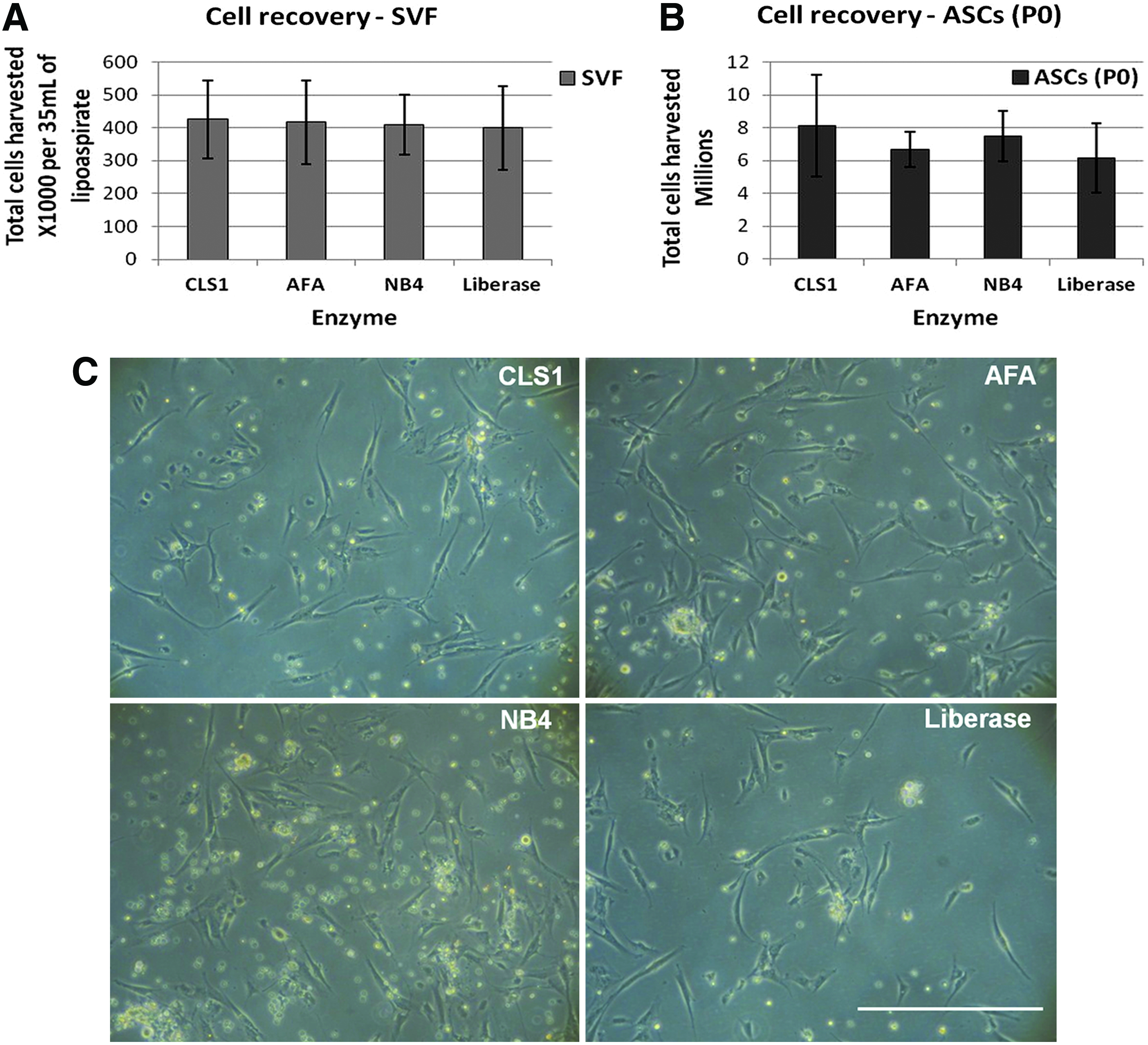
Our first assessment compared the yield and viability of total nucleated cells recovered from the SVF and human ASC cultured for 7 days, until reaching 80%–90% confluence, after enzymatic digestion of lipoaspirate samples with each collagenase product (total of five donors). The number of recovered cells and their viability were not significantly different among any of the products (Fig. 1A, B). Concurrently, the cell morphology was observed by light microscopic examination (Fig. 1C). No morphologic differences were found in cells processed with different collagenase products throughout the entire culturing time.
Immunophenotype
Next, studies evaluated the immunophenotype of ASCs cultured after being processed with each of the collagenase products. A subset of well-characterized human ASC cell surface markers (Fig. 2) was examined by flow cytometry.21,23,27 Consistent with previously published results, 23 the human ASCs displayed strong positivity for adhesion markers CD29 (β1 integrin), CD105 (endoglin), CD73 (5′ecto-nucleotidase), and the extracellular matrix protein CD90 (Thy-1). More important, we could not find any significant difference in the expression of any of the analyzed markers among cultured ASCs independent of the collagenase products used for the initial tissue digestion.
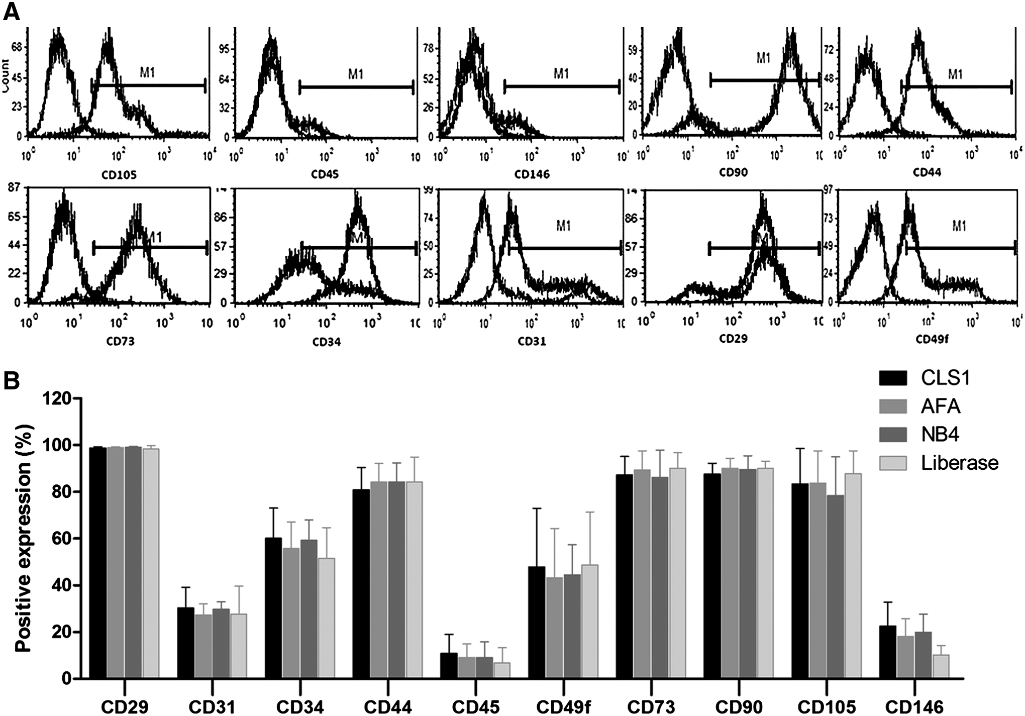
Immunophenotype by flow cytometry analysis. Top panel
Differentiation potential
We evaluated the differentiation potential of ASC isolated by tissue digestion with each collagenase product, by staining with Oil Red O (adipogenic), Alizarin Red (osteogenic), Safranin O, and Alcian Blue/Neutral Red (chondrogenic). None of the enzymatic products used had any effect on the ability of ASC to differentiate along the adipocyte, osteoblast, or chondroblast lineage pathways (Fig. 3). All observations, both microscopic and macroscopic, were conducted by the same person (PPC) to minimize the variability in the analysis.
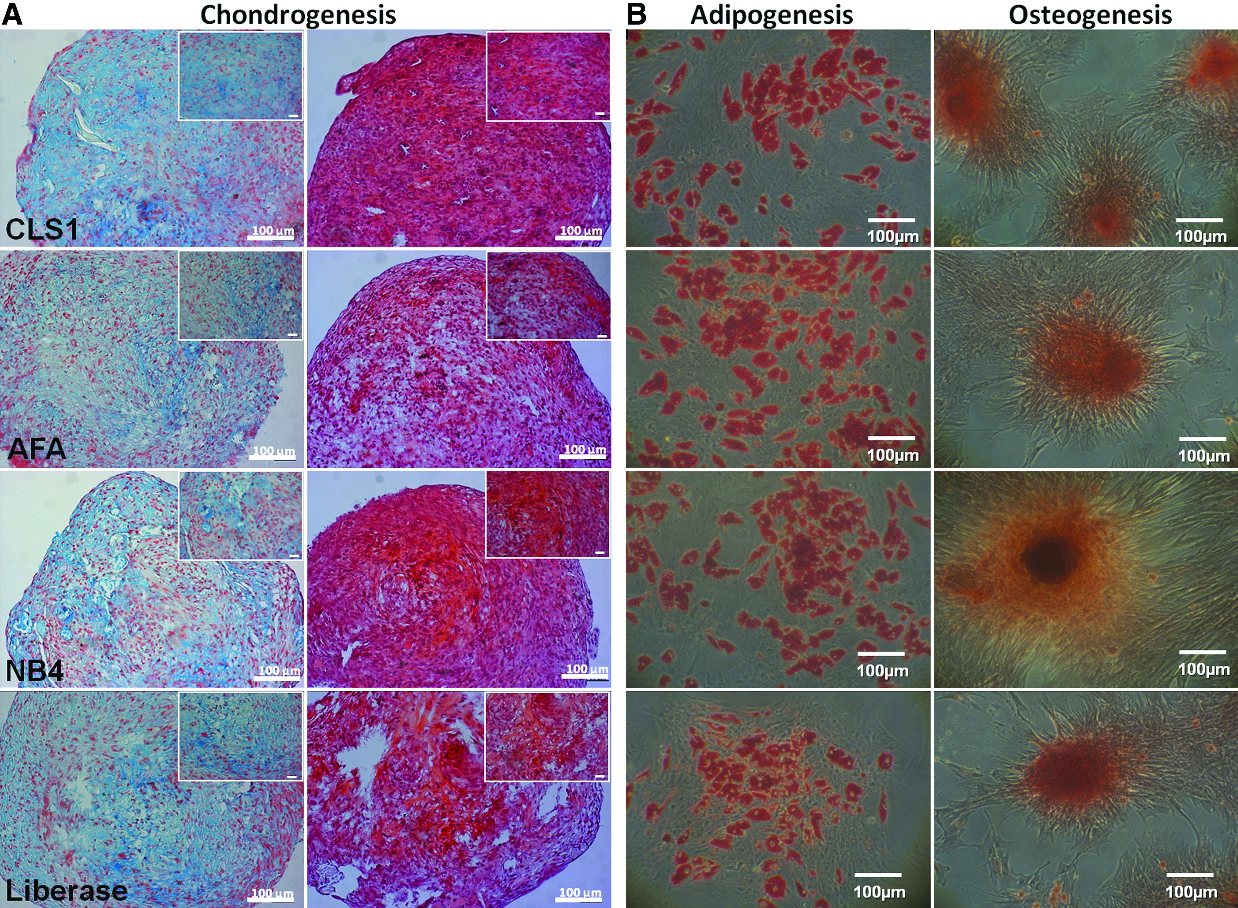
Light microscopy photographs of ASCs induced to differentiate into different lineages.
Discussion and Conclusion
To use adult stromal/stem cells in human subjects for regenerative medicine therapies, we need to take into consideration several criteria, namely the safety, efficacy, reproducibility, and quality control. Consequently, clinical cell-based therapies that employ xenogenic reagents may prove to be an unsuitable option with respect to patient safety and represent a high risk of infection and severe immune reactions in the patient.
The results obtained from the present study indicate that human lipoaspirate tissue samples can be successfully processed using different enzymatic products, without substantial differences in the yield of both SVF cells and ASCs. The total number of recovered cells and their viability were not significantly different among the different products used to digest adipose tissue samples. Although a culture medium containing FBS was used to conduct this study, previous work 28 reported comparable results when using serum-free and xenofree culturing conditions. Therefore, we believe that the FBS-containing medium had no effect on ASC behavior after the isolation process. Furthermore, the assessment of the total nucleated cells present in the SVF established the efficacy of the enzymatic products used to digest lipoaspirate samples.
Realizing that any of the products was equally effective retrieving ASCs from lipoaspirate samples, we needed to evaluate if cells would be altered by any of these products and the enzymatic digestion process itself. Flow cytometry assessed the immunophenotype of ASC obtained with the different enzymatic products, through a panel of cell surface markers. The human ASC displayed strong positivity for the stromal markers CD29 (β1 integrin), CD73 (5′ ectonucleotidase), CD90 (Thy-1), and CD105 (endoglin) when processed with any of the enzymatic products. Moderate expression of CD 44, CD49f, and the hematopoietic progenitor cell marker CD34 was found in all ASCs. Consistently with published reports, 23 low expression of CD31, CD 45, and CD146 was found. The results were consistently similar among all products.
Further experiments were carried out for evaluation of the differentiation potential of ASCs obtained upon tissue digestion with each product being assessed, by staining with Oil Red O (adipogenic), Alizarin Red (osteogenic), and Alcian Blue/Neutral Red and Safranin O (chondrogenic). The product used had no effect on the ability of ASCs to differentiate along any of the different lineage pathways analyzed.
Extensive characterization of stem cells is essential before their clinical use, as different culture conditions may change the characteristics of the cells. The results obtained under the present study show that ASCs isolated after enzymatic digestion using different collagenase products display similar phenotypes based on their differentiation ability and surface marker expression, in conformity with previously published reports.21,29
These results show that highly purified and nonanimal sources of collagenase suitable for Current Good Manufacturing Process (cGMP) perform as effectively as our routine laboratory research-grade products. Thus, these clinical-grade reagents can be substituted into existing protocols without loss of yield or function of human ASCs during isolation from adipose tissue samples. Furthermore, a more intensive production of these products due to their generalized use would provide more suitable cost-effective alternatives for stem cell research laboratories. To use adherent human stromal/stem cells clinically, it will be advantageous to eliminate all potential sources of xenoprotein contamination. These studies will have relevance for optimizing cGMP methods as the use of human ASC in tissue engineering and regenerative medicine grows.
Footnotes
Acknowledgments
P.P.C. acknowledges the Portuguese Foundation for Science and Technology (FCT) for a Ph.D. scholarship (SFRH/BD/44128/2008) and financial support of the project MIT/ECE/0047/2009. The authors thank the following individuals for their assistance: Ana Frias for her help with the flow cytometry; Alessandra Zonari for her help preparing histological samples. The authors would also like to thank Worthington Biochemical Corp, USA; SERVA Electrophoresis GmbH, Germany; and Roche Applied Sciences, Portugal, for kindly providing samples of their products for this study.
Disclosure Statement
No competing financial interests exist.