
Abstract
Efficient transport of stem/progenitor cells without affecting their survival and function is a key factor in any practical cell-based therapy. However, the current approach using liquid nitrogen for the transfer of stem cells requires a short delivery time window is technically challenging and financially expensive. The present study aims to use semipermeable alginate hydrogels (crosslinked by strontium) to encapsulate, store, and release stem cells, to replace the conventional cryopreservation method for the transport of therapeutic cells within world-wide distribution time frame. Human mesenchymal stem cell (hMSC) and mouse embryonic stem cells (mESCs) were successfully stored inside alginate hydrogels for 5 days under ambient conditions in an air-tight environment (sealed cryovial). Cell viability, of the cells extracted from alginate gel, gave 74% (mESC) and 80% (hMSC) survival rates, which compared favorably to cryopreservation. More importantly, the subsequent proliferation rate and detection of common stem cell markers (both in mRNA and protein level) from hMSCs and mESCs retrieved from alginate hydrogels were also comparable to (if not better than) results gained following cryopreservation. In conclusion, this new and simple application of alginate hydrogel encapsulation may offer a cheap and robust alternative to cryopreservation for the transport and storage of stem cells for both clinical and research purposes.
Introduction
Mesenchymal stem cells (MSCs) are multipotent, with the potential to differentiate into bone, cartilage, and muscle-like cells when cultured under defined conditions. 10 Examples of their clinical application include cardiac repair 4 and improving the engraftment of hematopoietic stem cells. 11 Embryonic stem cells (ESCs) are highly proliferative and retain pluripotency after extended periods of in vitro expansion. 12 As ESCs can give rise to any adult cell type, they present great potential for therapeutic use. Certainly, ESCs have been successfully used to treat severe degenerative diseases, including Parkinson's disease.13,14 ESCs are also powerful tools for the study of disease. Several congenital syndromes have been extensively studied through genetic modification of human ESCs (hESCs),15,16 such investigations of disease-specific hESCs may indicate appropriate treatment modalities. Therefore, ESCs and MSCs hold tremendous promise, not only as tools for understanding disease, but also as a basis for cell therapy. 17
Current approaches for stem cell therapy generally require their direct transplantation. 18 These approaches require some basic assumptions, including that stem cells must be delivered both alive and functional and in sufficient number to generate the required therapeutic response. 19 Improved tools for the preservation of these cells, prior to use, have the potential to greatly increase frequency and ease of use while providing predictable results. For example, correct storage allows the generation of quality-controlled stock of cells, cell transport between laboratory and hospital facilities, and removes the need for expensive and time-consuming continuous culture.20,21 However, strategies for stem cell storage that will overcome the time lag between their preparation at laboratories or good manufacturing practice (GMP) compliant facilities and their implantation at hospital units are required. Stem cells in suspension do not survive for extended time intervals during transfer under ambient condition22,23 requirements for cell storage and transportation within appropriate delivery time windows (typically, global delivery between different continents requires 3–5 days) and at a reasonable financial cost. Furthermore, cryopreservation of stem cells was previously demonstrated as unreliable due to the detrimental effects of dimethyl sulfoxide. 24 Therefore, specialized conditions for the preservation of transported stem cells are required. A hydrogel that is chemically inert, structurally uniform, and biocompatible, may be applicable to a simple and economical storage method, which subjects stem cells to minimal manipulation and preserves their viability and phenotype.
Cell encapsulation within semipermeable alginate hydrogels is well established in the fields of tissue engineering and regenerative medicine.25–31 The success of cell encapsulation using alginate is due to its biocompatibility and rapid gelation in the presence of live cells. The mechanical properties of alginate hydrogels are largely dependent on the ratio of component polysaccharides (1,4-linked β-D-mannuronic acid and α-L-guluronic acid residues) and the cationic cross linker used for gelation. 32 Although calcium is the conventional crosslinking ion for alginate gels, other cations, including strontium and barium, are suitable.33,34 Strontium alginate microbeads were found to be more chemically and physically stable than calcium alginate beads.34,35 Other approaches for controlling the mechanical properties of alginate gels in the presence of live cells, involve modification of the internal porosity of these gels. Recently, we discovered that the addition of hydroxyethyl cellulose to alginate, results in gels with a controllable pore size, which correlate with the improved viability of cells immobilized within this scaffold. 31
The present study aims to demonstrate that human and animal stem cells, encapsulated within alginate gels, can be stored successfully for 5 days under ambient conditions in an air-tight environment. Our methodology also aims to develop tools that permit the easy retrieval of cells, and maintain their viability and phenotype. We describe a versatile and practical technology for the transport of stem cells, which will benefit both clinical and research purposes.
Materials and Methods
Culture of mouse ESCs
Mouse ESCs (mESCs) [CGR8, Health protection agency culture collections (ECACC), United Kingdom] were cultured in the Dulbecco's modified Eagle's medium (DMEM) (Stemcell Technologies), supplemented with 10% fetal bovine serum (FBS) (Stemcell Technologies), 0.2% 2-mercaptoethanol (Life Technologies), 1% nonessential amino acid (Stemcell Technologies), 10 ng/mL leukemia inhibitor factor (Stemcell Technologies), 100 IU/mL penicillin, and 100 mg/mL streptomycin (Invitrogen), in 0.1% gelatin (Life Technologies)-coated T75 flasks (Greiner CellStar
Culture of human MSCs
Human mesenchymal stem cells (hMSCs) (Tulane Medical Center) were cultured in the low-glucose DMEM (Life Technologies), supplemented with 10% FBS (Life Technologies), 100 IU/mL penicillin, and 100 mg/mL streptomycin (Life Technologies), at 37°C under 5% CO2 and 95% humidity. Cells were replenished with a fresh medium every 3 days and grown to 70%–80% confluence.
Passaging cells
Passaging both hMSCs and mESCs were carried out using the DMEM (Life Technologies). When cells reached 70%–80% of confluence, 10 mL phosphate-buffered saline (PBS) was added and followed by 5 mL 0.5% trypsin (Life Technologies). Detached cells were then centrifuged at 300 g for 5 min and seeded into new tissue culture flasks containing 20 mL of fresh DMEM; the medium was changed every 3 days.
Preparing sodium alginate solutions
Sodium alginate (Sigma Aldrich) powder was sterilized by UV-light (30W, 250 nm) treatment for at least 30 min. 0.048g of sodium alginate was suspended in the 10 mL DMEM (Life Technologies). These 4.8% (w/v) sodium alginate solutions were kept in 4°C as stock solutions.
Encapsulation of mesenchymal and embryonic cells in strontium alginate gel discs
Mixing suspended cells and sodium alginate stock solution
The cell encapsulation method was as previously reported, 31 with some modifications. About 360 μL of 3×10⁵ (viable cells) of either MSCs or ESCs in the low-glucose DMEM was mixed with 120 μL of the 4.8% (w/v) sodium alginate solution, the final concentration of sodium alginate solution was 1.2%, before gelling into discs using 102 mM SrCl2.
Alginate disc-shaped hydrogel formation
Gel discs were formed by pipetting 480 μL sodium alginate cell solutions into circular molds made from chromatography paper (Whatman) saturated in 10 mL 102 mM SrCl2. The paper molds were prepared thus a paper ring with a 3 cm diameter and 1 cm annulus was placed over a 3-cm-diameter paper disc. A nylon mesh square with dimensions of 1.5 cm×1.5 cm was placed in the center of the paper ring (to avoid breakup of gel during subsequent storage). The paper mold and nylon mesh were saturated with 102 mM SrCl2 before alginate alone containing hMSCs or mESCs, respectively, was pipetted into the space within the ring. A second 3-cm-diameter paper disc saturated with 102 mM SrCl2 was placed over the alginate/paper assembly. The formed/gelled alginate discs containing cells were removed from the paper mold and further exposed (1 min) to an excess of 102 mM SrCl2 for 5 min to complete gelation of a disc 2 cm in diameter and 2 mm in thickness.
Alginate hydrogel storage
The subsequently formed strontium crosslinked alginate discs containing cells were suspended in the 1.5 mL supplemented DMEM medium within a parafilm-sealed, capped cryovial. The gels were then stored at room temperature (18°C–22°C, atmospheric CO2) avoiding direct sunlight for 5 days without further interference (number of independent experiments for both mESCs and hMSCs was >9).
Cryopreservation of mESC and hMSC
Viable mESCs and hMSCs were centrifuged at 1500 rpm for 5 min and then resuspended in a freezing medium (50% supplemented DMEM, 40% fresh DMEM, and 10% dimethyl sulfoxide) (Fisher Scientific). mESCs and hMSCs were aliquoted equally into cryogenic storage vials (Fisher Scientific), with a final concentration of 3×10⁵ cells per vial (n>6). Cryogenic vials were transferred into Mr. Frosty (Fisher Scientific) containing of 100 mL of Isopropyl alcohol (Fisher Scientific) and placed into −80°C freezer overnight to allow cells to freeze at a rate of 1°C/min. Finally, cryogenic vials were transferred into liquid nitrogen for 5 days of storage.
Analysis of cell distribution in strontium alginate gel discs
hMSCs and mESCs were encapsulated in strontium alginate (1.2% alginate) gel discs. Gels were subsequently embedded in OCT (TissueTek), frozen, and cryosectioned (10 μm). Transverse sections of gels were mounted on glass slides with Vectorshield containing propidium iodide (PI) fluorescent stain to visualize cell nuclei. Sections were observed by fluorescence microscopy (Carl Zeiss Meditec).
Extraction of cells from storage gel and subsequent cell viability analysis
Cells were extracted from alginate gel discs by incubation for 4 min in the alginate-dissolving buffer (0.15M NaCl, 0.055M sodium citrate) with gentle agitation. A Trypan blue exclusion assay was performed by mixing a 10 μL of the resulting cell suspension with a 10 μL Trypan blue dye solution (v/v), before counting live (unstained) and dead (stained blue) cells using a hemocytometer.
Microscopy
Microscopy images of cells in culture were obtained with a Nikon Eclipse TE200-U (Nikon) color and florescence camera with magnification of 10x.
mESC and hMSC growth rate
The mESCs and hMSCs extracted from alginate after 5 days of storage were cultured in the supplemented DMEM for 9–10 days to monitor the ability of these cells to attach, form colonies postextraction, and compare growth rates with cryopreserved cells. Data from individual growth curves were recorded and used to calculate the population doubling time (using algorithm provided by
Isolation of RNA and cDNA synthesis
Total RNA was isolated using the TRI reagent (Sigma) from both mESCs and hMSCs either stored by alginate gel encapsulation or cryopreserved in liquid nitrogen for 5 days according to the manufacturer's protocol. Total RNA was quantified spectrophotometrically (NanoDrop 2000; Lab-Tech), and 1 μg RNA was reverse-transcribed using the Revert Aid H Minus First Strand cDNA synthesis Kit (Fermentas), following the manufacturer's protocol.
Gene expression analysis
Expression levels of mouse Oct-3/4 and SSEA-1, human CD90, CD73, and STRO-1 selected genes were determined along with mouse/human GAPDH as a reference gene. Sequences of primers used are listed in Table 1. Primers for all the genes were designed using sequences obtained from the public domain. Quantitative polymerase chain reaction (QPCR) was carried out in triplicate with input of 10 ng cDNA per reaction using SYBR Green Dye (QIAGEN) chemistry on the ABI 7900 (Applied Biosystem) sequence detection system. Total reaction volume was 14 μL. Preincubation and initial denaturation of the template cDNA was performed at 95°C for 10 min, followed by amplification for 40 cycles at 95°C for 15 s and 60°C for 1 min. The test genes were normalized relative to the mean threshold cycle (Ct) value of the reference genes. Expression levels of the test genes were calculated relative to their expression in cells stored in gel. Gene expression calculations were done using standard and established methods to ascertain the fold change in expression patterns. 36
Flow cytometry analysis for mESC markers
The percentage of mESCs expressing lineage-specific markers was determined using the mESC multicolor flow cytometry kit (R&D System), according to the manufacturer's protocol. In brief, mESCs were harvested following four different treatments. (1) Immediately after extraction from alginate gel following 5 days of storage; (2) Immediately after defrosting following liquid nitrogen storage; (3) After 10 days in standard culture conditions (37
Statistical analysis
Unpaired Student's t-tests were performed using Microsoft Excel. QPCR and flow cytometry results are presented as the mean of three experimental repeats (with three replicates within each experimental repeat) with standard deviation and p-value≤0.05 considered significant.
Results
Cells encapsulated inside alginate gel
In this present work, we developed a composite gel, comprised of an alginate gel for cell encapsulation containing an inert nylon mesh to improve the gels mechanical properties (Fig. 1A and B). hMSC and mESC density and distribution immediately following encapsulation within the alginate–nylon gels were shown to be randomly distributed (Fig. 1C–F).
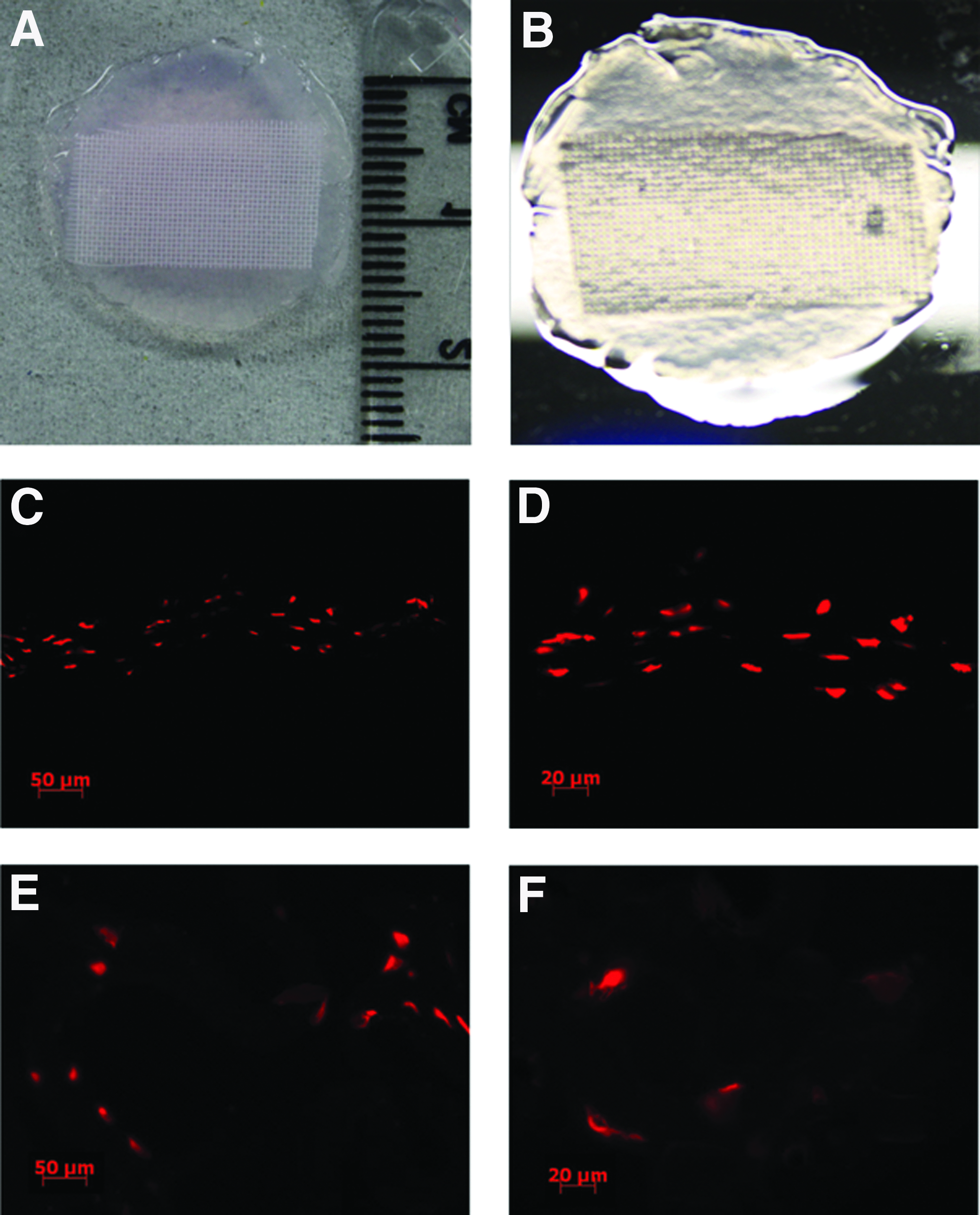
A photograph of an alginate gel disc (1.2%) with nylon mesh inside. The gel disc is ∼20 mm in diameter
The survival status of hMSC and mESC inside alginate–nylon gel discs
To examine the suitability of an alginate–nylon gel disc for preservation and storage of hMSCs and mESCs, the proportion of viable cells retrieved (relative to the initial amount encapsulated i.e., total cell survival) from the gel was investigated.
After a 5-day storage period, the proportion of total live cells retrieved following alginate encapsulation or cryopreservation was compared between hMSCs and mESCs (Fig. 2A). The number of viable hMSCs retrieved from alginate gel discs (2.46×10⁵, 82.22%) was slightly higher than those retrieved following cryopreservation (2.33×10⁵, 77.78%), but not statistically significant; in contrast, the viability of mESCs stored inside alginate gel (1.33×10⁵, 44.44%) was significantly lower than from cells retrieved following cryopreservation (2.0×10⁵, 66.67%, Fig. 2A).

The percentage of total
In terms of the proportion of relative cell survival (i.e., not including cells that have been lost during the storage process), the two different storage conditions for both hMSCs and mESCs showed a statistically similar level. Approximately, 80% (∼1.97×10⁵ cells) hMSCs retrieved from alginate–nylon gels were viable, 5% lower than the cryopreserved hMSCs. The percentage of retrieved mESCs (from inside alginate gel discs) was similar (74%, ∼0.93×10⁵) to those recovered following cryopreservation (69%) (Fig. 2B).
Survival status of hMSC and mESC postextraction
To further evaluate the cells' growth following extraction from strontium alginate–nylon gel storage, morphological assessment and cell doubling assays were performed on the retrieved hMSCs and mESCs for up to10 days following extraction. Postextracted culture of both mESCs (Fig. 3A and B) and hMSCs (Fig. 3C and D) at 37°C under 5% CO2 and 95% humidity demonstrated that cells from alginate gel discs were still capable of assembling into colonies. More importantly, extracted cells not only survived, but also maintained similar proliferation rates. Cell doubling measurements clearly showed no significant difference between untreated cultured cells and those extracted from calcium alginate gel discs (Fig. 3E) using the same passage number and initial seeding density.

Comparison between mESC and hMSC before encapsulation and following extraction from alginate gels after 5 days encapsulation. mESC colonies were found under both conditions before encapsulation
Expression of common cell markers following extraction from strontium alginate–nylon gels after 5 days of storage.
To validate the cells phenotype after storage within alginate–nylon gel at room temperature in a sealed container, a number of robust stem cell markers were examined. Quantitative PCR analyses were performed on hMSCs and mESCs before and after 5 days in storage within either alginate gel discs or cryopreserved. Both mesenchymal and embryonic cell markers were examined, respectively. The QPCR results showed no sign of decreasing levels of common hMSC and mESC markers (Fig. 4). Interestingly, mRNA levels of both hMSC (CD90, CD73, and STRO-1) and mESC markers (Oct-3/4 and SSEA-1) increased following storage. mRNA levels of hMSC markers from cells stored inside alginate gels were very close to cells stored in liquid nitrogen. Surprisingly, for mESCs stored inside alginate gel, both Oct-3/4 and SSEA-1 mRNA levels were twofold higher than from cryopreserved mESCs upon extraction.
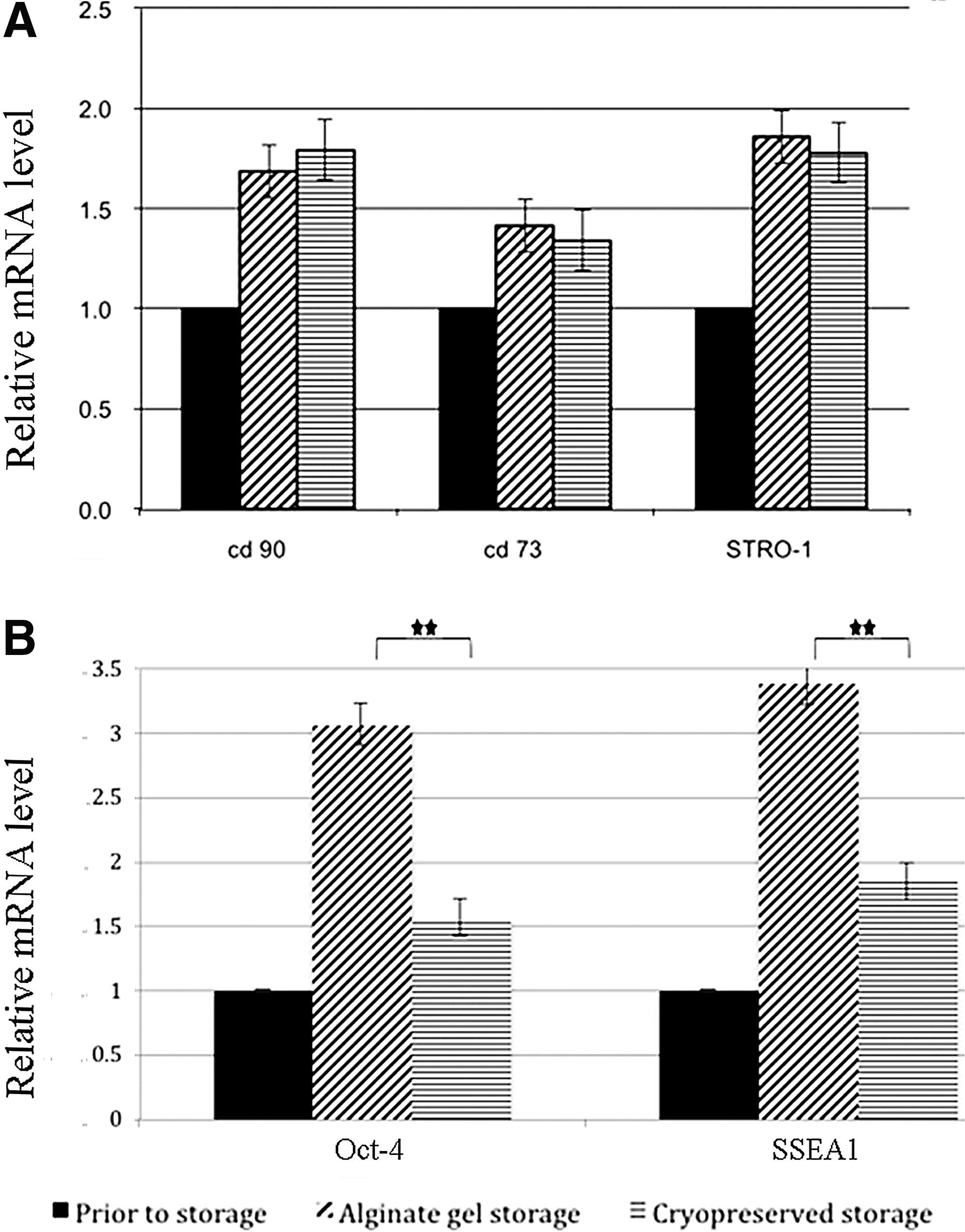
Detection of hMSC or mESC markers under optimal culture condition, before or following alginate gel encapsulation or cryopreserved. All three MSC markers showed increased mRNA levels after alginate and cryopreservation storage
This unexpected finding in increased mRNA levels within mESCs following alginate storage was explored at a protein level by flow cytometry. Flow cytometry was performed against four specific mESC markers following either storage for 5 days (in either alginate gel or liquid nitrogen) only or 5 days (in either alginate gel or liquid nitrogen) plus 10 days subsequent incubation at 37°C under 5% CO2 and 95% humidity.
Following storage, expression of Oct-3/4 and SSEA4 was significantly decreased following cryopreservation, but after 10 days in optimal culture conditions, they returned to levels similar to those seen in the alginate-encapsulated cells. Interestingly, the expression of each marker remained stable following gel encapsulation, suggesting cells could be used immediately following release from the gel, unlike those stored by cryopreservation (Fig. 5).

Expression of mESC markers between mESC encapsulated within alginate gel and cryopreserved in liquid nitrogen by flow cytometry. mESCs were studied immediately following release from a 5-day storage (alginate or cryopreserved). Cells were also studied following 10 days in standard culture media poststorage. Cryopreservation resulted in a considerable, although temporary loss of OCT3/4 and SSEA4 expression. No difference was seen between alginate-encapsulated cells
Discussion
As the need for stem cells to investigate and treat human diseases continues to grow, the necessity for effective and clinically safe methods for transporting them will also increase.
Although cryopreservation is still undoubtedly the best present method for long-term preservation of stem cells, its disadvantages have driven research in to alternatives. Cryopreservation has long been associated with cell membrane damage and apoptosis.37,38 Furthermore, cryopreservation is labor intensive and operator dependent and typically uses equipment that is not suitable for cell therapy such as liquid nitrogen, which is not GMP compliant. To circumvent these problems, costly alternatives are proposed that both prevent cells becoming into direct contact with the coolant and are able to maintain frozen cells for transportation. While these approaches remain in development, a cheap and simple robust alternative method for preserving cells for transportation is timely. Increasingly, the uses of hydrogels as cell carriers for delivery of stem cells are now challenging conventional cryopreservation methods.39,40
Previously, we have shown how both the macro- and microstructure of an alginate hydrogel can be modified to support the short-term preservation of corneal epithelial cells.
31
We continued this study further by investigating the use of a composite gel comprised from strontium alginate and nylon mesh as a robust method for the storage (and transport) of both hMSCs and mESCs at room temperature in an air-tight container with minimal media content. We compared their survival rate and maintenance of the stem cell phenotype to conventional cryopreservation and found similar levels of cell viability, expression of stem cell markers, proliferation rates, and morphology between the stored live cells. These results confirmed that specifically modified alginate gels are suitable to serve as a stem cell carrier. As further proof of the concept, we have also investigated a variety of other somatic cell types each showing a similar level of total cell viability following encapsulation and storage (Supplementary Fig. 1; Supplementary Data are available online at
Having found that calcium alginate gels were too fragile, and agitation would release the encapsulated cells resulting in their death, we surmised that the transport of cells would cause significant agitation to the gels resulting in their disintegration. Therefore, to avoid possible alginate hydrogel fragmentation during long-distance transportation, strontium was chosen to crosslink with alginate gels in this study. Previously, strontium was found to have a greater affinity to alginate than calcium, resulting in alginate hydrogels with improved stability. 34 Strontium alginate hydrogels have successfully been used in bone tissue engineering.33,41 We further improved the gels overall mechanical strength by encapsulating within it an inert nylon mesh. Together, the composite material, comprised of strontium alginate gel and nylon mesh, produced an extremely robust and effective carrier for hMSCs and mESCs.
Previously, alginate gels have been used as effective vehicles for stem cells, for example, in the treatment of spinal diseases.42,43 However, the ability of alginate to preserve stem cells in an undifferentiated state for a period commensurate with global distribution (5 days) has not been shown previously and is crucial to any future application in regenerative medicine. Our results clearly show that both mESC and hMSC markers were positive following extraction from the alginate gels remarkably even after 5 days at ambient (room temperature) in a sealed cryovial.
This evidence is consistent with that of previous studies, which have investigated alginate hydrogels as cell encapsulation modules for ESCs. Alginate (1.1% (w/v) calcium alginate)-encapsulated human ESCs cultured in a basic maintenance medium for up to 260 days retained their pluripotency, 44 and these cells were shown to self-renew in composite chitosan alginate gels. 45 Interestingly, hESCs stored in alginate microcapsules and cryopreserved in stirred tank bioreactors retained their stem phenotype. 46 Therefore, alginate-based methodology for preserving and transporting ESCs may range from basic strategies under ambient conditions (e.g., the methods described in the present study) to strategies involving composite alginate gels together with conventional cryopreservation approaches. The typical applications for alginate-encapsulated MSCs in alginate are different from those for ESCs immobilized in these scaffolds. MSCs are mainly cultured in alginate gels to drive osteogenic or chondrogenic differentiation of these cells.47–50 The finding in this study showing that under ambient conditions alginate gels are able to prevent the differentiation of MSCs suggests, however, that at certain gel concentrations, gel volumes, and temperatures, the multipotent nature of MSCs can be retained in alginate hydrogels. The flow cytometry results showed a surprisingly large difference in the expression of Oct-3/4 and SSEA-4 between freshly thawed cells and cells 10 days in culture (Fig. 5), whereas only very small differences were seen from cells following alginate encapsulation. It is well reported that freezing can cause loss of the secondary and tertiary protein structure that would subsequently affect antibody labeling,51,52 possibly suggesting that these proteins are more susceptible to denaturation during freezing or thawing, but that under optimal culture conditions, normal levels can return after 10 days. However, another possible explanation might be that alginate encapsulation is positively selecting for these cell types, which then proliferate within the gel resulting a in a proportional increase in Oct 3/4 and SSAE1. Certainly, the QPCR results (Fig. 4) show a threefold increase in Oct 3/4 and SSEA1 following storage within the alginate gel and are both important to the proliferative capabilities of mESCs.53,54 Furthermore, we have previously shown that adult (limbal) stem cells are able to proliferate, while encapsulated within an alginate gel. 31 Therefore, it is probably that a proportion of the viable cells extracted from the alginate gel are the progeny of those initially encapsulated, this of course is not possible during cryopreservation.
The viability of extracted cells from strontium alginate gels were all over 80%, which is consistent with previous findings using bovine corneal epithelial cells. However, despite also having shown that an optimized geometry and increased pore size of alginate gels can significantly increase cell viability following storage, the percentage of mESCs lost following storage in alginate gels was high (66%) in comparison to the hMSCs (18%) (Fig. 2B). This could be due to the relatively small size of the mESC. In general, the average size of the mESC is 8 μm, whereas hMSCs are 20–30 μm. The pore size of our alginate gels averages around 5 μm. 31 Therefore, we suspect that mESCs were less stable within alginate gels due to their smaller size (relative to pores) and were not properly retained within the gel during the 5-day storage. To investigate this, we compared mESC retention following cell encapsulation within alginate gels with and without the use of the live cell porogen hydroxethyl cellulose (HEC). We have previously shown that the pore size within an alginate gel can be controlled by the addition of HEC and that changes in the pore size affect overall cell viability of the encapsulated cells. 31 Our results (Supplementary Fig. 2) appear to support this hypothesis as the alginate gel with increased mean pore size (+HEC) retained a significantly lower number of viable cells (42.5% vs 35%) following 5 days of storage. However, more work is required to fully elucidate the mechanism behind the effect of both gel encapsulation and pore size on the viability of these cells.
Finally, we set out to test our gel encapsulation technique in a real-world scenario. mESCs were encapsulated in alginate and placed in a sealed cryovial (as described above), and then two such tubes were placed into a paper envelope and mailed (by second class post) to ourselves (Supplementary Fig. 3). Five days later, the envelope containing the encapsulated cells was received. Measured levels of cell viability and phenotype gave comparable levels to those obtained under laboratory conditions.
In conclusion, this technical study provides evidence that both human and animal stem cells can be immobilized within strontium alginate gels, and stored for prolonged periods at ambient temperature. More importantly, the subsequent easy retrieval of these cells with high levels of viability and maintenance of their stem cell phenotype offers a new and simple method for the transport of stem cells for both clinical treatment and research purposes.
Footnotes
Acknowledgments
The authors thank Prof. Bing Song and Mr. Stephen Swioklo for kindly providing mESCs and hMSCs for this study.
This study was financially supported by United Kingdom's Medical Research Council (G/0900877) and Biotechnology and Biological Sciences Research Council (BB/I00985X/1).
Disclosure Statement
No competing financial interests exist.
References
Supplementary Material
Please find the following supplemental material available below.
For Open Access articles published under a Creative Commons License, all supplemental material carries the same license as the article it is associated with.
For non-Open Access articles published, all supplemental material carries a non-exclusive license, and permission requests for re-use of supplemental material or any part of supplemental material shall be sent directly to the copyright owner as specified in the copyright notice associated with the article.