
Abstract
Bone tissue engineering (TE) aims to develop reproducible and predictive three-dimensional (3D) TE constructs, defined as cell-seeded scaffolds produced by a controlled in vitro process, to heal or replace damaged and nonfunctional bone. To control and assure the quality of the bone TE constructs, a prerequisite for regulatory authorization, there is a need to develop noninvasive analysis techniques to evaluate TE constructs and to monitor their behavior in real time during in vitro culturing. Most analysis techniques, however, are limited to destructive end-point analyses. This study investigates the use of the nontoxic alamarBlue® (AB) reagent, which is an indicator for metabolic cell activity, for monitoring the cellularity of 3D TE constructs in vitro as part of a bioreactor culturing processes. Within the field of TE, bioreactors have a huge potential in the translation of TE concepts to the clinic. Hence, the use of the AB reagent was evaluated not only in static cultures, but also in dynamic cultures in a perfusion bioreactor setup. Hereto, the AB assay was successfully integrated in the bioreactor-driven TE construct culture process in a noninvasive way. The obtained results indicate a linear correlation between the overall metabolic activity and the total DNA content of a scaffold upon seeding as well as during the initial stages of cell proliferation. This makes the AB reagent a powerful tool to follow-up bone TE constructs in real-time during static as well as dynamic 3D cultures. Hence, the AB reagent can be successfully used to monitor and predict cell confluence in a growing 3D TE construct.
Introduction
To bring TE constructs and their processes closer to GMP requirements, tissue engineers aim to characterize and follow-up parameters related to the environmental three-dimensional (3D) tissue culture condition and the physiological state of the growing TE construct.5–8 Currently, these characteristics are analyzed by integrating appropriate biosensor technology in bioreactor systems to monitor parameters such as the pO2, pH, glucose, and lactate concentration.5,9–11 In addition to the established methods for cell count in which cells and TE constructs are only tested at predefined phases of manufacturing or in a destructive way,12,13 the real-time monitoring tools can be used for a nondestructive, real-time cell characterization in nontransparent 3D constructs. In addition, authorities are inquiring these novel alternative methodologies to demonstrate equality, consistency, and safety of ATMPs, and reviewing them on a case-by-case basis. 1
The overall metabolic cell activity is a widely characterized parameter and serves multiple purposes: evaluation of cytotoxicity and drug effects,14,15 assessment of biocompatibility, 16 quantification of viability, and proliferation of a cell population.17,18 Hence, this study explores the use of a metabolic assay to monitor in real-time the evolution of bone TE constructs during in vitro culture.
Of the variety of metabolic assays that are available on the market, the alamarBlue® (AB) assay is nontoxic to cells19,20 and has shown a good correlation with other metabolic activity assays such as the XTT assay, 21 the 3H-Tdr assay,19,20 and the MTT assay. 22 AB contains the nonfluorescent resazurin as a primary constituent, which is reduced to the fluorescent resorufin by different oxidoreductases that use NAD(P)H as a primary electron donor. 14 Resazurin as redox indicator is used in assays for cell proliferation, 23 cell viability,14,24 and mitochondrial respiratory activity. 25
The AB assay has already been applied to follow up 3D cell cultures under static conditions or 3D dynamical cultures.26–28 However, limitations of the AB assay were reported and related to the diffusion properties of AB into the 3D TE constructs.27,29 Because a fluid flow will enhance the transport of AB in and out of the TE constructs, this study investigated the feasibility to use the AB assay as a noninvasive, real-time monitoring technique to evaluate the in vitro metabolic activity, viability, and/or cellularity of 3D TE constructs after seeding as well as during static cultures and dynamic cultures by integrating the AB assay in a bioreactor-driven culture process.
In this study, bone TE constructs were produced by a two-step seeding procedure, followed by a culturing step under static or dynamic conditions. With the aid of the AB assay, the metabolic activity of each individual bone TE construct was monitored after seeding, and during bioreactor culturing. The TE constructs were additionally characterized in a destructive way by a combined and validated end-point analysis as an established reference for the AB measurements: a qualitative live/dead staining to assess the viability and homogeneity, and a quantitative DNA analysis technique to assess the total cell number.30,31 Resazurin based real-time analyses were already performed on 3D cell cultures but so far not correlated to established quantitative techniques for the determination of cell number. 28
This study evaluated three different aspects of the AB metabolic assay to assess its potential as noninvasive, real-time monitoring tool for bioreactor culture of TE constructs: (1) the influence of the AB incubation time on the measured metabolic activity, (2) the correlation between the metabolic activity and the cellularity upon cell seeding, and (3) the correlation between the metabolic activity and the cellularity upon TE construct culturing.
Materials and Methods
Cells and scaffolds
A pool of primary human periosteum-derived cells (hPDCs), being a clinically relevant and multipotent cell source with proven osteogenic potency, were isolated and cultured as described previously.32,33
To evaluate the use of AB for the characterization of 3D distributed cells within a carrier with a minimal influence of the carrier structure itself, an inert titanium alloy biomaterial allowing cell attachment, proliferation, and differentiation was used as raw material to produce, by the additive manufacturing technique of selective laser melting (SLM), highly porous Ti6Al4V scaffolds (Ø=6 mm, h=6 mm), further referred to as Ti scaffolds.34,35 The scaffolds were designed based on the unit cell represented in Figure 1A, thereby generating a highly open and regular pore structure with measured permeability coefficient kscaffold (mean±standard deviation) 1.69×10⁻⁹±1.78×10⁻¹⁰ m2 (Fig. 1B–D).34,36

The Ti scaffolds were produced and consequently analyzed as described by Pyka et al. and had a porosity of 73%±1%, a surface area of 6.5±0.2 cm2, a strut size of 245±2 μm and a pore size of 455±3 μm.37,38
Before use, the scaffolds were cleaned ultrasonically, immersed 10 min in acetone, 10 min in ethanol 70%, and 10 min in distilled water, subsequently oxidized for 12 h at 60°C in a 5 M sodium hydroxide (Sigma-Aldrich) solution, rinsed with distilled water, and finally sterilized in a steam autoclave. Before cell seeding, all scaffolds were prewetted with a cell culture medium consisting of Dulbecco's modified Eagle's medium with high-glucose (Invitrogen) containing 10% fetal bovine serum (BioWhittaker), 1% sodium pyruvate (Invitrogen), and 1% antibiotic–antimycotic (100 units/mL penicillin, 100 mg/mL streptomycin, and 0.25 mg/mL amphotericin B; Invitrogen) by vacuum impregnation in the cell culture medium for 2 h in a humidified incubator at 37°C, and drying overnight in a nonhumidified incubator. 31
TE construct seeding
Bone TE constructs were produced by a two-step seeding procedure. Prewetted scaffolds 31 were positioned in 1.5 mL eppendorf tubes (Invitrogen) that serve as seeding vessels. The scaffolds were drop-seeded with a 100 μL cell suspension (12,500–400,000 hPDCs for the investigation of the seeded TE constructs and 200,000 hPDCs for the investigation of subsequently cultured TE constructs), correlating with approximately half of the free scaffold volume. After 1 h of incubation in a humidified incubator (37°C, 5%CO2 and a relative humidity of 95%), 1 mL of the cell culture medium was added. The seeding vessels were placed on a rotator (Mini LabRoller™; Sigma-Aldrich), and further incubated overnight at a rotational speed of 24 rpm.
TE construct culturing
Ti scaffolds seeded with 200,000 hPCDs were cultured for 3, 7, 10, or 14 days under static or dynamic culture conditions. (i) For static culturing, bone TE constructs were either positioned in 12-well plates containing 1 mL of the cell culture medium, as generally reported in the literature, 39 or in 50 mL falcon tubes (disposable 50 mL Falcon tubes; BD Biosciences) containing 10 mL of cell culture to resemble the dynamic culturing conditions more closely, and incubated at 37°C in a humidified and CO2-controlled incubator. (2) For dynamic culturing, bone TE constructs were cultured in an in-house developed perfusion bioreactor equipped with seven parallel perfusion circuits (Fig. 2). Each bone chamber, containing a single scaffold press-fitted in a silicon tube to fix the position of the scaffold in the bioreactor, is connected to an individual medium reservoir (disposable 50 mL Falcon tubes; BD Biosciences) containing 10 mL of the cell culture medium via a Tygon® (Cole Parmer) tubing and a two-stop tubing (BPT; Cole Parmer) connected to a peristaltic pump (IPC-24; Ismatec SA), generating a fluid flow of 0.2 mL/min.

Peristaltic perfusion bioreactor composed of a medium reservoir, a peristaltic pump, and a bone chamber, housing a tissue engineering (TE) construct.
TE construct evaluation
The overall metabolic cell activity of the TE constructs was assessed after seeding, at day 3, 7, 10, and/or 14. After measurement of overall metabolic cell activity, the TE constructs were either cultured in the refreshed cell culture medium or harvested to evaluate the cellularity, viability, and/or homogeneity.31,40
Overall metabolic cell activity
The overall metabolic cell activity of the TE constructs was monitored using the nontoxic AB assay for all above-mentioned culture conditions. The AB solution (Invitrogen) was prepared by diluting 1 mL AB reagent in the 10 mL cell culture medium.41,42 To monitor the overall metabolic activity after seeding or during static 3D cell culturing, the 3D TE constructs were transferred into 48-well plates (Greiner) for the static AB protocol a, or into 50 mL falcon tubes (disposable 50 mL Falcon tubes; BD Biosciences) for the static AB protocol b containing, respectively, 1 and 3 mL of the AB solution. To monitor the metabolic activity during dynamic 3D culturing, the assay was integrated in the perfusion bioreactor in a minimally invasive way by changing the cell culture medium by 10 or 3 mL AB solution for the dynamic AB protocol a and b, respectively. The 3D TE constructs were incubated for 1, 2, 4, 7, 9, 11, 20, 21, and 25 h in static and dynamic AB protocol a and for 1 h in the static and dynamic AB protocol b. The fluorescence of 100 μL aliquots was measured in triplicate by an automated microplate fluorometer (SerColab systems) using an excitation wavelength of 544 nm and an emission wavelength of 590 nm. The intensity of the fluorescent signal was expressed in arbitrary fluorescence units (FU). For each measurement a blank was measured under identical conditions as the TE constructs but without cells or scaffold present to determine the baseline fluorescent signal of the solution. The blank value was subtracted from the TE construct values to obtain the fluorescent signal originating from the cellular activity.
Cellularity
The cellularity was obtained by performing a highly quantitative and selective DNA assay (Quant-iT™ dsDNA HS kit; Invitrogen). 40 Bone TE constructs and—as a reference—100 μL cell suspensions (125–4000 cells/μL) were washed with phosphate-buffered saline (PBS) and lysed in 350 μL RLT lysis buffer (Qiagen). The lysed TE constructs were stored at −80°C before analysis, thawed at room temperature, and spun down for 1 min at 13,000 rpm. The DNA concentrations of the supernatants were quantified with a Qubit®Fluorometer (Invitrogen).30,31,40
Viability and homogeneity
The viability and homogeneity of the cells in the 3D bone TE construct were assessed by a LIVE/DEAD® viability/cytotoxicity assay (Invitrogen) according to the manufacturer's instructions. For each TE construct a staining solution containing 1 mL PBS, 0.5 μL calcein acetoxymethyl ester, and 2 μL ethidium homodimer was prepared. The TE constructs were rinsed with 1 mL PBS before transfer to the staining solution and incubation for 20 min at 37°C. Before imaging, the constructs were transferred to a PBS-containing well plate. Fluorescent images were taken by a digital camera mounted on a stereomicroscope (Discovery V8 Microscope; Zeiss) to qualitatively evaluate the viability and homogeneity of hPDCS in the bone TE constructs.
Statistics
All experimental conditions represent three to five TE constructs and were plotted in point diagrams, representing individual points or averages±the standard deviation. The obtained results were set to be significantly different when p-values were<0.05, by performing an unpaired Student's t-test.
Results
AB incubation time
The overall metabolic activity of 45 bone TE constructs, dynamically cultured for 7 days, was assessed by increasing the AB incubation time in the initial bioreactor protocol from 1 up to 25 h to identify optimal and saturation conditions. The resulting fluorescence signal increased with increasing incubation time (Fig. 3). The fluorescence signal reached a plateau value around 4000 FU. The overall metabolic activity values obtained by incubating over 20 h did not significantly change anymore, while this was still the case at shorter incubation times (1–11 h). The linear correlation coefficient R2 of the overall metabolic cell activity in function of the incubation time between 1 and 11 h was 0.97, indicating a strong linear correlation up to 3000 FU, but decreased when fluorescence reached the maximally obtained value around 4000 FU.

Overall metabolic activity of TE constructs dynamically cultured for 7 days. The values at different incubation times were obtained by integrating the initial alamarBlue® (AB) assay (10 mL) in the bioreactor. The linear fit of the time points below 11 h is represented. Error bars show the standard deviation of three measurements of the same TE construct.
To evaluate the use of a static and bioreactor AB assay to monitor the overall metabolic activity of statically as well as dynamically cultured 3D TE constructs, the metabolic activity was assessed after seeding, at day 3, 7, 10, and 14. To investigate the potency of the AB assay, the incubation time of the AB solution (static condition 1 mL, dynamic condition 10 mL) was increased from 1 to 4 h (Fig. 4). The 4 h timepoint for the static condition (Fig. 4A) resulted in fluorescent values above 3500 FU, which we showed earlier to be out of the linear range of the assay (Fig. 3). All timepoints for the dynamically cultured and measured TE constructs were within the linear range resulting in an R2 value of >0.99 on average. In addition, dynamically cultured TE constructs showed a significantly increased σ from 1 to 4 h of incubation. This was not observed in the static TE constructs since the 4 h timepoint was close to the maximal intensity of the signal. In order to perform all measurements in the linear range of the assay with minimal standard deviation, an incubation time of 1 h was considered to be optimal and was applied in all further experiments in the static setup.
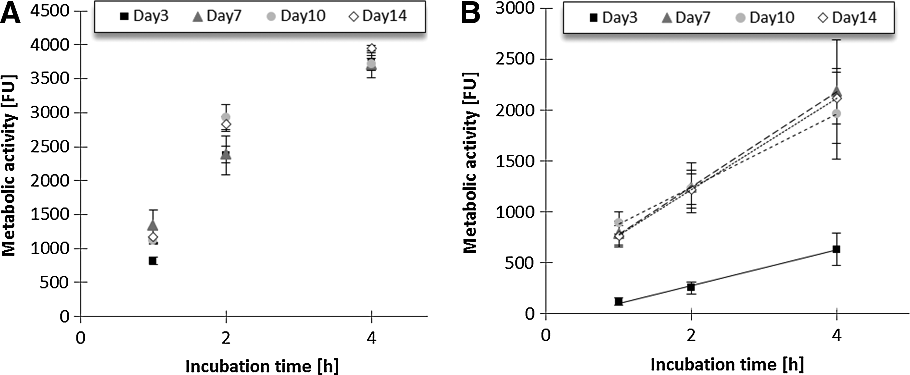
Effect of the incubation time of the initial AB assay on the obtained metabolic activity of
AB in cell seeding process
The overall metabolic cell activity of 18 TE constructs seeded with 12,500 to 400,000 hPDCs was evaluated by incubating the seeded scaffolds for 2 h using the static AB protocol a. The metabolic activity values are presented in function of the number of seeded cells (Fig. 5A) and in function of the total DNA content (Fig. 5B). The results show a good linearity resulting in R2 values of, respectively, 0.95 and 0.88 (Fig. 5A, B).
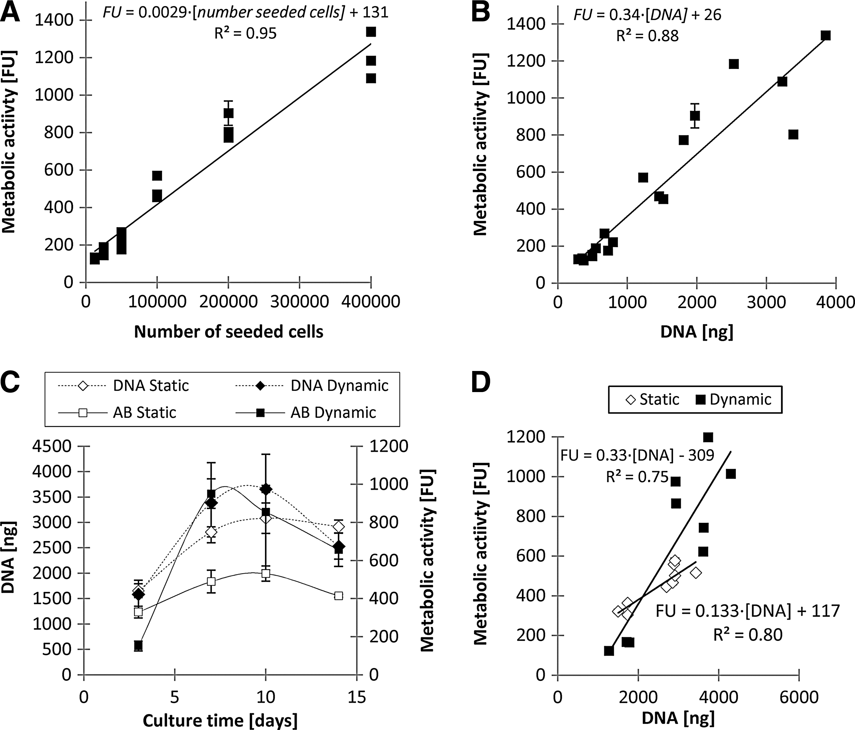
Correlation of the metabolic activity with the cellularity of the
AB in cell culturing process
To evaluate the use of AB to monitor and follow-up growing bone TE constructs, the total DNA content and overall metabolic activity of 24 bone TE constructs was monitored/assessed over 14 days of static or dynamic culturing. The bioreactor AB protocol a for the measurement of the overall metabolic cell activity was adjusted for the dynamically cultured 3D TE constructs to minimize the use of AB solution to 3mL (instead of 10 mL) referred to as bioreactor AB protocol b. Consequently, the same amount of AB solution was used for analysis of the overall metabolic cell activity in statically cultured 3D TE constructs (static AB protocol b). The AB and DNA results indicate, except for the metabolic activity of dynamically cultured TE constructs, that all parameters showed a similar tendency in function of the culture time: maxima were reached at day 10 (Fig. 5C). Also, two other observations were made: (1) there was a significant difference (p<0.05) in the AB activity and DNA content between day 3 and all later time points, while no statistical significant differences were observed between the values obtained at day 7 and 10; (2) the total DNA content was not significantly influenced by the culture conditions (static versus dynamic), while the overall metabolic cell activity was significantly different (lower at day 3 and higher at day 7, 10, and 14) between the static and dynamic culture conditions at all investigated time points (p<0.05).
To evaluate whether the overall metabolic cell activity can predict the cellularity, the values shown in Figure 5C were presented in function of the total DNA content. However, no strong correlation was observed between the cellularity and the metabolic activity of the TE constructs cultured for 3, 7, 10, and 14 days. The obtained R2 values were 0.69 for the dynamically cultured TE constructs and 0.61 for the statically cultured TE constructs (data not shown). A significant decrease between day 10 and 14 of the DNA content in the dynamic culture conditions and of metabolic cell activity in the static culture condition is suggesting over confluency and cell death. Therefore, the values of 14 days were not taken into account in Figure 5D, which hence depicts an increased linearity, resulting in R2 values of, respectively, 0.75 for the dynamically cultured and 0.80 for the statically cultured TE constructs (Fig. 5D).
After 7 days of 3D culturing, viable hPDCs were homogeneously distributed throughout the Ti scaffolds (Fig. 6). The cells reached confluence between day 7 (Fig. 6A) and day 14 (Fig. 6B, C), thereby confirming the AB and DNA data, which were maximal at day 10. The fluorescent images suggest that the culture conditions (static versus dynamic) affected the cell distribution at the bottom surface (Fig. 6B, C). The difference in cell distribution at the radial outer surface of statically and dynamically cultured TE constructs is most likely related to the presence of the silicone tubing in the dynamically cultured TE constructs since the constructs were press-fitted in the tubing to ensure perfusion through and not around the TE construct.

Green fluorescent images of the top, side, and bottom views of the live/dead-stained bone TE constructs after
Discussion
The quality and reproducibility of ATMPs, in particular their cell viability, identity, potency, and purity, must be guaranteed in order to bring these products to the market. Hence, there is a growing need to monitor and control noninvasively the TE constructs, and to unravel the complex cellular processes.2,7 The gained knowledge would enable an early detection of irregularities in the production process of a bone TE construct. 8
The AB assay, commonly used for cytotoxicity, biocompatibility, viability, as well as proliferation studies,15,16 is widely applied in two-dimensional (2D) in vitro cultures, and more recently in 3D cell cultures.9,17,27 However, because the assay itself is usually performed under static conditions, it implies that dynamically cultured TE constructs need to be transferred from the bioreactor into, for example, culture well plates, analyzed, and reintegrated in the bioreactor system. Such an invasive, multistep procedure increases the risk for scaffold contamination and negatively affects GMP compliance. To avoid such an invasive manipulation consisting of several manual handlings to characterize the metabolic activity when culturing under perfusion conditions in this work, the AB was refreshed by using the same noninvasive procedure as during medium refreshment. To our knowledge, only Gloeckner et al. reported AB results performed under dynamic 3D conditions, thus without interfering with the dynamic culture conditions. 42 By circulating an AB solution (2.5% v/v) for 24 h in a hollow fiber bioreactor, fluorescent analyses of medium samples could distinguish between 105, 106, or 107 proliferating leukemic HL-60 cells.
This study evaluated and validated for the first time the AB assay as a noninvasive tool for bone TE constructs and in particular investigated the following aspects: (1) the influence of the AB incubation time on the fluorescence signal, (2) the correlation between the metabolic activity and the cellularity upon cell seeding, and (3) the correlation between the metabolic activity and the cellularity upon TE construct static and dynamic culturing.
Our results showed that the AB assay could be used for the determination of the metabolic activity of bone TE constructs as long as the signal was ≤3000 FU. The fluorescence values reached a plateau near 4000 FU, which was also reported by Al-Nasiry et al. They observed that the fluorescent signal from the AB dye increased linearly up to the point where ∼80% of the AB dye was reduced, and a plateau value was reached. 17 This could be explained as followed: (1) resazurin, the active component of the AB solution, could be fully reduced, thereby resulting in a maximum fluorescence value; (2) O'Brien et al., on the other hand, reported that highly metabolically active cells may induce over-reduction of resazurin, resulting in a subsequent reduction of the fluorescent resorufin into the uncoloured and nonfluorescent hydroresorufin, hence resulting in an underestimation of the overall metabolic activity of the TE construct. However, this over-reduction would result in a signal decrease in function of time after the maximal intensity was reached rather than resulting in a plateau value 14 ; (3) Although the AB dye was often reported to be nontoxic for cells, even after longer incubation times,19,43,44 several groups reported a negative effect of the AB assay on cell growth and cell viability.28,42,45 However, the toxicity is dependent on the cell type, the AB concentration, and exposure time. Gloeckner et al. and Erikstein et al. therefore suggest to adjust the AB protocol in function of the cell type, and to make use of a reduced incubation time followed by replacing the AB solution by a fresh cell culture medium.42,45 Our experiments suggest an incubation time of 1 h to be optimal for the tested bone TE constructs (Fig. 4). Since toxicity is only reported after longer incubation times and the AB solution was replaced after each measurement, potential AB toxicity is not expected to have influenced our experiments. 45
To investigate the correlation between the metabolic activity and the cellularity upon cell seeding, the overall metabolic activity and the total DNA content of the seeded TE constructs were compared. Although the correlation of the metabolic activity expressed in function of the number of seeded cells (Fig. 5A) was higher (R2=0.95) than when expressed in function of the total DNA content (R2=0.88), the linear correlation remained fairly high (Fig. 5B). This indicates that for the cellularity of each individual, seeded scaffold can indeed be reliably and noninvasively evaluated with the AB assay when fluorescence values were ≤3000 FU.
Based on the obtained results depicted in Figures 3 and 4, and the data published by Erikstein et al. and Gloeckner et al., the initial AB assay conditions were adjusted. In the final experiments (Fig. 5C, D), comparing the DNA content and AB of both statically and dynamically cultured bone TE constructs, a reduced AB incubation time of 1 h and in 3 mL of AB solution was used, the latter being the lowest volume still resulting in a complete filling of the bioreactor circuit.
Although the AB assay correlates with the cell number in 2D cultures of different cell lines,14,24,46 our analysis only resulted in a low linear correlation between the overall metabolic activity and the DNA content of a TE construct during static as well as dynamic culture conditions (R2<0.70, data not shown). Hence, the obtained results imply that the AB assay is not a suitable analysis technique to monitor the cellullarity under the given conditions. However, when only taking the proliferative stage into account (day 1–10, Fig. 5D), the linear correlation was enhanced for both the statically and dynamically cultured TE constructs (R2 of, respectively, 0.80 and 0.75, Fig. 5D). This suggests that during the initial proliferative stage of a growing 3D TE construct, the AB activity can be used to estimate the cellularity. Wilson et al. reported a similar correlation between the cellularity and metabolic activity and stated that the AB followed cell proliferation. 29
The DNA and metabolic activity of statically and dynamically cultured TE constructs (Fig. 5C) showed comparable trends in function of the culture duration (Fig. 5C): the AB and DNA values of the statically cultured as well as for the dynamically cultured TE constructs increased until day 10 and decreased after reaching cell confluence.
In 2D as well as in 3D cell cultures the increase in cellularity was followed by a plateau, that is, when cells reach their maximal density in the used culture setup. At this point cells can differentiate or also enter apoptosis.47,48 The results presented in Figure 5C suggest that the AB assay is a potent, noninvasive parameter to estimate the time point at which maximal cell density is reached. This information is highly valuable when producing bone TE constructs by procedures that distinguish between cell proliferation and cell differentiation or that depend on cells reaching a certain critical cell density. 49
The DNA results of the cultured 3D TE constructs were not influenced by the culture condition (static versus dynamic), while the metabolic activity of the static cultured TE constructs was significantly different from the dynamically cultured scaffolds (Fig. 5C). The differences in the overall metabolic activity of statically and dynamically cultured TE constructs are thought to be partially related to the observed differences in cell distribution and matrix deposition (Fig. 6), but might also be caused by the difference in fluid flow conditions. As reported in literature, the scaffold permeability potentially affects the diffusion of the AB dye into the 3D scaffold and, hence, the outcome of the AB assay.27,29 Moreover, the diffusion characteristics of the AB dye in the TE construct will change over time due to the progression of the cellularity and the matrix deposition, making the AB assay semi-quantitative rather than quantitative.27,29 By integrating the AB assay in the TE construct containing an open porous Ti scaffold in perfusion bioreactor, the diffusion kinetics were enhanced in this dynamic culture system compared to static incubation. 36 Analyzing and comparing the overall metabolic activity of statically and dynamically cultured TE constructs (identical AB solution volume and incubation time, Fig. 5C), indeed indicated higher AB values at day 7, 10, and 14, while no statistical differences were observed for the DNA content of statically and dynamically cultured scaffolds (Fig. 5C). When further interpreting these results, it should thus be taken into account that the higher fluorescence values could be related to the higher overall metabolic activity of the dynamically cultured scaffolds, to the higher diffusion kinetics, or a combination of both. One should thus consider the 3D nature of the scaffolds and changes in the diffusion kinetics due to the proliferation of the TE constructs. 29
One of the important advantages of the AB assay is its versatility. The relation between the incubation time and the total volume allows optimization of the assay to provide an optimal measuring window. An increase in incubation time will cause the observed differences to be more distinct but will also increase the absolute fluorescent signal which should remain within the linear range of the assay. A similar result can be achieved by decreasing the volume and keeping the incubation time constant. A combined decrease in incubation time and volume enables a similar signal in a shorter timeframe and will also reduce the standard deviation on the signal as shown earlier (Fig. 4B).
In comparison with other methods that correlate metabolic-related parameters such as pO2, glucose, and lactate concentration to cell proliferation,5,9–11 the versatility of AB to monitor cell proliferation has an important advantage. Santoro et al. 2012 recently showed that the decrease in oxygen tension over a TE construct could be used to monitor cell proliferation. 10 However, the magnitude of this decrease is dependent on both perfusion flow characteristics and cell number. Using the model developed by Truscello et al., 50 we calculated the difference in oxygen tension across the TE construct in our bioreactor system. Based on our in-house hPDC database of standard-curves correlating DNA content with cell number, we calculated an average DNA content of 8.9 pg DNA/cell (n=24) for hPDCs, which corresponds well with the 9.3 pg/cell reported in literature for human mesenchymal stem cells. 51 Using this value and an average oxygen consumption rate of 98 fmol/(cell·h), 52 a DNA content of 3500 ng would result in 400,000 cells, implicating a difference in oxygen saturation of 1% between the in and outlet of the TE construct. While the proliferation is apparent based on the AB measurement, it would not be measurable using the difference in oxygen tension. Similar to the measurement of the oxygen consumption, monitoring cell proliferation based on glucose consumption or lactate accumulation is also limited by the volume of medium used and the total number of cells present. Pattappa et al. reported a glucose consumption rate of 342 fmol/(cell·h). 52 Applying this value to our system resulted in a decrease in glucose concentration of 1% in a period of 24 h. Although this difference can be measured using novel glucose sensors, 53 the low decrease in glucose concentration makes the glucose approach not suited to monitor cell proliferation accurately in this setup. In addition, when measuring glucose or lactate separately, the potential of cells to metabolize lactate instead of glucose at low glucose concentrations has to be taken into account, which complicates the processing of glucose and lactate based data. 11
Although monitoring of low cell numbers might not be feasible using only glucose or oxygen measurements in this setup, these methods still hold potential as complementary read-out to the AB assay for manufacturing of ATMPs. While we showed that the AB assay can be used to monitor proliferation in a perfusion bioreactor system, and that the versatility of the assay allows adjusting the parameters to provide an optimal measuring window, the high cell numbers required for the development of an ATMP can result in a saturated signal. Therefore, the AB assay could be used in the initial stages of this process, while, once a certain critical cell number is reached, oxygen and glucose measurements could provide a continuation in the monitoring of proliferation in the later stages.
The main advantage in using glucose or oxygen monitoring compared to the AB assay is the proven noninvasiveness of this method. While the AB assay requires the perfusion of a solution through the TE construct with the possibility of leaving remnants of the solution in the system afterward, glucose and oxygen monitoring require the use of one or multiple sensors that can be placed in the medium flow circuit, before and/or after the TE construct. For the use of the AB assay in the manufacturing of ATMPs, the protocols should ensure that no remnants can be present in the final construct.
Nonetheless, this study suggests that the AB assay is, within defined and validated conditions, a useful technique to follow-up the production process of 3D TE constructs.
Conclusions
Integrating the AB assay in 3D cultures to follow-up and monitor the overall metabolic cell activity within 3D bone TE constructs is a promising strategy, as the AB assay is a relatively simple, reliable, noninvasive, and useful technique that can be used real-time under static as well as dynamic conditions. Moreover, within the investigated ranges, the metabolic activity linearly correlates with the cellularity of the 3D TE construct upon seeding and during the initial TE construct culture, and allows to identify the time point at which cell confluence is reached. As part of the AB protocol validation and TE construct customization, the incubation time, volume, and concentration of the AB solution should be carefully selected to ensure read-outs within the broad, but linear range of the assay.
Footnotes
Acknowledgments
This work is part of Prometheus, the Leuven Research, and Development Division of Skeletal Tissue Engineering at the KU Leuven (
Disclosure Statement
No competing financial interests exist.