
Abstract
Objective:
To assemble a biohybrid cardiac patch consisting of a large (5×5 cm) elastomer scaffold whose pores are filled with a self-assembling peptide (SAP) gel entrapping adipose stem cells, to be used as a novel implant in a big animal model (sheep) of myocardial infarction. The study focuses on the way to determine optimal procedures for incorporating the SAP solution and the cells in the patch to ensure cell colonization and a homogeneous cell distribution in the construct before implantation. The problems associated with the scale-up of the different procedures raised by the large size of the construct are discussed.
Materials and Methods:
Experiments were performed to choose between different assembling alternatives: incorporation of the SAP gel before cell seeding or simultaneous SAP and cell loading of the scaffold; surface seeding of cells or cell injection into the scaffold pores; dissemination of the cells throughout the scaffold before incubation by gentle shaking or by centrifugation. Immunocytochemistry techniques and confocal and scanning electron microscopies were employed to assess and quantify cell colonization of the material and early cell distribution. Cell concentrations and the uniformity of cellular distribution throughout the scaffold were taken as the main criteria to decide between the different alternative procedures.
Results:
The combination of peptide preloading, cell injection, and shaking before incubation yielded the best results in terms of greater cell density and the most uniform distribution after 24 h of culture compared with the other methods. These techniques could be scaled-up to obtain large biohybrid cardiac patches with success.
Conclusions:
The results obtained after the different seeding methods allowed us to establish an effective protocol for the assembly of large biohybrid patches for their subsequent implantation in the heart of a big animal model of myocardial infarct in the context of a preclinical study.
Introduction
A
Alternative tissue-engineering strategies8,9 combine cells with three-dimensional scaffolds or patches that host them and improve their survival, inducing the formation of new blood vessels and extracellular matrix and at the same time mechanically assisting the host tissue. The polymers employed for this purpose include collagen, gelatin, fibrin, hyaluronic acid, and alginate,10–13 or the synthetic polylactide acid, polylactide-co-glycolic acid, polycaprolactone, poly(ethylene glycol), polypropylene, and poly(glycerol sebacate), with different architectures and combined with a variety of cells.14–17 As an example, Chachques et al. 18 implanted collagen sponges seeded with bone marrow cells onto the postischemic myocardial scar of a series of patients in a clinical feasibility study and observed an increase of the thickness of the infarct scar with a viable tissue as well as the normalization of the cardiac wall stress. The main disadvantages of those scaffolds were the low mechanical characteristics of the materials employed and the fast bioresorption rate of the sponges, which completely degraded before the end of the study.
The concept of the 7th FP project RECATABI, where this study is framed, is the design of a biohybrid cardiac patch consisting in an elastomeric polymer scaffold whose pores are filled with the self-assembling peptide (SAP) hydrogel RAD16-I, which at the same time encapsulates adipose tissue-derived stem cells (ASCs). A first series of these patches employ poly(ethyl acrylate) (PEA), to develop scaffolds with interconnected spherical pores. This polymer is compatible with the myocardial tissue in terms of mechanical properties, is easy to process, and has an excellent cell compatibility ( 19 and references cited there).
SAPs are resorbable nanomaterials that mimic the structure of the extracellular matrix, promoting and modulating cell functions such as adhesion, proliferation, and migration. Thanks to these properties, they have been used for a variety of in vitro applications with different types of cells (osteoblasts, embryonic stem cells, adult neural stem cells, endothelial cells, among others)20–22 and for regenerative strategies in animal models.23–26 One commonly employed SAP, RAD16-I,18–20 is an hydrogel consisting of simple repeated amino acid sequences of RADA with alternating hydrophobic and hydrophilic lateral groups. It is injectable in an aqueous solution, and forms percolating β-sheet nanofibers when exposed to a saline solution or physiological media.21,27,28 At low concentrations (0.15–0.25%), the gel is soft and fragile, resulting in poor manageability; on the contrary, at an elevated concentration (from 0.5% to 1%), the gel becomes tough and impedes an adequate cellular ingrowth. 21 In our design, the elastomer scaffold membrane provides the three-dimensional context and the mechanical integrity, whereas the peptide gel RAD16-I filling the scaffold pores is expected to act as an encapsulating medium for the cells, improving their survivability and retaining them inside the membrane, while allowing proper permeability to cellular metabolites and wastes, and likely improving vascularization throughout the scaffold.
ASCs are a convenient cell source for cardiac regenerative purposes since they can be easily harvested from the patient, have an elevated proliferation rate in vitro and are nonimmunogenic; besides, some studies and clinical trials have demonstrated their potential to improve the ventricular function,29–32 which could lead to their use for cardiac clinical application.33,34 Since a direct cell graft into the infarcted myocardium resulted in poor results, with the rapid dissemination of cells to other sites and a low rate of cell survival of the effectively engrafted cells, 35 the transplantation of the cells inside a physical support may help improve over those results: the scaffold can cover the damaged area, can protect the grafted cells by offering a cellular niche, and thus prolong their survival and paracrine effect, while impeding their migration from the site of interest. An increased localized activity of the cells could lead to a greater wall thickening and neovasculogenesis, eventually improving the heart function.
In the context of a preclinical study in a big animal model (Ille de France sheep), additional factors that could condition the design of the cardiac patch must be taken into account. The most relevant ones are the size of the scaffold, the number or concentration of cells to be transplanted, the timing of preimplant seeding, and in vitro culture of the cells in the patch. The dimensions of the patch designed for this study (5×5 cm) are related to the size of the infarct model created by a surgical procedure (coronary artery branch ligations) in adult sheep,19,36 and the number of cells to be transplanted and the preimplant culture time have followed previous experience. 18 Previous work showed the relevance of a complementary postseeding dynamic procedure, to achieve a better cellular distribution within the pores 19 compared with static seeding. Altogether, these circumstances impose stringent requirements on the design of the patch: it must be able to lodge the maximum number of cells to obtain the best results possible with this experimental therapy, and at the same time, these cells must be seeded and distributed uniformly across the patch in 24 h before implantation (as the medium timeframe for a clinical procedure of this kind). Afterward, the whole patch must be rapidly vascularized once implanted onto the myocardium to keep the seeded cells alive. In the present work, we address these questions, related to the design and implementation of a biohybrid patch for use in an infarcted sheep heart. 37
Materials and Methods
Preparation of the scaffolds
Scaffolds of PEA with interconnected spherical pores were prepared following a porogen template leaching method as described in. 38 Briefly, poly(methyl methacrylate) microspheres (PMMA; Colacryl dp 300) of known size, 130±20 μm, were sintered between two plates to obtain a porogenic template. A monomer solution was prepared by mixing ethyl acrylate (EA, 99%; Sigma-Aldrich) with 2 wt% ethyleneglycol dimethacrylate (EGDMA, 98%; Sigma-Aldrich) as a crosslinker and 1 wt% benzoine (98%; Scharlau) as an initiator, stirred, and injected into the porogenic template. The filled template was then placed between two glass plates, polymerized under a UV source for 24 h, and postpolymerized in an oven at 90°C for another 24 h. The template was removed by soxhlet extraction for 24 h with acetone (Scharlab). Afterward, a gradual solvent exchange to water was performed to avoid the collapse of the obtained scaffolds due to the fast evaporation of acetone. Finally, the 1 mm-thick PEA scaffolds obtained were dried under vacuum at 40°C until constant weight, and cut as small discs of 8 mm diameter for all the in vitro assays. The obtained samples were sterilized with a 25 kGy dose of gamma irradiation in a 60Co source (Aragogamma) before use.
SAP preparation and filling of the scaffolds
The SAP RAD16-I solution (PuraMatrix™ 1% (w/v); BD Biosciences) was employed as a filler hydrogel in the PEA scaffold pores. The concentrated stock solution was sonicated for 30 min at 25°C applying 30 W in a Bandelin bath, diluted with water (extra pure; Scharlau) up to 0.3% (w/v), and vortexed (Elmi SkyLine) to ensure its homogenization. In one series of scaffolds, the SAP solution and the cells were simultaneously incorporated to the scaffolds; these will be hereafter called the 1-step loaded scaffolds (Fig. 1a). In a second series of scaffolds (hereafter referred to as 2-step loaded, Fig. 1b), the SAP was incorporated as a 0.15% (w/v) solution with the help of some vacuum, as in19,39; more precisely, the 8-mm scaffold discs were placed (folded if necessary) in a 50-mL sterile syringe, then the aqueous SAP solution was loaded, and the air removed. Next, maintaining the Luer taper of the sealed syringe, the peptide solution was forced to penetrate throughout the scaffold by repeatedly pulling the syringe plunger until the samples were completely wet.

Scheme with the three steps required to produce the biohybrid patches, including all the variants for this study. I consists in either a pre-encapsulation of the cells into the self-assembling peptide (SAPs) solution
Seeding of ASCs and preculture of the biohybrid patches
Adipose tissue-derived stem cells (ASCs) of subcutaneous fat tissue biopsies were obtained from the mediastinal fat tissue of female Ille de France sheep and isolated according to previous work. 12 The adhered cells were incubated at 37°C in a humidified atmosphere with 5% CO2 in the minimum essential medium alpha (α–MEM) supplemented with fetal bovine serum (FBS; 10%), L-glutamine (2 mM), penicillin (10 U/mL), streptomycin (10 mg/mL), gentamicin (10 mg/mL) (all products from Gibco/Invitrogen), and plasmocin (5 μg/mL, ant-mp; Invivogen) to avoid mycoplasma contamination. The cells were allowed to proliferate in culture flasks until passage 6 to obtain a high enough number of cells, then harvested by trypsinization (0.25% Trypsin-EDTA; Gibco/Invitrogen), and resuspended in a 10% sucrose (Sigma-Aldrich) aqueous solution (instead of a culture medium, to avoid the SAPs gelling upon their contact) at densities of 5 and 10×106 cells/mL to be used in the two series of scaffolds.
About 105 cells were seeded in each 8-mm-diameter, 1-mm-thick scaffold disc, which corresponds to a density of 2×10⁶ cells/cm3 of scaffold. All the discs were placed in 48-well tissue plates without plasma treatment. In the 2-step loaded scaffolds, the 105 cells were seeded in a 20 μL aqueous droplet after the vacuum-assisted incorporation of the 0.15% (w/v) SAP solution. For the 1-step loaded scaffolds, the initial 0.3% (w/v) SAP solution was half diluted with the cell suspension in 10% sucrose, and then 20 μL of the resulting 0.15% (w/v) SAP solution containing the 105 cells was incorporated at once.
The cell seeding was performed by two different methods: in a subset of samples, a droplet of 20 μL cell suspension (with or without 0.15% SAP) was seeded onto the upper surface of each scaffold, and in the other, the droplet was injected inside each scaffold, approximately in the center of the disc, making use of a 50 μL, 22s gauge Hamilton™ 700 series syringe (Hamilton Co.).
Two dynamic postseeding methods were also compared: half of the scaffolds were smoothly shaken (25 rpm, 30 min) after the seeding in a Titramax 101 shaker (Heidolph instruments), which was previously sterilized with ethanol 70%(aq) and placed inside an incubator, while the other half were centrifuged (600 rpm, 5 min) on the flat bottom of 30-mL closed sterile tubes in a 5804 Eppendorf device centrifuge. Next, 300 μL of fresh culture medium was carefully added to each well to gel the peptides and entrap the cells, and then all samples were incubated at 37°C in a humidified atmosphere under 5% CO2 for 24 h.
Each one of the eight resulting experimental groups (1-step or 2-step loaded scaffolds, either seeded on their surfaces or by internal injection, and shaken or centrifuged before culture, see Fig. 1) consisted in three 8 mm-diameter disc replicae.
Biological characterization of the biohybrid patches
After 24 h of culture, the biohybrid patches were processed for fluorescence staining. Samples were rinsed with 0.1 M phosphate-buffered saline (PBS; pH 7.4) and fixed for 15 min in 4% paraformaldehyde (Panreac). After 30 min of permeabilization with 10% FBS and 0.1% triton X-100 in PBS, samples were incubated for 60 min in the F-actin selective stain Phallacidin Bodipy FL (Invitrogen) at a dilution of 1:200 in 0.1% BSA-PBS at room temperature (A21236; Invitrogen). The samples were then rinsed in PBS and then stained for 5 min with 10 μg/mL DAPI (4′,6-diamidino-2-phenylindole, 1:5000; Sigma). Afterward, the samples were cryoprotected by immersion in 0.1 M PBS at pH 7.5 containing 30% sucrose and included in OCT. Fifty-micrometer-thick sections of the whole discs were obtained by using a cryostat (Leica; CM 1900), collected onto Superfrost™ slides (Thermo Fisher Scientific), and rinsed with PBS. The slices were mounted on glass slides using Fluoromount-G™ (F4680; Sigma-Aldrich), and examined to collect fluorescent images under an epifluorescence (Leica DM6000) and confocal laser scanning microscope (CLSM; FV 1000; Olympus).
Image processing
For cell quantification, DAPI-labeled cell nuclei were counted in three images taken under the fluorescence microscope before the scaffolds were cut, each image corresponding to 0.004 cm2 per experimental group; the number of cell nuclei per unit area was obtained from these quantifications. To further evaluate the spread and attachment of the ASCs throughout the scaffolds, the expression of F-actin (filamentous actin) in the cytoskeleton was quantified together with the DAPI labeling to determine the percentage of image area covered by cells. The mean cell surface area gave an idea of the cell spread and attachment in each case. All image processing and analysis were performed using an in-house software developed under MATLAB R2006a (The MathWorks, Inc.).
Scale-up and characterization of large biohybrid patches
To set up the assembly of the biohybrid patches in the final dimensions following the best conditions found in vitro, large 5×5×0.1 cm3 PEA scaffolds were first prepared following the fabrication methodology explained above. To validate such protocol for large patches and ensure homogeneous high porosity and pore interconnectivity, bare scaffolds were examined by scanning electron microscopy (SEM) in a JSM 6300 (JEOL Ltd.) device, previously sputter coated with gold, at 15 kV of acceleration voltage and 15 mm of working distance. The scaffolds were fractured in liquid nitrogen to obtain surface and transversal images. The proposed SAP injection protocol and their gelling inside the pores upon the addition of the culture medium was checked in large scaffolds by Congo red 0.1% (w/v) aqueous solution (Fischer Scientific) staining by immersion of the patch for 20 min followed by 30 min of dilution with abundant water, and then by a macroscopical observation.
The scaffold+SAP+ASC assembly protocol that gave the best results in terms of invasion and homogeneous distribution of the cells in small-scale biohybrid patches was translated to the large patches; the scaffolds were preloaded with the peptide solution, next seeded internally with a Hamilton syringe, smoothly shaken for 30 min at 80 rpm, and finally cultured for 24 h before analysis. After expansion and trypsinization, ASCs were resuspended in a 10% sucrose aqueous solution at a density of 40×106 cells/mL. About 100×106 cells were seeded per SAP preloaded scaffold, distributed in 50 uniformly spaced injections of 50 μL each.
After a 24-h culture, slices of the patches were analyzed by immunocytochemistry and confocal microscopy as described above, and by scanning electron microscopy in a SEM Hitachi S-4800 device. Briefly, the samples were rinsed in 0.1 M PBS at pH 7.5 and fixed in a 2% paraformaldehyde and 2.5% gluteraldehyde solution. The samples for SEM were postfixed with 1% OsO4 (Aname; 19112) and dehydrated in serial ethanol (30%, 50%, 70%, 96%, and 100%); next, they were dried using liquid CO2 (critical point values: 328°C, 1100 psi; Autosambri 814) and coated with gold before observation.
Statistical analysis
All values are expressed as mean±standard deviation (SD) and analyzed statistically using a two-tailed Student's t-test. The level of significance was set at p<0.05. All studies were made in triplicate.
Results
Scale-up of the fabrication procedures to implantable large biohybrid patches
Figure 2 shows both macroscopical (Fig. 2a, b) and SEM (Fig. 2c, d) images, of 5×5 cm2 PEA scaffolds, manufactured with the same methods previously employed for small scaffold discs, but with greater control over the quality and uniform sintering of the porogenic templates and also over the handling of the swollen scaffolds during the rinsing of the porogen. The obtained scaffolds show interconnected spherical pores with pore diameters of sizes corresponding approximately to those of the porogen beads and to the throats generated by the sintering of the beads. In fact, the expected diameters of the spherical pores are somewhat smaller than the original bead diameter, since some monomers penetrate the beads before polymerization. The bulk porosity of the scaffolds was 80.8%±3.5%. They were flexible and adaptable to curved surfaces such as the myocardium.
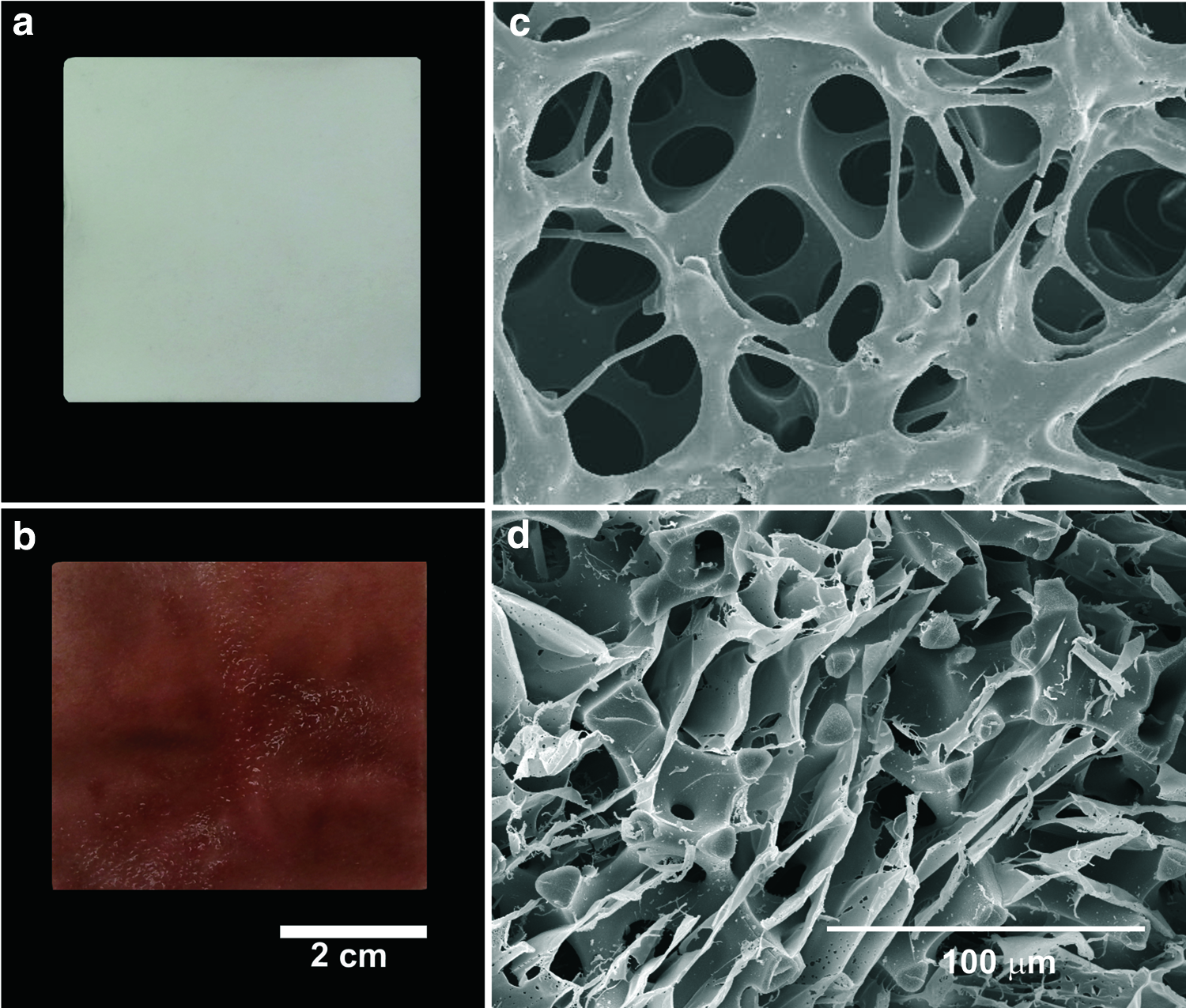
Myocardial patches. Macroscopic image before
The SAP filling procedure employed for the smaller samples was also valid for the large scaffolds; their elastomeric nature allows their folding inside a syringe and successive pulls of the syringe plunger force the viscous peptide solution to penetrate into the pores. Figure 2d shows the SAPs filling in the scaffold pores under cryoSEM, which appears as stretched-out fibers formed as water sublimates. The peptide solution successfully gels in situ within the pores of large scaffolds when in contact with the culture medium, and the β-sheet structures are positively stained with Congo red throughout the entire patch (Fig. 2b).
Cell viability and distribution after the different seeding procedures in small biohybrid patches
The effectiveness of the eight different seeding procedures was assessed after 24 h of culture. Figure 3 shows the distribution of cell nuclei (stained with DAPI) under the fluorescence microscope. In the 1-step loaded and internally seeded scaffolds, the majority of the cells were concentrated in the vicinities of the injection point in all samples, while in the case of those seeded on the upper surface in the same conditions, few cells were able to migrate through the scaffold pores and the nonattached leftover was lost (almost 90% of the cells seeded in all cases). Neither of the homogenization methods (shaking or centrifugation before culture) resulted in differences in the distribution of cells within the scaffold pores. Contrarily, when cells were seeded after SAP preload (2-step loaded scaffolds), they were able to diffuse from the seeding point, and more cells penetrated the scaffold when cells were injected than when just seeded on the surface. This method permitted a colonization of the whole available volume in the scaffold of all discs tested. Both dynamic dispersion methods were equally effective in achieving rapid cell diffusion through the peptide filler.
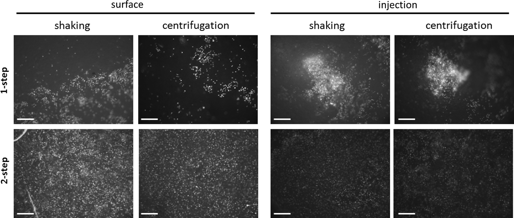
Nuclear staining (DAPI) fluorescence microscopy of small biohybrids after the different seeding procedures tested: adipose tissue-derived stem cells (ASCs) were seeded together with the SAP solution (1-step seeding) or after preloading the SAP solution (2-step seeding) onto the surface of the PEA scaffold or in its interior, and helped to invade the whole three-dimensional structure by shaking or centrifugation. Scale bars: 200 μm.
Next, a more thorough study was undertaken of the following factors: (1) the effect of the SAP solution on the efficiency of cell seeding, (2) the seeding points of the cells, either internal or superficial, and (3) the dynamic conditions to enhance cell diffusion before incubation.
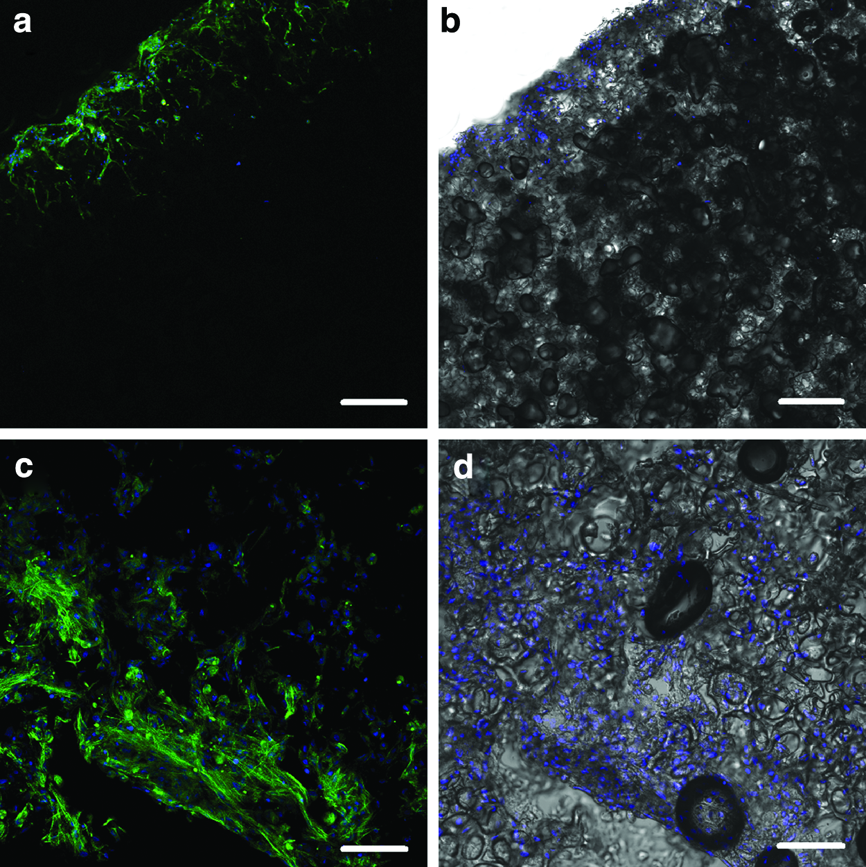
The 1-step and 2-step loaded scaffolds, both seeded in their core with a Hamilton syringe and shaken before incubation, were compared to understand the role of SAPs during the seeding. The confocal microscopy images of longitudinal slices of such scaffolds after immunocytochemistry assays (Fig. 4) confirm that when SAPs and cells are incorporated simultaneously, the cells are retained in the peptide solution at the site of injection (Fig. 4a, b, taken at the injection site); whereas when cells are seeded following the SAP loading, they are able to invade the scaffold and distribute homogeneously throughout it (Fig. 4c, d, representative of the whole scaffold). Interestingly, once lodged within the pores (24 h), the cells seem to attach to the PEA hydrophobic surface rather than remain suspended in the SAP hydrogel (see the detail, Fig. 4e, f). In 1-step loaded scaffolds, the cells maintained a spherical morphology, whereas in 2-step loaded ones, the cells appeared more elongated, which is an indicative of the adhesion to the material surface (Fig. 4e, see arrows).
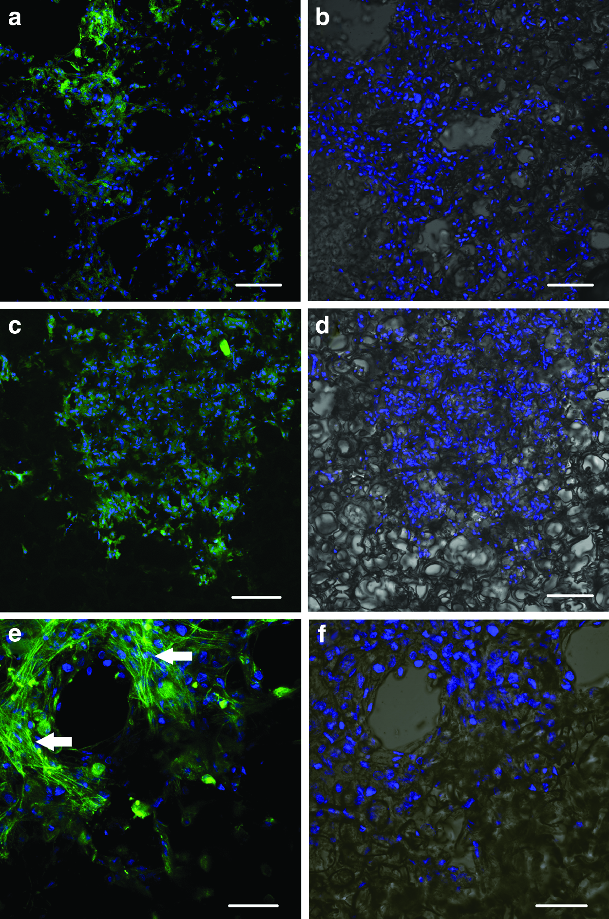
Confocal laser scanning microscope (CLSM) images of 1-step
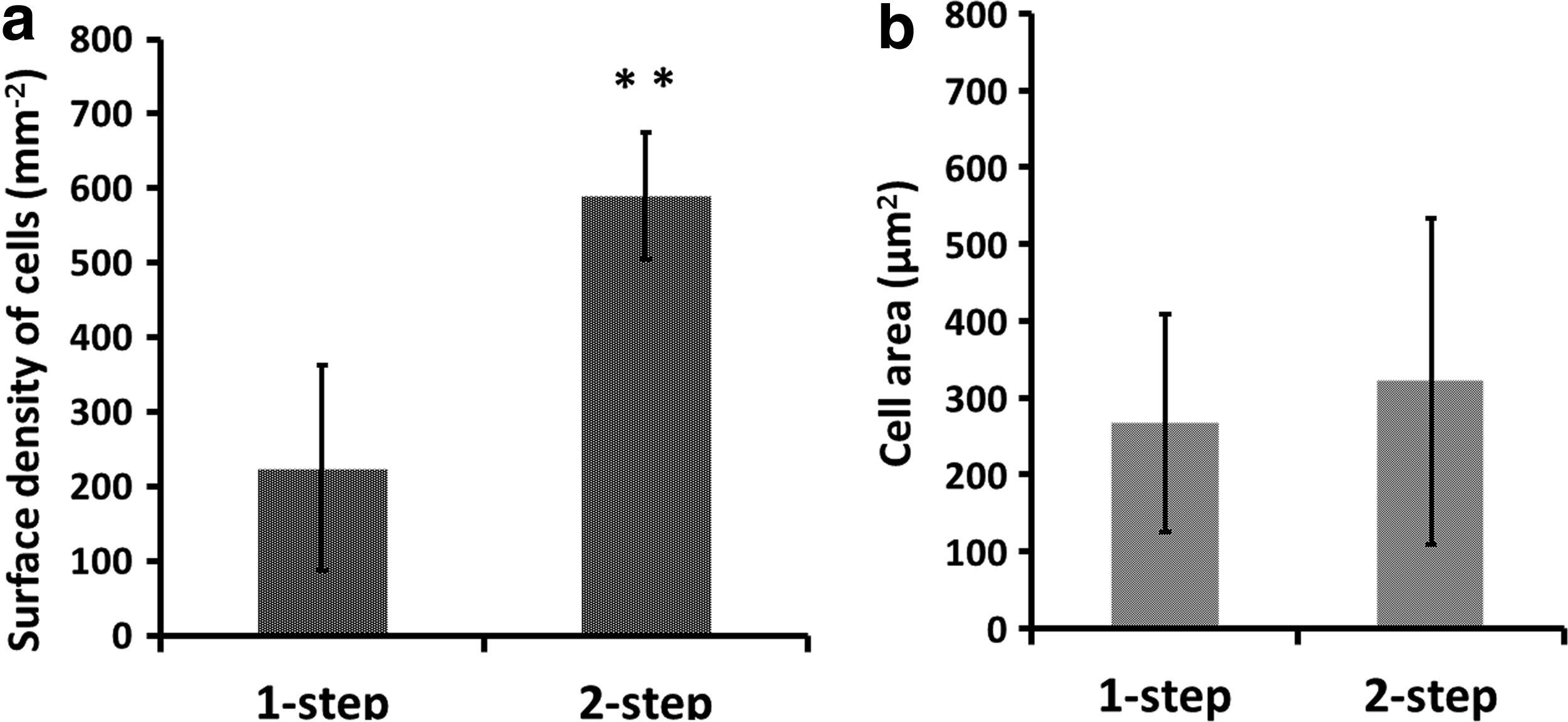
The number of cells in both 1-step and 2-step loaded scaffolds was quantified afterward by image analysis at 24 h of culture. The DAPI-labeled cell nuclei yielded the number of cells per square millimeter, which increases up to three times when the cells are incorporated after the peptide solution has been loaded in the inner pores, (Fig. 5a). The F-actin staining with phalloidin allowed to define the area covered by the cytoskeleton and the mean cell surface area, calculated as the area covered by the cells divided by the number of cell nuclei (Fig. 5b). The first parameter was considerably larger in 2-step loaded scaffolds, although the cytoskeleton area per cell was approximately the same for the two analyzed procedures, in spite of the different cell morphology observed between them. The seeding efficiency percentage as quantified by this technique was 32±4.3 in the case of nonencapsulated cells and 12.2±6.9 in the case of encapsulated cells (independently of the other combined methods used).
Two-step loaded scaffolds seeded either internally or on the upper surface and shaken before incubation were compared by immunocytochemistry and confocal microscopy to determine the depth to which cells diffuse through the peptide solution before being entrapped upon gelation. The images of transversal cuts (Fig. 6) show that few of the cells seeded on the surface were able to penetrate the porous structure, reaching only a distance 50 μm inward the scaffold (Fig. 6a, b), whereas a large proportion of the initially seeded cells remained on the surface trapped by the gelled SAPs and were lost in the sample processing. By contrast, those cells injected internally are well distributed throughout the scaffold pores (Fig. 6c, d).
CLSM images of 2-step loaded scaffolds seeded with ASCs, either onto the surface
In 2-step loaded scaffolds seeded internally, no significant differences were found between the two dynamic (shaking and centrifugation) seeding protocols proposed, insofar as both were equally effective to evenly distribute the cells (fig. not shown). It must be remarked that the application of some cell-dispersing protocol before the SAP gelling and consequent cell entrapment is crucial, as was demonstrated in 19 against a conventional (static) seeding.
Scale-up of the assembly protocol to large biohybrid patches for their implantation in a sheep model
The best results produced by the culture of small discs were those of the 2-step loaded scaffolds, seeded internally with a Hamilton syringe and smoothly shaken (or centrifuged) before incubation; such small format samples hosted in their pores the highest and most uniformly distributed cell populations. These same methods were then translated to the large patches, but with a double cell concentration, 4×10⁶ cells/cm3 of scaffold, to achieve a final cell concentration closer to the desired one. Thus, 100×106 cells, suspended in 2.5 mL of sucrose supplemented aqueous medium, were incorporated to large 5×5×0.1 cm3 scaffolds previously loaded with the SAP solution, with 50 equally spaced injections of 50 μL each, which correspond to 1 internal injection per 0.5 cm2 of external surface (as in the small discs, where they had proved to spread over such an area). Next, the biohybrid patches were shaken horizontally at 80 rpm for 30 min at 37°C and incubated. After 1 day, they were analyzed to find out their state at conditions corresponding to just before a hypothetical implantation in an infarcted myocardium. Under confocal microscopy (Fig. 7), it was possible to observe that the ASCs were homogenously distributed throughout the entire scaffold and tended to attach to the PEA trabeculae establishing cell–cell contacts, rather than remaining suspended and isolated within the gel. Similar results were found by SEM (Fig. 8), although the preparation procedure was quite disruptive and a non-negligible fraction of cells was lost from each slice, reducing the amount observed. However, cells were seen accommodated on the available PEA three-dimensional struts establishing intimate cell–cell interactions.
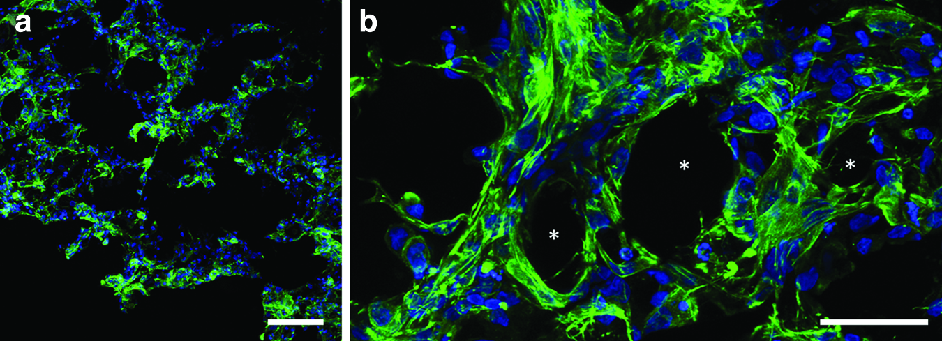
Large patches cultured 24 h: CLSM images of DAPI (nuclei) and phalloidin (actin filaments) stainings in blue and green, respectively
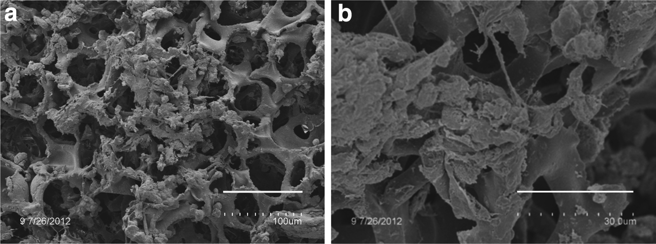
SEM images at different magnifications of large patches seeded with ASCs after 24 h culture, showing a high number of hosted cells in the scaffold structure. Scale bars: 100 μm
Discussion
Heart failure leads to myocardial wall weakening, and consequently to a ventricular chamber enlargement and ventricular wall thinning. This raises the need to physically assist the infarcted heart to decrease the ventricular wall deterioration. Previous attempts in this direction included the permanent implantation of cardiac wrapping devices of synthetic materials (polyester and nitinol meshes),40–42 but these failed in the improvement of systolic function and showed additional adverse effects like restriction in the diastolic function and additional fibrosis. On the other hand, the association of stem cells and polymeric scaffolds raises the expectations of achieving the repair of the myocardial tissue and avoiding ventricular chamber dilation. These biohybrid patches could provide a supporting band-aid effect, limiting the spread of the infarcted areas and reinforcing the ventricular wall to yield stress tolerance by a passive girdling effect of the patch and improving strain distribution along the ventricular wall, while reducing cell apoptosis by the paracrine effect of the grafted stem cells.
To progress from bench to bedside, studies on small animal models must be followed by large preclinical animal ischemic models. This raises specific readjustments to scale-up the implants, and the need to assess the effect of implant size on factors such as the SAP distribution within the elastomeric membrane as well as on the viable concentration of seeded stem cells. In the future, devices will be designed for left ventricular and/or right ventricular support and regeneration, including different sizes for partial (patch) or complete ventricular wrappings, with implant characteristics (mechanical, physical, chemical, and biological) adapted for the left or the right ventricle geometry, physiology, and pathology.
The purpose of the present study was to establish a methodology to determine an effective way to build-up large implantable biohybrid patches, choosing among different possible strategies available to combine their three components: scaffold, SAP gel, and cells. The variables under study were as follows: the sequence for the incorporation of the peptide solution and the cells, the method to seed the cells in the scaffold, and the subsequent dynamic conditions before incubation to achieve the greatest cell invasion and most uniform cell distribution within the construct. The alternative choices were first studied in small-sized samples, and the selected methods were then scaled-up to establish a protocol to prepare and assemble the large biohybrid patches for their subsequent implantation in the infarcted sheep.
The need for a large patch uniformly colonized by a high number of cells, 24 h before surgery as a hypothetical clinical situation, determined the main design options for the biohybrid construct, which would consist in a filler hydrogel inside the pores of a patch with regular pore architecture, produced by a template. The seeded cells in the implant could be viable after surgery only if the pore sizes of the scaffold allow for a rapid ingrowth of microcapillaries, and it was judged that only a template-based manufacture of the scaffold, using a sintered template made of regular-sized spherical microbeads, could ensure the necessary control over pore size and distribution. Other porogenic techniques lead to less controllable porous structures that cannot ensure perfect connectivity and size of the pores. Furthermore, it was thought that a soft hydrogel filling the pores of the scaffold would constitute a medium for an improved cell spreading throughout the scaffold, thus permitting a fast and uniform cell colonization of the implantable structure. These basic hypotheses were confirmed by the results of our study. As several works have remarked,43–45 the microstructure of the scaffolds for tissue engineering is critical to ensure the hosting of a high enough cell density, while allowing for cell migration, diffusion of nutrients and metabolites, new tissue growth, and vascularization.
The SAP solution proved to be an excellent retention medium for the cells, provided that it was incorporated within the scaffold pores before the seeding (2-step loaded scaffolds). In the 24 h following the seeding, ASCs first migrate through the hydrogel and then attach to the PEA hydrophobic trabeculae in an extended conformation with numerous cell–cell contacts. When cells were seeded simultaneously with the peptide in a specific location of the scaffold (1-step loaded scaffolds), the cells were only able to colonize the vicinities of the spot where they had been seeded.
The internal seeding of the cells (injected with a Hamilton syringe) into the scaffold gave much better results than seeding on top of the surface of the scaffold, because in this latter case, those cells that have not been able to invade the three-dimensional structure (especially if they have been seeded simultaneously with the peptide solution) are dragged away when the culture medium is added and are consequently lost (up to a 90% of the initial cells, approximately). Before the gelling of the peptide solution, upon the addition of the culture medium and the consequent entrapment of cells, the mechanical assistance to improve cell spreading by shaking or centrifuging contributes to the uniform distribution of the cells throughout the construct, as has already been established in previous work, 19 when compared with a static seeding. The results obtained here reveal that such mechanical assistance can be performed by different means (use of shaker or centrifuge, in our case) without significant differences in the outcome, at least in the conditions tested here. We conclude that a smooth shaking in incubating conditions for 30 min is able to achieve a uniform distribution of the cells throughout the patch.
These observations, gained from the experiments on small scaffold samples, resulted in a biohybrid patch assembly method consisting of the following steps: (1) a preloading of the scaffold with the peptide solution with the help of vacuum to force the viscous solution to penetrate into the pores of the hydrophobic scaffold, (2) the injection of the cell suspension within the filled pores of the scaffold, and (3) a smooth shaking of the biohybrid patch to distribute the cells before the peptide solution was gelled by adding the culture medium, and (4) a subsequent incubation of the construct.
Next, this assembly protocol was scaled-up to the needs of a large 5×5 cm2 scaffold for a myocardial patch in the context of a preclinical study.
The transition from the manufacture of small scaffolds to that of large format ones posed specific technical problems. Achieving a uniform sintering of the microbeads throughout the large porogenic template is a critical step to guarantee the homogeneity of the porous structure of the ensuing scaffold, especially in what refers to the pore interconnectivity. Furthermore, large scaffolds require a much more careful handling when swollen in solvents during the washing procedures to obtain one-piece nondefective structures. A quality check to control these aspects had to be introduced, based on the inspection of SEM images of the structures obtained in different, randomly selected spots of the large patches.
Regarding cell seeding, these large scaffolds required multiple, uniformly distributed, injection sites with a Hamilton syringe. To translate the results obtained from the experiments on the smaller samples, the area of the large patch was divided into units of the same area as the smaller samples, where it had been established that uniform cell distribution could be achieved in 24 h with the selected protocol. This resulted in a number of 50 injections uniformly spaced through the large scaffold. Once the cells were seeded, a mechanically assisted dispersion greatly helped the uniform colonization of the construct. Both dispersion methods studied here were equally effective with the small samples; nonetheless, in view of the technical limitations facing centrifugation of large biohybrids, the simpler shaker-assisted protocol was finally selected. After 24 h of culture, cells were observed throughout the material, adhered to the PEA walls and establishing cell–cell interactions.
In our later preclinical studies, the bioactive implants were successfully attached by surgery to the infarcted areas. 37 The application of the bioactive implants on ischemic sheep hearts showed enhanced parameters of systolic and diastolic functions in a 6-month trial. The LV chamber dilatation was reversed and infarct size was reduced in treated animals. In addition, Magnetic Resonance Imaging (MRI) showed in the biohybrid scaffold-treated group, a significant reduction of the infarct volume related with the LV myocardial mass.
Ventricular support bioprostheses should avoid heart transplantation or offer a relatively secure mid- to long-term bridge to heart transplant allowing critically ill patients to significantly improve their quality of life while waiting for a heart donor.
Conclusions
An effective protocol for assembling large biohybrid patches consisting in an elastomeric scaffold, with a peptide gel filling entrapping ASCs, has been established. The alternative options open to achieve this goal have been settled with different experiments, and specific parameters related to the fabrication of large uniform scaffolds, with a highly interconnected porosity, have been discussed, including their manipulation in the different stages, and the incorporation of the peptide solution and a high concentration of homogeneously distributed cells. The assembly protocol of these large biohybrid patches consisted in the incorporation of the peptide solution into the scaffold pores, the injection of the cells in different and evenly spaced locations, their dispersion by shaking before the gelling of the peptide hydrogel, and the incubation of the hybrid construct. This sequence ensures that after a 24-h in vitro culture, such biohybrid patches host a large number of viable cells uniformly distributed throughout the whole volume of the patch, and that they are ready to be implanted onto the infarcted myocardium.
Footnotes
Acknowledgments
The authors thank Dr. Carolina Soler-Botija and Dr Antoni Bayés-Genis for her generous contribution in the in vitro cell expansion of ASCs. The authors acknowledge the financing from the European Commission through the “Regeneration of cardiac tissue assisted by bioactive implants” (RECATABI) FP7 NMP3-SL-2009-229239 project. MMP acknowledges support of Instituto de Salud Carlos III with assistance from the European Regional Development Fund through CIBER-BBN initiative.
Disclosure Statement
No competing financial interests exist.