
Abstract
The field of tissue engineering is rapidly evolving, generating numerous biodegradable materials suited as regeneration platforms. Material sterility is of fundamental importance for clinical translation; however, a few studies have systematically researched the effects of different sterilization methods on biodegradable materials. Here, we exposed a novel bioabsorbable nanocomposite based on a poly(ɛ-caprolactone urea) urethane backbone integrating polyhedral oligomeric silsesquioxane nanoparticles (POSS-PCLU) to autoclave, microwave, antibiotics, and 70% ethanol sterilization and systematically correlated differences in material characteristics to the attachment, viability, proliferative capacity, and shape of human dermal fibroblasts (HDFa). Nanotopographical profiling of autoclaved or microwaved surfaces revealed relatively deep nano-grooves, increasing total surface area, roughness, and hydrophobicity, which resulted in significantly fewer adherent cells. Antibiotics or 70% ethanol-treated surfaces displayed shallower nano-grooves, a more hydrophilic character, and significantly greater cellular adhesion (p<0.05). In fact, relative cell proliferation on ethanol-treated films surpassed that of cells grown on every other surface by a factor of 9 over 7 days. Filamentous actin staining demonstrated spindle-like morphologies characteristic of HDFa when grown on ethanol-treated films as opposed to cells grown on other films that were significantly more spread out (p<0.05). We argue that treatment with 70% ethanol serves not only as a laboratory-based sterilizing agent but also as a postproduction processing tool to enhance cytocompatibility of tissue engineering scaffolds.
Introduction
T
Polyhedral oligomeric silsesquioxane-modified poly(ɛ-caprolactone urea)urethane (POSS-PCLU) is a novel bioabsorbable nanocomposite material suited as a platform material for tissue regeneration applications.2,5–7 POSS-PCLU scaffolds can be fabricated in a number of ways, including casting, coagulation using a salt leaching method, three-dimensional (3D) printing, and electrospinning to suit individual applications. This versatility renders bioabsorbable POSS-PCLU a highly desirable platform material for tissue engineering of temporary implantable devices. However, a frequently underestimated, although highly relevant aspect in the development of implantable devices is the ability to ensure impeccable sterility. As micro-organismal contamination tends to falsify in vitro studies as well as elicit a potentially exuberant immune reaction toward such devices in the human, sterility remains a fundamental prerequisite for the transition of tissue engineered scaffolds into the clinical arena. Sterilization is defined as the destruction of all forms of life by heat, chemical or radiation treatment. 8 Conventional laboratory-based sterilization techniques include high pressure steam sterilization (autoclaving) and exposure to microwaves. While effectively removing surface-adherent bacteria, these methods often irreversibly damage hydrolytically unstable materials, leading to material melting, collapse of internal porous architectures, unpredictable changes in mechanical and surface properties, as well as altered rates of biodegradation. Chemical means of microbial removal such as exposure to 70% ethanol or antibiotics, while strictly speaking not considered sterilizing agents, have been routinely adopted as antimicrobial agents for laboratory cell studies. 9
Besides the sterilizing effect, exposure to heat sources or potentially abrasive chemicals influences surface topographies (e.g., surface roughness), thereby significantly changing cell–substrate interactions and cell function.10–12 Changes in cellular attachment, motility, proliferation, and differentiation 13 have been shown to be brought about by modulating material surface tension and chemical and mechanical properties. 14 It is a recognized fact that 3D topographical features of polymers, 15 oxides, 16 carbon nanofibers, 17 and metals 18 are related to cyto- and biocompatibility of the respective materials. Surface topographies ranging within the nanometer scale are thought to facilitate cell–substrate interactions by mimicking the biological cues present on extracellular matrix components of natural tissues.19–22 Since focal adhesion proteins located on the outer surface of the cell membrane (e.g., integrins) have a diameter measuring ∼10–15 nm, cell–substrate adhesion is enhanced by nanoscaled surface topographies. 23
The aim of this work was to systematically investigate (1) the effectiveness of four different, easily accessible sterilization methods for bioabsorbable POSS-PCLU material, and (2) the influence of these sterilization methodologies on surface topography and chemistry as well as cell–substrate interactions.
Materials and Methods
POSS-PCLU nanocomposite material synthesis
The polymer was prepared according to a previously published method. 24 In brief, dry polycaprolactone diol (Mw 2000) and trans-cyclohexanechloroydrinisobutyl-POSS were placed in a reaction flask equipped with a mechanical stirrer and nitrogen inlet. The mixture was heated to 135°C to dissolve the POSS nanocage and then cooled to 60°C. To this, dicyclohexylmethane diisocyanate (Desmodur W; Bayer) was added and reacted under nitrogen at 70°C for 90 min to form a prepolymer. Then, N,N-dimethylacetamide (DMAC) was added and the mixture was cooled to 40°C. A mixture of ethylenediamine and diethylamine in DMAC was added to allow chain extension of the prepolymer. 1-Butanol in DMAC was added to the mixture to form an 18% POSS-PCLU solution. All chemicals and reagents were purchased from Sigma-Aldrich Ltd. Figure 1 schematically represents the reaction steps involved in the synthesis of a polyurethane incorporating POSS nanoparticles.
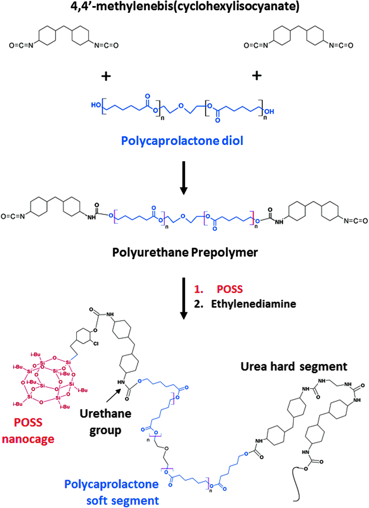
Preparation of POSS-PCLU polyurethane by reacting polycaprolactone diol and trans-cyclohexanechloroydrinisobutyl-POSS with two diisocyanates and chain extending the resulting polyurethane prepolymer with ethylenediamine. POSS-PCLU, polyhedral oligomeric silsesquioxane-modified poly(ɛ-caprolactone urea)urethane. Color images available online at
Manufacturing of scaffolds
Samples were fabricated by either casting or coagulation/phase inversion method to obtain non-porous films or porous sponges, respectively. Cast sheets were prepared by pouring diluted 15 wt.% POSS-PCLU polymer solution (∼8 mg) into a clean Petri dish (Ø 10 cm) and allowing solvent evaporation overnight at constant temperature (65°C). Coagulated scaffolds were prepared by a sacrificial porogen leaching technique combined with a phase inversion coagulation method. The polymer solution was supplemented with sodium bicarbonate (NaHCO3) particles (Brunner Mond) and surfactant (Tween 20) to obtain a viscous slurry. An excess of NaHCO3 particles were dissolved in de-ionized water and poured over the stainless steel plate. The liquid phase was evaporated overnight at 65°C to leave a thin layer of particles over which the slurry was then poured. Top surface modification by sprinkling of NaHCO3 particles was applied to avoid skin formation and increase percentage porosity. The polymer mixture was then extruded according to a protocol developed in-house. Overnight extrusion in sterile de-ionized water resulted in solvent exchange and NaHCO3 leaching and the fabrication of porous scaffolds. Washing over a period of 48 h using regular changes of sterile de-ionized water completely removed all remaining salt particles and DMAC, as confirmed by inductively coupled plasma optical emission spectrometry (Warwick Analytical Services).
Sterilization techniques
Autoclave sterilization
Samples were exposed to saturated steam at 121°C for 15 min at pressures of 115 kPa (Classic Prestige Medical autoclave; Prestige Medical Limited). Before use, samples were cooled to room temperature for 12–24 h.
Microwave sterilization
Samples were completely submerged in de-ionized water, placed in an unmodified domestic microwave, and exposed to highest power radiation (750 W) for a total of 30 s. 25 Samples were allowed to cool down before use.
Antibiotic treatment
Samples were placed in a 5% (v/v) antibiotic antimycotic solution (10,000 U/mL penicillin G, 10 mg/mL streptomycin sulfate, and 25 mg/mL amphotericin B [Sigma-Aldrich]) in phosphate-buffered saline (PBS) for 24 h at 4°C. Samples were washed thrice with PBS under agitation before use.
Ethanol treatment
Samples were ethanol-treated according to a method published by Kweon et al. 26 Samples were soaked in two changes of 70% ethanol over 30 min under agitation, and subsequently washed in four changes of PBS solution over 2 h.
Control
Non-sterilized samples served as a control for all tests.
Sterility testing
Sterility testing was performed according to British Standards (ISO 11737-2:2009) on cast and coagulated samples immediately after sterilization. The samples were immersed in normal cell culture medium to cultivate micro-organisms and maintained at 37°C for 7 days. Untreated scaffolds were used as the positive control, and sterile medium alone served as the negative control. Samples were assessed daily for any turbidity that denotes infection and inefficient sterilization. All experiments were conducted in triplicate.
Scaffold characterization
Scaffold shrinkage
Changes in scaffold diameter poststerilization were used as an indicator for sterilization-induced scaffold shrinkage. Diameters were measured both pre- and poststerilization using a calliper.
Scaffold surface micro- and nanotopography assessment
Scanning electron microscopy (SEM): Differences in scaffold thickness, porosity, pore interconnectivity, and wall smoothness poststerilization were evaluated using SEM digital photography at magnifications of ×40,×320, and ×640. Sterilized scaffolds were washed with PBS and fixed in 3% (w/v) glutaraldehyde for 1 h at room temperature before being dehydrated with ethanol/distilled water (10% ethanol increments) at 41°C. Samples were then mounted on aluminum stubs for conductivity and sputter coated with a thin film of gold (A SC500 [EM Scope]) before sample analysis with a Philips 501 scanning electron microscope. ImageJ (freeware) was used to analyze the specified parameters.
Attenuated total reflectance Fourier transform infrared spectroscopy: Fourier transform infrared spectra (FTIR) were obtained on a Jasco FT/IR 4200 Spectrometer equipped with a diamond attenuated total reflectance (ATR) accessory (Diamond MIRacle ATR; Pike Technologies). A total number of three pieces (1.5×1.5 cm per piece) per sterilization technique or control and three points on each scaffold (at 4 mm intervals along the midline of the specimen) were analyzed. Spectra were produced from an average of 20 scans at 4 cm−1 resolution over a range of 600–4000 cm−1 wave numbers. A background scan was performed before each sample measurement.
Atomic force microscopy: Surface nanotopographies of sterilized and control cast polymer samples were measured using a non-contact mode atomic force microscope (AFM; Bruker Dimensions 3100). Evaluation of the average surface roughness (Ra), root mean square roughness (Rq), and correlation length (ξ) were carried out on a surface area of 20×20 μm using Nanoscope Analysis software (Bruker Corporation). Commercially available AFM tips (radius of curvature 7 nm, PPP-NCH; Windsor Scientific) were used with intermediate contact mode.
Surface wettability measurement using contact angle (θ)
Contact angle (θ) measurements were obtained using a goniometer (EasyDrop DSA20E; Kruss) that was equipped with a digital camera and image analysis software (DSA1 version 1.80; Kruss). De-ionized water was used as the wetting liquid that was deposited onto the samples using an automated syringe. Sessile drop method was used to analyze contact angles of the air-water-substrate interface of control and sterilized cast samples that were measured thrice in three samples of every group.
Mechanical properties of scaffolds
Tensile stress-strain properties were assessed according to British Standards (BS ISO 37:2005). Cast or coagulated POSS-PCLU samples were cut into dumbbell-shaped pieces type 3 (shaft length 20 mm, width 4 mm, n=5) using a cutting press (Wallace Instruments). Thickness was measured using a digital electronic outside micrometer. Uniaxial tension was applied to either ends of the scaffolds until failure using an Instron-5565 tensile tester (Instron Ltd.) that was equipped with a 500 N load, pneumatic grips with 1 kN capacity and at a rate of 100 mm/min. Bluehill software was used to analyze the scaffold's tensile strengths at room temperature. Stress (MPa) was calculated by dividing the force generated during stretching by the initial cross-sectional area. Strain was calculated as the ratio of the change in length in reference to the original sample length (%).
Cell cultures
Primary fibroblasts were isolated from human dermis (human dermal fibroblasts [HDFa]) and cultured in Dulbecco's modified Eagle's medium (DMEM) supplemented with 1 mM L-glutamine, 10% fetal bovine serum (FBS), 1% penicillin/streptomycin, and 1% Fungizone (Invitrogen). HDFa were cultured to 80–90% confluency, trypsinized, and either re-plated or stored in 10% DMSO/10% FBS in −80°C until required. For our studies, HDFa between passages 4–8 were used.
To allow for isolated assessment of sterilization-induced variations in surface topography and the influence on cell adhesion and proliferation, HDFa cells were seeded onto DMEM presoaked and non-porous cast POSS-PCLU scaffolds (1.9 cm2) at a density of 5×10⁴ cells/150 μL/scaffold and incubated at 37°C, 5% CO2 for 2 h. Then, 1 mL culture medium was added and cells were incubated until the predetermined time points. Previous tests determined 2 h sufficient for cell attachment (results not shown). A staggered seeding technique was utilized to ensure cell adhesion onto scaffolds rather than tissue culture plastic (TCP). 27 TCPs containing no scaffolds were seeded with an equal amount of cells (positive control) or medium only (negative control). Medium-soaked POSS-PCLU scaffolds not seeded with cells served as background noise, which was subtracted from final results of cell-seeded scaffolds. Cells were incubated at 5% CO2 in air at 37°C, and culture medium was changed every 2–3 days for a total of 7 days.
To demonstrate the material's suitability as a potential tissue engineering platform for skin regeneration, 3D porous scaffolds were seeded with HDFa at a density of 5×10⁴ cells/150 μL/scaffold and incubated at 37°C, 5% CO2 and cellular viability, proliferative capacity, as well as migration into the scaffold's internal architecture were analyzed.
On days 1, 3, and 7, cell morphology was visualized using the filamentous actin (F-actin) stain fluorescein isothiocyanate (FITC)-phalloidin and nuclei counterstained with propidium iodide (PI) (both Sigma-Aldrich). Cell viability and proliferative capacity were assessed using Live/Dead® stain and AlamarBlue® assay, respectively.
Assessment of cell viability using AlamarBlue cell proliferation assay
Cell metabolism and proliferative capacity were assessed using AlamarBlue assay (Invitrogen). HDFa were grown on cast or coagulated POSS-PCLU scaffolds and incubated for 1, 3, and 7 days. At each time point, media and non-adherent cells were removed with sterile PBS (0.1 M, pH 7.4). AlamarBlue solution in normal culture medium (ratio of 1:10) was added to each scaffold and incubated at 5% CO2 and 37°C for 4 h. Fluorescent readings were recorded by Fluoroskan Ascent Fluorescence Microplate Reader (Thermo Scientific™) with excitation at 530 nm and emission at 590 nm. Fluorescence readings for culture medium only were used for baseline corrections. Cell viability in terms of % of control cells grown on TCP is derived from the following equation
28
:
where Ftest is the fluorescence of cells grown on sterilized or non-sterilized surfaces, Fblank is the fluorescence of medium only (no cells), and Fcontrol is the fluorescence of cells grown on TCP.
Live/Dead cell survival assay
Cell viability on sterilized scaffolds was assessed at days 1, 3, and 7 using the Live/Dead fluorescence assay (Invitrogen) according to the manufacturer's instructions and the number of attached live and uniformly green stained and non-viable/dead, pyknotic red cells was assessed immediately on staining using an Eclipse C1 Plus Confocal microscope (Nikon) with a ×10 objective lens.
Cytoskeletal staining
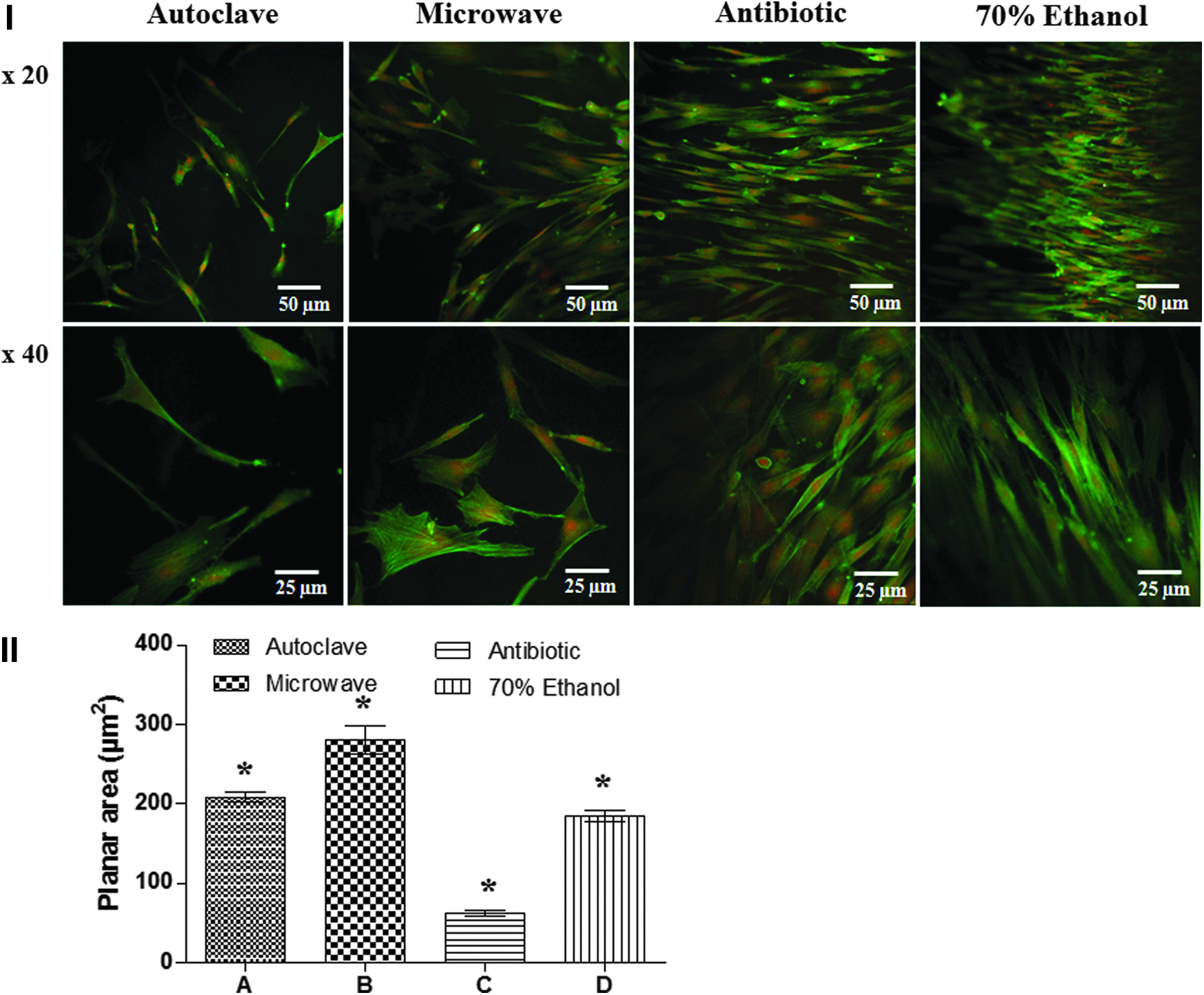
Cell attachment to differently sterilized scaffolds and their morphology were studied using FITC-labeled phalloidin according to the manufacturer's instructions. Cells grown on sterilized samples for 1, 3, and 7 days were fixed using 4% paraformaldehyde in PBS, permeabilized with 0.1% Triton-X 100 in PBS for 20 min, and then blocked with 1% bovine serum albumin (BSA) in PBS for 45 min. Cells were exposed to a 1:40 ratio FITC-phalloidin mixture in PBS supplemented with 0.1% (w/v) BSA for 45 min for visualization of F-actin. Nuclei were counterstained using PI stain. Images were taken using an Eclipse C1 Plus Confocal microscope (Nikon) with a ×20 and ×40 objective lens. Cell attachment and spreading to each surface were quantified by measuring the planar area of cells using ImageJ (freeware). The average planar area was determined from 30 cells per sample.
Potential application of POSS-PCLU as skin tissue engineering scaffolds
The potential for cellular movements across 3D POSS-PCLU scaffolds was assessed on 70% ethanol-treated porous scaffolds and compared with the migratory capacities of HDFa in commercially available Integra® Dermal Regeneration Template (Ethicon Endo-Surgery, Inc.), which consists of a porous coprecipitate of bovine tendon type I collagen and shark glycosaminoglycan (chondroitin-6-sulfate). At day 7, cellular nuclei were visualized using PI stain and confocal imaging in conjunction with z-stacking.
Statistics
All data are presented as mean±standard deviation. Experiments were repeated six times unless stated otherwise. Data comparisons were carried out by one-way ANOVA analysis of variance. Significant differences between experimental groups were determined using Bonferroni's test of multiple comparisons or Dunn's multiple-comparison post-test in the case of surface roughness analysis. A p-value of <0.05 was considered statistically significant.
Results
Effectiveness of different sterilization methods by immersion in culture medium
Table 1 summarizes the results of the sterility tests. An incubation period of 7 days allowed sufficient time for any potential microbial migration. After 7 days, all non-sterilized cast and coagulated control samples showed signs of infection and bacterial growth, whereas medium-only controls did not exhibit turbidity, thus proving the feasibility of this testing technique. Autoclave sterilization (high pressured steam) is known to be effective and rendered both cast and coagulated samples sterile. However, extensive polymer deformation and shrinkage due to melting of the heat-sensitive POSS-PCLU were observed in the coagulated samples. Exposure to microwaves for 30 s sterilized cast samples but not coagulated sponges, likely due to deflection of microwaves by the polymer material and subsequently insufficient penetration into the core. Antibiotic treatment resulted in unreliable sterilization and clouding of the incubation medium for both cast and coagulated samples. Immersion of samples in two changes of 70% ethanol over half an hour and subsequent washing with four changes of sterile PBS over 2 h resulted in effective removal of micro-organismal contaminants from both cast and coagulated samples.
Cast samples were successfully sterilized using autoclave, microwave, and 70% ethanol treatments but still harbored micro-organisms after soaking in antibiotic solution. Coagulated sponges were less effectively sterilized with remaining micro-organisms within the porous structure after microwave and antibiotic treatment. High pressure, high temperature autoclaving effectively sterilized the sponges but led to significant polymer melting. Finally, treatment of porous sponges with 70% ethanol effectively removed micro-organisms while preserving the internal porous architecture of the scaffolds. Experiments were carried out in triplicate.
−ve control, medium only; +ve control, non-sterilized sample; x, no growth present; ✓, growth present.
Sterilization induced microscopic changes in scaffold architecture and topography
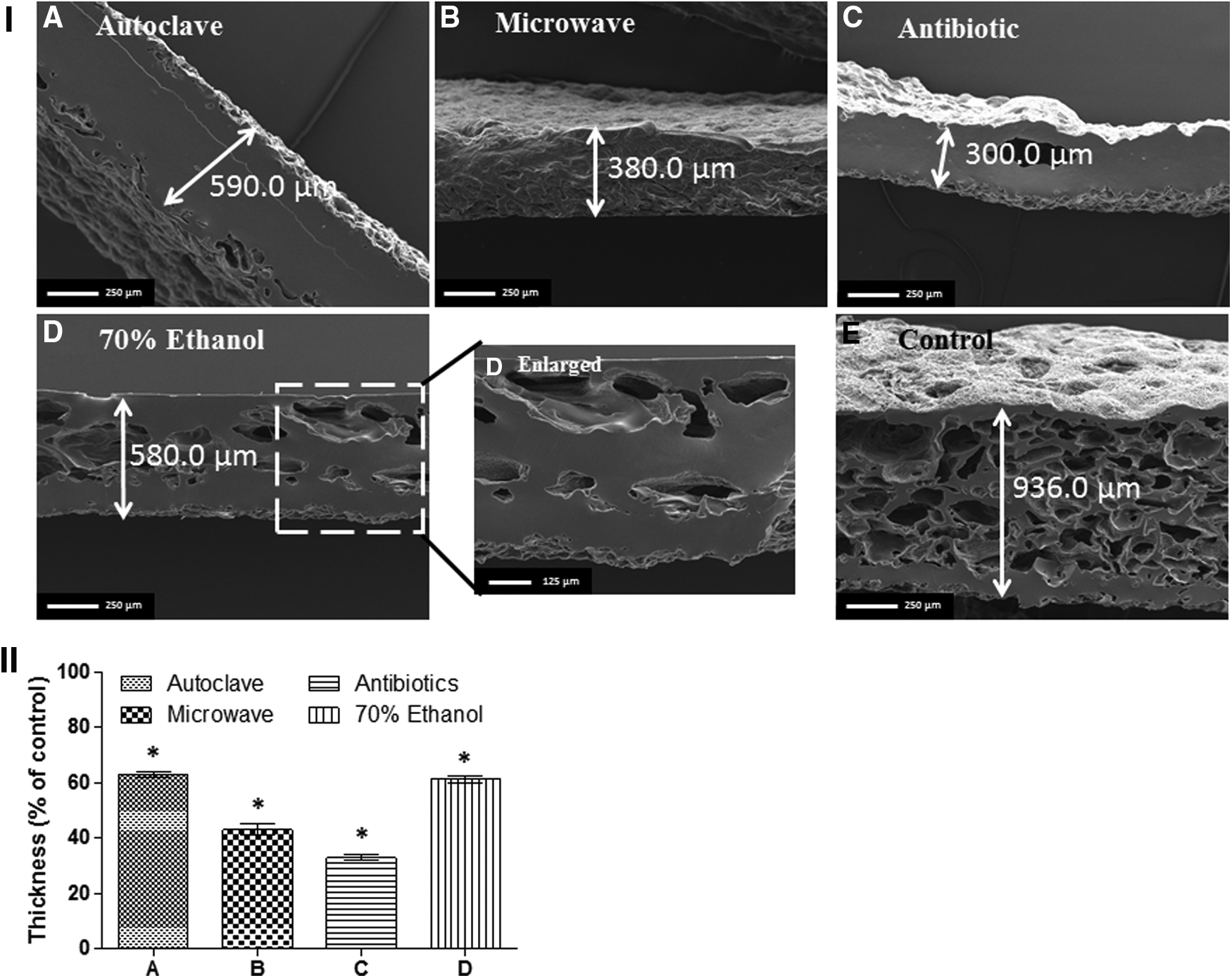
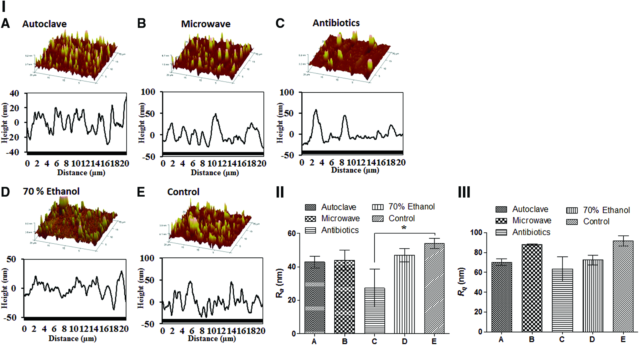
Surface micro- and nanotopographies of unsterilized and sterilized POSS-PCLU sheets were analyzed using SEM (Figs. 2 and 3), FTIR spectroscopy (Fig. 4), goniometry (Fig. 5), and AFM (Fig. 6). The SEM image of an unsterilized coagulated sample surface (Fig. 2E) exhibits sub-micron scale tufts of polymer imprinted during the surface modification step of scaffold synthesis. Such tufts were lost after using autoclave, microwave, and 70% ethanol sterilization with polymer melting and irregular re-formation into larger and flatter ridges (Fig. 2A, B, D). Antibiotic treatment seemed to have the least effect on surface melting from a microscopic point of view (Fig. 2C). SEM images of vertically cut edges of an unsterilized coagulated scaffold revealed a spongy internal architecture characterized by numerous interconnected pores [Fig. 3 (I) E], while all sterilization methods resulted in a reduction [Fig. 3 (I) D] or complete loss of the porous internal architecture [Fig. 3 (I) A–C]. Scaffolds treated with 70% ethanol were found to retain the porous internal architecture best compared with other sterilization methods [Fig. 3 (I) D]. The reduction in scaffold thickness was statistically significant for all sterilization methods [Fig. 3 (II), *p<0.001]. Scaffolds treated with 70% ethanol retained their thickness best along with autoclaved scaffolds.
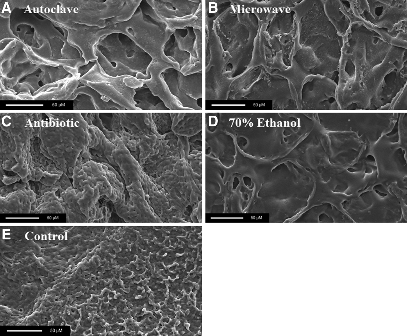
Representative SEM images of coagulated sterilized
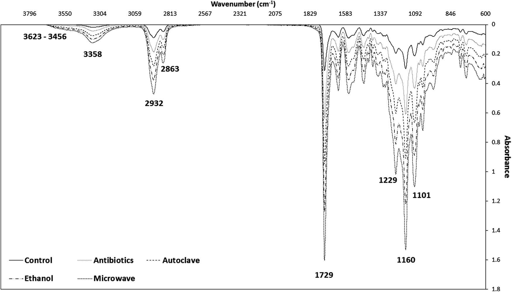
Fourier transform infrared spectra spectrum of cast sterilized and control samples. Particularly high temperature steam sterilization (autoclaving) induced a broadening of the OH peak at around 3550 cm−1. No reductions in peak intensity were observed at the methylene bridge at 2932 cm−1 or the carbonyl region (1729 cm−1), indicating the presence of both ester and urethane linkages and an intact hard segment.
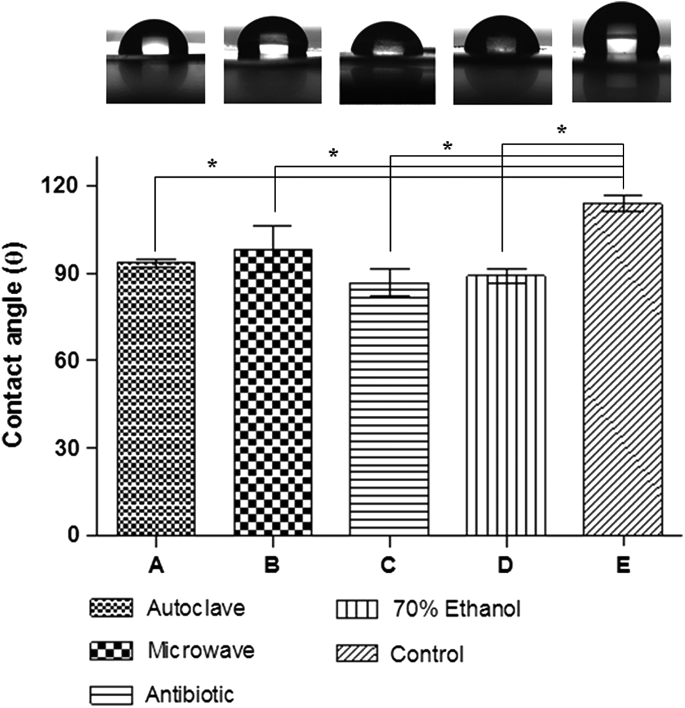
Contact angle (θ) measurements on cast sterilized
Chemical bond scission demonstrated by ATR-FTIR
Material surface chemistry and sterilization induced changes were analyzed using FTIR spectroscopy. Figure 4 shows an FTIR spectrum overlay of individual representative cast POSS-PCLU surfaces sterilized using each technique. All sterilization methods and, in particular, high temperature steam sterilization (autoclaving) induced an increase and broadening of the OH absorption peak at 3550 cm−1 that corresponds to hydrolytic degradation of the soft segment of PCLU component of this composite polymer. Multiple cycles of high temperature steam sterilization confirmed a gradual increase in height in the OH band, indicative of successive hydrolysis of the polymer and accumulation of OH-capped degradation products such as carboxylic acids (results not shown).29,30 Hydrogen bonding between these carboxylic OH moieties influenced peak shape, causing significant peak broadening. 31 Furthermore, peak shifting toward higher wavenumbers was observed at around 3550 cm−1, which indicates degradation with mass loss. Since higher vibrational frequency is inversely proportional to mass of vibrating molecules, the lighter the molecule, the higher the vibration frequency and the higher the wavenumber. Increases in peak intensity were observed at the methylene bridge at 2932 cm−1 and the carbonyl region at 1729 cm−1 as well as at peaks around 1632 (urethane carbonyl), 1560–1530, 1350, 1233, 992, and 793 cm−1. Such increases in intensity may be correlated to the loss in thickness after sterilization with subsequent compaction of the polymer sample.
Surface roughness changed significantly as demonstrated with contact angle and AFM
Sterilized and non-sterilized cast samples were also characterized in terms of wettability and nanotopography (Table 2). Contact angle measurements of non-sterilized control samples demonstrated an inherent hydrophobic character of the POSS-PCLU polymer surface (θ=114°) All sterilization methods significantly reduced the hydrophobicity compared with the control (Fig. 5, p<0.05). Polymers treated with antibiotics and 70% ethanol exhibited the most extreme loss in θ (mean loss of 23.7% and 21.9%, respectively), while exposure to microwaves had the least effect on contact angle (mean loss of 13.9%). Treatment with antibiotics and 70% ethanol resulted in the most hydrophilic surfaces with θ of 87° and 89°, respectively. The results obtained by goniometry were further corroborated by surface roughness measurements using AFM [Fig. 6 (II), (III) and Table 2]. Figure 6 (I) shows the scaffold surfaces on a nano-level as well as the roughness profiles of sterilized and control surfaces. All surfaces exhibited a random pattern of peak heights with antibiotic-treated surfaces demonstrating an overall smoother appearance of grooves compared with the other surfaces. Ra as well as Rq were the highest in non-sterilized samples while ξ of unmodified surfaces was in the mid-range (ξ=14.6 nm) (Table 2). Based on the AFM data, Ra and Rq decreased in a parallel fashion to the reductions observed in contact angle measurements. Although one-way ANOVA did not yield a significant difference among most data sets of Ra and Rq, samples exposed to antibiotic solution experienced the most pronounced and statistically significant loss in Ra (50% reduction compared with control, p<0.05). Rq, while generally following a falling trend on sterilization, did not exhibit statistically significant reductions in values compared with control surfaces. ξ of sterilized surfaces differed significantly from control surfaces with microwave and antibiotic solution-exposed samples exhibiting a significantly increased ξ value (+213.6% and +117.8%, respectively) and autoclaved or 70% ethanol-treated surfaces demonstrating a significant reduction in ξ (−71.2% and −62.3%, respectively). In summary, changes in contact angles corresponded to nanotopographical changes in roughness as demonstrated by AFM.
All sterilization methods resulted in a significant reduction in contact angle, which correlated to a reduction in mean surface roughness (Ra) and root mean square roughness (Rq). Correlation lengths (ξ) were also significantly affected by all sterilization methods (p<0.05), most notably on cast microwaved surfaces. Contact angles were measured at 24 h after sterilization. Values are given as mean±one standard deviation.
Mean surface roughness (the arithmetic average of the absolute values of all points of the profile.
Root mean square roughness (RMS, the root mean square of the values of all points of the profile.
Correlation length (ξ, measure of how microscopic variables at different positions are correlated).
Statistically significant differences compared with control.
θ, contact angle; AFM, atomic force microscopy; POSS-PCL, polyhedral oligomeric silsesquioxane-incorporated poly(epsilon caprolactone urea)urethane.
Mechanical characteristics of sterilized scaffolds
No differences in tensile strengths were observed between all but antibiotic-sterilized scaffolds fabricated using the same technique [Fig. 7 (I) cast samples: top; coagulated samples: bottom]. Coagulated samples expectedly had significantly lower tensile properties compared to cast samples (Table 3; p<0.05). Maximal tensile strength was found to be slightly reduced in cast samples treated with all sterilization methods with antibiotic treatment resulting in the most pronounced and statistically significant reduction in maximal tensile strength [Fig. 7 (II), p<0.05]. Stiffness and ductility of both cast and coagulated scaffolds were not affected by any treatment as evidenced by measured Young's moduli and strain at failure. In general, coagulated POSS-PCLU was demonstrated to be more elastic than human skin, which may have important implications for tissue coverage of highly mobile areas such as above joints.
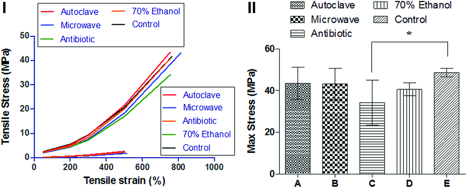
Loss of scaffold diameter was assessed in porous samples. The most significant loss was observed in autoclaved samples, indicative of severe polymer degradation during high temperature steam sterilization. Mechanical properties of cast sheets were analyzed to eliminate any potential variations induced by random pore distribution of the coagulated scaffolds, thus highlighting the mechanical properties of the pure polymer. Sterilization did not induce any significant changes neither in cast nor in coagulated samples. Compared with human skin, coagulated scaffolds were significantly more elastic (E=0.01 vs. E=0.003, respectively; p<0.05, paired t-test). Sample number=5.
Statistical significance at p<0.05.
E, Young's modulus.
In vitro cell viability and proliferation
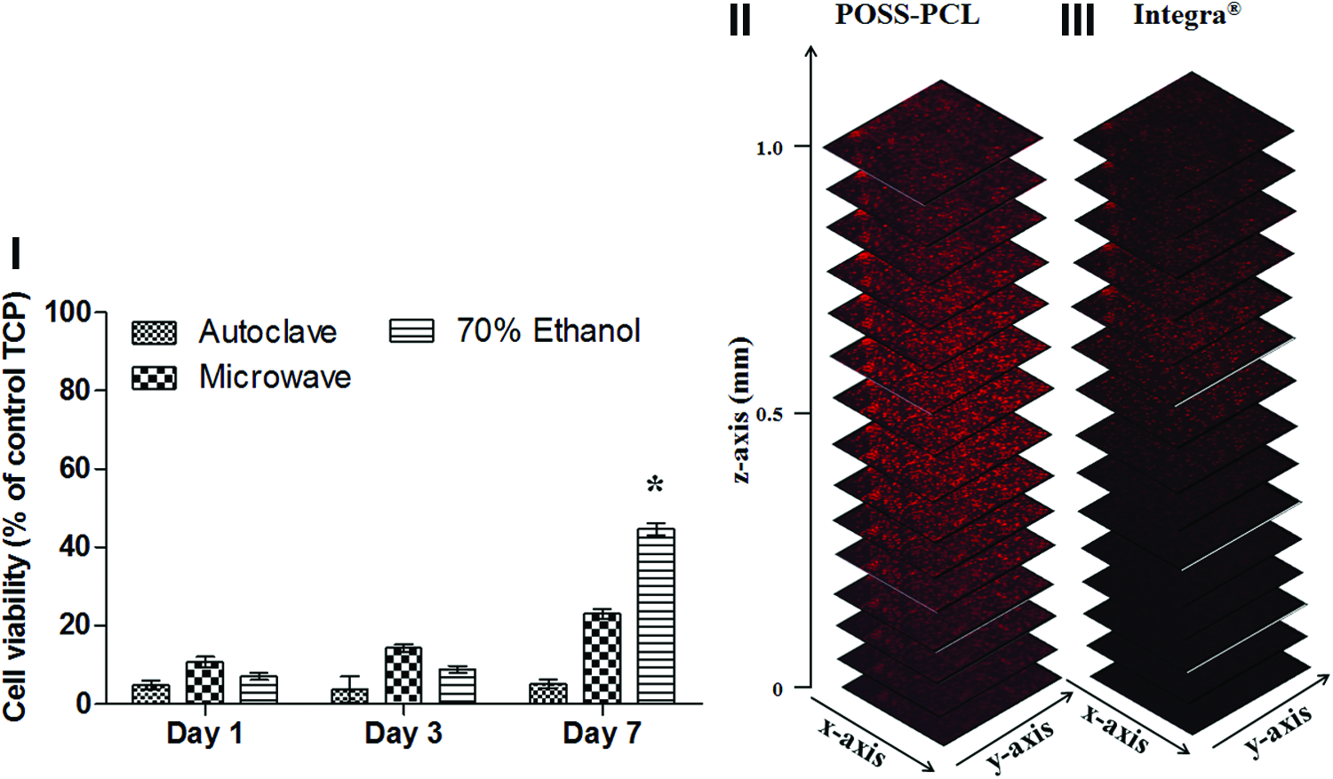
The percentage cell viability versus time and sterilization condition is represented as a bar chart in Figure 8. Due to sterility issues, AlamarBlue absorbance values of cells cultured on sterilized POSS-PCLU surfaces could not be normalized to values of absorbance of cells grown on untreated POSS-PCLU surfaces. Thus, viability of cells grown on normal TCP serves as the control total viability (i.e., 100%). The general trend demonstrated increased cell adhesion and metabolic viability at each consecutive time point. However, AlamarBlue metabolic assay demonstrated an initial lag in percentage viability of cells grown on 70% ethanol-treated films, which is compensated for on day 7. In fact, relative cell viability on ethanol-treated films surpassed that of cells grown on every other surface (apart from TCP) by a factor of 9 over a period of 7 days. Similarly, cell viability was found to be well supported by films treated with antibiotic solution. Autoclaved samples exhibited minimal to no cell adhesive capacity and significantly reduced cell viability. Exposure of films to microwaves supported cellular viability but significantly less so compared with treatment with both antibiotic and 70% ethanol solution (p<0.05). Live/Dead images of cells cultured on sterilized cast films are shown in Figure 9. Films exposed to any of the sterilization methods demonstrated acceptable initial cell attachment (day 1), but cell adhesion and viability was visibly reduced on day 3 on autoclaved surfaces, which was in accordance with AlamarBlue studies (Fig. 8). All except autoclaved surfaces supported cell attachment and viability throughout the entire study period of 7 days. Autoclaved surfaces displayed the lowest capacity for cell adhesion and viability throughout the entire study period. In terms of morphology, cells grown on antibiotic or 70% ethanol-treated films exhibited spindle-like morphologies typical for fibroblasts, similar to cells grown on control TCP, while cells cultured on autoclaved or microwaved films arguably exhibited more spread-out shapes. Generally, few or no dead cells (red) were visible on all sterilized or control surfaces. Films treated with 70% ethanol supported cell adhesion and viability best.
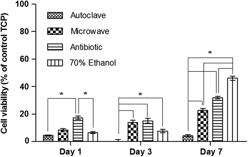
Percentage viability of HDFa grown on sterilized cast discs compared with cells grown on TCP over a 7 day period assessed using AlamarBlue® cell viability assay. *p<0.05. Values are given as mean±1 SD. HDFa, human dermal fibroblasts, TCP, tissue culture plastic.
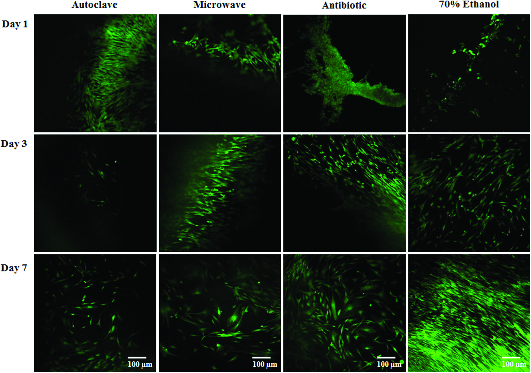
Representative laser scanning confocal microscopy images of HDFa cells grown on cast-sterilized POSS-PCLU discs stained with Live/Dead® stain. The overwhelming majority of cells remained alive throughout a culture period of 7 days with the highest proliferation rate observed on discs sterilized with 70% ethanol. Cell growth gradually diminished on autoclaved and microwaved samples, indicative of an unfavorable adhesion surface. These results are in accordance with surface roughness and wettability data. Color images available online at
Cell morphology and attachment
Confocal laser scanning microscopy images indicated that fibroblasts developed different morphologies when grown on differently sterilized surfaces [Fig. 10 (I)]. Cells grown on microwaved or autoclaved surfaces exhibited a more spread-out phenotype compared with cells grown on surfaces exposed to antibiotics and 70% ethanol solution that demonstrated an aligned and elongated spindle-like character. These results were not congruent with those predicted by peak-to-peak distance measurements and thus suggest that factors other than ξ may be involved in determining cell spreading behavior such as surface energy, surface charge, or mechanical properties.
POSS-PCLU scaffolds as tissue engineering platforms
The ability of this novel polymer system to act as a tissue engineering scaffold was subsequently analyzed on coagulated spongy POSS-PCLU scaffolds (Fig. 11). Scaffolds were sterilized with 70% ethanol and subsequently seeded with HDFa at a density of 5×10⁴ cells/scaffold. % cell viability of control cells (grown on TCP) was derived from AlamarBlue metabolic assay and was found to steadily increase over a period of 7 days [Fig. 11 (I)]. To visualize cellular penetration depth into the porous architecture of scaffolds, cells were cultured for 7 days on 70% ethanol sterilized sponges and nuclei were visualized with PI. Confocal imaging in conjunction with z-stacking demonstrated excellent cellular penetration into the internal architecture of the porous POSS-PCLU sponges [Fig. 11 (II)]. A preliminary comparative study investigating different cell behaviors on POSS-PCLU scaffolds and Integra dermal regeneration template (bovine collagen matrix, current clinical gold standard) demonstrated better adhesion and deeper migration of HDFa into POSS-PCLU scaffolds [Fig. 11 (II) vs. (III)].
Discussion
We have developed and patented two families of nanocomposite materials for the development of organs and tissues. The non-biodegradable family is based on POSS-PCU and has already been translated into the clinical setting with in-human applications, including transplantations of synthetic trachea, bypass grafts, tear ducts, and stents. The bioabsorbable material, on the other hand, is based on POSS-PCLU and demonstrates tunable degradation rates. Tissue engineering of implantable biodegradable devices is increasingly advocated as an elegant solution to restore the diseased or imperfect human body to a premorbid state.32–34 In particular, the paediatric population is thought to benefit from absorbable devices as is the field of skin reconstruction.
Tissue-engineered devices are often highly porous and therefore, by nature, display large surface areas with known incidences and histories for clinical infections. 35 Further, it has been shown that bacteria can remain dormant within the internal scaffold structure, only to manifest as pathogenic biofilms after implantation. 36 Material sterility is of fundamental significance for all implantable biomedical devices and is a particularly important consideration for all bioabsorbable materials. 37 Here, we introduced a novel biodegradable nanocomposite polymer based on a polyurethane hard segment and a poly(urea urethane)caprolactone soft segment incorporating POSS nanoparticles for use as a tissue engineering scaffold. Major advantages of using a PCLU backbone as tissue engineering platforms include its inherent biocompatibility, non-immunogenicity, and slow degradation into non-toxic metabolites. Unmodified POSS-PCLU, however, has a relatively hydrophobic nature that increases non-specific protein adsorption, leading to poor cell attachment and proliferation. 38 Several researchers investigated surface modification strategies including collagen and acrylic acid functionalizations to enhance cell adhesion and improve biocompatibility of PCL.38–40 However, one fundamental aspect that most studies relating to biodegradable scaffolds fail to mention is the necessity for a terminal sterilization step. Depending on the sterilization method used, the biomaterial surface may undergo different degrees of modifications, which may influence cell–biomaterial interactions.41,42 This may render any prior surface modifications obsolete, resulting in unpredictable material properties. In addition, surface wettability/chemical changes, while desirable in certain cases such as ours, may not be beneficial in other cases. This makes sterilization of biodegradable polymers problematic due to the inherent influence on hydrolytically and/or heat labile surfaces. In this study, we present a one-step method to achieve both sterilization and enhance biocompatibility after the initial synthesis of the scaffold, a technique termed postproduction processing.
Sterilization efficiency
Sterility results predictably showed efficient microbial removal on non-porous, cast films using autoclave, microwave, and 70% ethanol sterilization, all of which have been proved effective in numerous studies.37,43,44 Porous scaffolds, however, were only inconsistently sterilized using microwave irradiation, retaining contaminants within some core structures that only surfaced days later to cause infection. Such delayed infection is indicative of incomplete microbial eradication due to microwave scattering along the rough polymer surface and wave reflection due to incomplete air elimination within the internal structures. 45 The presence of such attenuating factors reducing microwave intensity has been confirmed in previous studies.46,47 It has been suggested that the presence of water vapor favors microwave absorption, thereby attenuating its intensity. Conventionally, microwaves have been used in the disinfection of denture base resins, 43 and the sterilization effect is thought to be due mainly to the heating effect. Studies investigating microwave sterilization of glass culture tubes demonstrated microbial growth when tube stoppers were purposefully opened to the ambient pressure, indicating a major role of pressurized steam in the killing of micro-organisms. 48 Exposure of both cast films and porous scaffolds to antibiotic solution resulted in ineffective sterilization. To the best of our knowledge, antibiotic antimycotic solutions have only been used as sterilizing agents in a study by Shearer et al., who recommended exposure over a period of 24 h. 9 Contrary to Shearer's results, our investigation showed inefficient sterilization using antibiotic solutions.
Exposure to 60–80% ethanol is considered disinfectant rather than sterilizing due to its inability to destroy hydrophilic viruses or bacterial spores. 37 However, ease of use and apparent effectiveness make ethanol a favorable tool for in vitro studies. Further, it has been demonstrated that ethanol treatment of a poly-L-lactide-co-glycolide polymer did not affect the molecular weight (Mw) of the polymer. 37
Scaffold microstructures
While all sterilization methods bar antibiotic solution had sterilizing effects, structural damage was evident in all cases. SEM images showed minor structural variations on all polymer surfaces due to processing, but the observed significant changes in surface microtopography poststerilization are suggestive of damages induced by these treatments. Exposure to high pressure steam sterilization and microwaves expectedly resulted in extensive morphological damage of hydrolytically unstable POSS-PCLU 49 indicative of partial dissolution of the polymer surface tufts due to hydrolysis. Subsequent fusion of polymer tufts into larger and flatter ridges is thought to be mainly driven by high polymer concentration at the polymer-moisture interface. 9 Scaffolds treated with antibiotic solution appeared the least destructive in terms of surface microtopography as seen on SEM images. However, the complete loss of internal porous structure observed on transverse cut SEM images was unexpected as antibiotic solution reportedly does not damage morphological or chemical characteristics of scaffolds. 50 Despite similar surface deformations of scaffolds exposed to 70% ethanol, no significant morphological damages were incurred to the internal 3D scaffold architecture, which is in accordance with the literature.37,51 Preserving internal porosity poststerilization is of fundamental importance for the in- and efflux of nutrients and waste products to and from the cells. 27
Chemical bond scission
All sterilization methods and, in particular, high temperature steam sterilization (autoclaving) induced an increase and broadening of the OH absorption peak at 3550 cm−1 suggestive of an accumulation of carboxylic and hydroxyl groups secondary to the hydrolytic degradation of the soft segment of PCLU (Fig. 4). Soft segment breakdown is known to play a major role in chemical aging of thermoplastic polyurethanes (TPUs) and negatively affects mechanical properties, including tensile stress and maximal strain. 52 Here, mechanical analysis postantibiotic treatment revealed significantly lower maximal tensile strength of cast polymers, indicative of soft segment scission. TPUs are phase separated block copolymers and owe their elasticity to their biphasic morphology, that is, a rigid and soft phase. While the soft segment provides the material with flexibility, the hard segment physically cross-links the phases to allow for elastic recovery. 52 Thermal oxidation of the methylene bridge of the methylene diphenyl diisocyanate (MDI) and subsequent breakage of the material's hard segment has been observed at temperatures above 150°C. 52 However, in our studies, temperatures did not exceed 121°C and, thus, no reductions in peak intensity were observed at the methylene bridge at 2932 cm−1 or the carbonyl region (1729 cm−1), indicating the presence of both ester and urethane linkages. Interestingly, we observed increases in peak intensities at 2932 and 1729 cm−1, which is likely to be correlated to the loss in thickness after sterilization with subsequent compaction of the polymer sample. Some evidence of hard segment exposure was evident with slight increases in peaks at 1632, 1560-1530, 1350, 1233, 992, and 793 cm−1 as similarly reported by Chan-Chan et al. 53
Surface nanotopography and impact on wettability
All sterilization methods induced an increase in surface wettability, which, according to the Wenzel phenomenon, is due to a reduction in surface roughness that results in a smaller surface area and thus increases hydrophilicity. 54 The initial surface roughness of non-sterilized control samples using Rq, was about 92 nm. Such nanoscale roughness is hypothesized to stem from the extrusion of POSS-nanocages (average diameter, including functional groups=3.5 nm) during the initial polymer synthesis step. All sterilization methods were found to reduce nanoroughness when compared with control surfaces, but only antibiotics induced a statistically significant drop in Ra of 49.4% (p<0.05). Such findings diverge from earlier studies into effects of different polymer sterilization methods, which found antibiotic treatment to significantly increase surface roughness of a poly(D,L-lactic-co-glycolic acid) flat sheet. 9 Such observations are suggestive of polymer-specific degradation modes toward the same treatment and thus display a non-predictability in terms of which sterilization method is the most effective while being least harmful. On comparison of SEM with AFM findings, it is surprising to find that antibiotic-treated surfaces appeared to differ least from controls on SEM images while demonstrating statistically significant changes in surface structures on analysis with AFM. This interesting phenomenon is likely due to different abilities of SEM and AFM to process vertical changes in topography. AFM can provide measurements in all three dimensions, including height information whereas the SEM cannot. This, essentially, results in a more detailed ability of the AFM to visualise differences in height on a nanometre scale.
As depicted in Figure 6, roughness profiles of sterilized and control surfaces exhibit a random pattern of peak heights. Due to only a small area being depicted, these roughness profiles are not representative of the overall surface structure. Instead, roughness profile measurements such as Ra represent a more accurate estimation (Table 2). Previous studies have shown that the topographical height threshold for fibroblast alignment lies at around 35 nm, below this value contact guidance no longer exists 20 giving rise to misaligned, broader cell morphologies. This differed from our results obtained on antibiotic-treated surfaces that exhibited the smallest Ra of 27.4 nm, yet displayed aligned and spindle-like fibroblast phenotypes.
Apart from pillar height, a further determining surface parameter is the correlation length (ξ). To quantitatively describe spatial variations of surface parameters at ultrastructural levels, ξ analysis was carried out. ξ, a characteristic parameter of the covariance function was employed to estimate the fluctuations in surface roughness induced by different sterilization methods. ξ describes how microscopic variations in relative positions change the correlation patterns between such positions. For example, if the distance between point A and point B is smaller than an arbitrary threshold ξ, then the interactions between A and B cause these points to be correlated. Peak-to-peak distance of surface micro- and nanostructures is known to influence cell–substrate behavior, cell adhesion and proliferation. Studies analyzing cellular responses to different surface structures demonstrated that smoother surfaces better supported cell attachment and subsequent viability compared with cells grown on nanorough surfaces.55,56 This is only partially in accordance with our cell adhesion and cell spreading results (Figs. 9 and 10). HDFa grown on surfaces treated with 70% ethanol demonstrated highest viability in accordance with surface topographical properties (θ=89°, Ra=47 nm, Rq=68 nm, ξ=5.5 nm). As can be seen in Figure 10, cell morphology was influenced by surface topography. However, cells grown on either microwaved or autoclaved surfaces exhibited a spread-out phenotype despite the fact that only microwaved surfaces displayed relatively high values for ξ whereas autoclaved surfaces had a low ξ (4.2 nm). This suggests that factors other than ξ influence the cell spreading behaviour. Different surface topographies have previously been shown to markedly alter cell morphology of fibroblasts with smoother surfaces being more favorable for fibroblast proliferation.57,58 Rough surfaces, on the other hand, were seen to provide multiple points of attachment. 59 Such conflicting results in terms of Ra and ξ indicate that cell behavior must be governed by a more complex array of parameters above and beyond surface roughness and correlation length. Further studies are required to investigate the effects of surface energy, surface charge, and surface mechanical properties.
Mechanical properties
None of the sterilization methods used in this study apart from antibiotic treatment significantly affected the overall tensile strength of cast or coagulated scaffolds, refuting earlier studies. 60 Ultimate tensile strength, however, was significantly reduced in antibiotic-treated samples, opposing a study conducted by Shearer et al., who exposed coagulated and electrospun poly(D,L lactic-co-glycolic acid) scaffolds to a 1% (v/v) antibiotic antimycotic solution. 60 The observed differences may be due to dosage-dependent effects of antibiotics and/or differences in susceptibility of different polymers.
Cytocompatibility and cellular morphology
Cell study results demonstrated that optimal adhesion and alignment of HDFa onto differently sterilized POSS-PCLU polymer occurs with surfaces showing a combination of moderate wettability and relatively short peak-to-peak distance. It has previously been demonstrated that the cut-off contact angle for good cell attachment and spreading is θ≤90°. 61 In terms of tissue engineering platforms, surface wettability of below 90° is regarded a highly important cue for the attachment of cells and the subsequent induction of tissue growth and regeneration.62,63 The enhanced adhesion of cells onto ethanol-treated samples can be ascribed to the different surface chemistries, indicating a preference of cells for hydrophilic structures as ethanol-treated films exhibited relatively more hydrophilic properties (θ<90° in both cases). Surface wettabilities of under 90° are known to promote cell adhesion and proliferation by increasing surface free energy.64,65 Zhao et al. previously demonstrated smaller water contact angles and higher values for surface free energy on sterilization with ethanol compared with autoclaving. 65 These results are corroborated here. FTIR results demonstrated that hydrolysis of the soft segments converted the ester groups on the surface to hydrophilic hydroxyl and carboxyl groups which were favorable for cell attachment. 66
Here, we demonstrated an effective routine for micro-organismal eradication that displayed the additional benefit of enhancing cell adherence onto a synthetic POSS-PCLU scaffold. It has previously been shown that 70% ethanol renders synthetic polymers such as PLGA as well as natural ones such as chitosan more hydrophilic, thus enhancing cell compatibility.9,37,67 While theoretically as well as in the laboratory setting, treatment with 70% ethanol is an acceptable means of microbial eradication, it is not easily transferrable to the clinical scenario where more stringent regulations set out by the General Medical Council (GMC) preclude the use of sub-optimally sterilized materials. This may potentially be a contributing factor for the relatively slow progression of biodegradable tissue engineering platforms from the bench to bedside.
Conclusion
Cast and coagulated biodegradable POSS-PCLU scaffolds were exposed to four different sterilizing methods, and their suitability as postproduction processing tools to enhance biocompatibility were assessed. Autoclave and microwave treatments successfully sterilized cast POSS-PCLU films but significantly damaged the 3D porous structures of coagulated scaffolds. Antibiotic antimycotic solution was ineffective in removing all micro-organisms and can therefore be excluded as a sterilizing agent. Exposure of both cast and coagulated scaffolds to 70% ethanol resulted in the best overall removal of infection-causing micro-organisms combined with the least destructive effect on porosity and enhanced cytocompatibility. Ethanol treatment induced favorable surface topographical changed which positively influenced cell attachment, morphology, alignment, as well as viability and proliferation. The present technique using 70% ethanol can pave the way for future tissue engineering using bioabsorbable POSS-PCLU nanocomposite materials as regenerative platforms by addressing two current obstacles at once.
Footnotes
Acknowledgment
This work was supported by the Medical Research Council Doctoral Training Grant and the Rosetrees Trust.
Disclosure Statement
No competing financial interests exist.