
Abstract
Conductive polymers (CPs) are organic materials that hold great promise for biomedicine. Potential applications include in vitro or implantable electrodes for excitable cell recording and stimulation and conductive scaffolds for cell support and tissue engineering. In this study, we demonstrate the utility of electroactive CP polypyrrole (PPy) containing the anionic dopant dodecylbenzenesulfonate (DBS) to differentiate novel clinically relevant human neural stem cells (hNSCs). Electrical stimulation of PPy(DBS) induced hNSCs to predominantly β-III Tubulin (Tuj1) expressing neurons, with lower induction of glial fibrillary acidic protein (GFAP) expressing glial cells. In addition, stimulated cultures comprised nodes or clusters of neurons with longer neurites and greater branching than unstimulated cultures. Cell clusters showed a similar spatial distribution to regions of higher conductivity on the film surface. Our findings support the use of electrical stimulation to promote neuronal induction and the biocompatibility of PPy(DBS) with hNSCs and opens up the possibility of identifying novel mechanisms of fate determination of differentiating human stem cells for advanced in vitro modeling, translational drug discovery, and regenerative medicine.
Introduction
T
Polypyrrole (PPy) is a CP that is easily synthesized and able to incorporate anionic biomolecules (dopants) to enhance biocompatibility and target cellular function. In addition, PPy allows spatiotemporal control of electrical stimulation and generates minimal inflammatory response beneficial for defined cell manipulation, regulatory approval, and clinical use. Several recent studies using rodent stem cells demonstrate that PPy doped with bioactive molecules can influence stem cell survival and differentiation.7–9 Zhang et al. investigated unstimulated PPy doped with peptides from extracellular matrix (ECM) protein laminin and suggested that PPy and other CPs have significant potential for engineering intelligent substrates for controlling stem cell growth. 9 Lundin et al. investigated the utility of CP substrates combined with electrical stimulation to regulate the rat neural stem cell (rNSC) state and fate. 8 More specifically, electrical stimulation of PPy doped with dodecylbenzenesulfonate (DBS) impaired fetal rNSC survival. Viability of rNSCs was rescued by thickly coating PPy(DBS) films with a gel layer composed of a basement membrane matrix, resulting in cell survival rates comparable to standard rNSC culture conditions (without CP and electrical stimulation).
Despite the evidence for CPs such as PPy supporting cell growth and differentiation,10,11 their compatibility with clinically relevant human neural stem cells (hNSCs), such as human brain-derived NSCs, and potential as electroactive substrates for controlling human stem cell fate remain undetermined. In this study, we investigated the differentiation of hNSCs on laminin-coated PPy(DBS) with electrical stimulation. To the best of our knowledge, this article is the first account of human brain tissue-derived neural stem cell differentiation and support using electroactive PPy.
Materials and Methods
Preparation of polymer films
The Pyrrole (Py) monomer was obtained from Sigma and distilled before use. All dopants used for PPy polymerization, including para-toluene sufonate (pTS), DBS, and the sodium salt of chondroitin sulfate (CS) A, were obtained from Sigma. All Py monomer and dopant solutions were prepared with distilled and deionized water (dH2O, Milli-Q). Gold-coated mylar (Solutia Performance Films) was prepared for polymerization by cleaning with isopropanol and dH2O and then drying under a N2 stream. Aqueous monomer solutions were prepared separately for each dopant consisting of 0.2 M Py, 0.05 M pTS, 0.05 M DBS, or 2 mg/mL CS. These aqueous solutions were degassed using N2 for 10 min before polymerization of the polymers. PPy films were polymerized galvanostatically at a current density of 0.1 mA/cm2 for 10 min in the aqueous monomer solution using an eDAQ EA161 potentiostat. Polymer growth was performed in a standard three-electrode electrochemical cell with the gold-coated mylar as the working electrode, a platinum mesh counter electrode (CE), and Ag|AgCl reference electrode (RE). After growth, the films were washed extensively with dH2O, gently dried with N2 gas, and stored under desiccated conditions until use.
Atomic force microscopy
Atomic force microscopy (AFM) imaging was performed using a JPK Biowizard II AFM (JPK Instruments). Polymer samples were prepared for characterization by washing with dH2O and drying with N2. AFM images were obtained in air at 25°C using a 0.42 N/m silicon nitride cantilever in the tapping mode with a scan rate of 0.5–1 Hz. Average roughness (Ra) and root mean square roughness (Rq) values were calculated from AFM height images using analysis software of the AFM.
Contact angle goniometry
Contact angle measurements were made with a 2 μL water droplet using the sessile drop technique and a DataPhysics OCA20 Goniometer, employing SCA21 software. At least five measurements were taken on each surface, and the average and standard deviation were calculated.
Cyclic voltammetry
Cyclic voltammetry (CV) was performed using a CH Instruments 660D Electrochemical Workstation. The three-electrode cell consisted of a 1 cm2 polymer-coated working electrode, a platinum mesh counter, and an Ag|AgCl RE. Measurements were performed in a phosphate-buffered saline (PBS) electrolyte at a scan rate of 10 mVs−1 over the potential range of −0.6 to +0.6 V.
Neural stem cell culture and differentiation
Working stocks of RenCell CX hNSCs (Millipore; approved for use by the University of Wollongong's Human Research Ethics Committee; HE14/049) were maintained by seeding at a density of 2–3×10⁶ cells into a low-attachment T-75 flask (Corning) containing a DMEM/F12 (Invitrogen)-based expansion medium supplemented with 2% B27 (Invitrogen), 2 μg/mL heparin (Sigma), epidermal growth factor (10 ng/mL; Peprotech), and basic fibroblast growth factor (20 ng/mL; Peprotech). Once hNSCs formed free-floating neurospheres after 7–14 days, they were passaged for subculture every 5–7 days by digesting in 1–2 mL TrypLE (Gibco BRL) for 5 min at 37°C. Digested neurospheres were triturated to single cells and plated at a density of 5×10⁴ cells/mL using ultralow attachment six-well plates (Corning).
hNSCs were differentiated by digesting neurospheres as above and plating 5×10⁴ cells/cm2 onto eight-well chamber slides (Nunc) or prepared polymers, coated with 20 μg/mL laminin (Life Technologies) containing two parts DMEM F-12: one part Neurobasal supplemented with 2% StemPro (Life Technologies), 0.5% N2 (Gibco), and 50 ng/mL brain-derived neurotrophic factor (Peprotech) up to 7 days. A half-volume medium change was performed every 2–3 days.
Electrical stimulation
For electrical stimulation experiments, cells were seeded at the same density (5×10⁴ cells/cm2) in two groups (one electrical stimulation group and one control group). In all groups, cells were seeded in differentiation media onto laminin-coated PPy(DBS) films, as described above, and allowed to adhere for 24 h before the media were topped up and electrical stimulation applied in accordance with the published method. 12 Cells were stimulated for 8 h per 24-h period for 3 days under 5% CO2 at 37°C, after which the cells were either fixed or allowed to differentiate for a further 3 days before fixing for immunostaining and other analyses. Electrical stimulation was performed through a two-electrode setup, whereby an auxiliary platinum mesh electrode was placed into the media at the top of each well and the PPy coated Au-mylar surface was used as the working electrode. The cells were stimulated at±0.25 mA/cm2 using a biphasic waveform of 100 μs pulses with 20 μs interphase open circuit potential and a 3.78 ms short circuit (250 Hz) using a Digital StimulatorDS8000 and A365 Isolator units (World Precision Instruments) interfaced with an e-corder system (eDAQ). The voltage waveform across the active electrode area in response to the current pulse applied was recorded, and using Ohm's law, total impedance (Zt) was calculated from the peak voltage (Vt) output divided by the applied current (i) (Zt=Vt/i), access resistance from the initial voltage drop (Ra=Va/i), and polarization impedance from the remaining voltage drop (Zp=Vp/i).
Immunocytochemistry and analysis
Cell samples were fixed by incubation with a 3.7% paraformaldehyde solution in PBS. Samples were incubated at room temperature (RT) for 10 min before rinsing with PBS. Cell samples were then blocked and permeabilized for 1 h at 37°C in 5% (v/v) goat serum in PBS containing 0.3% (v/v) Triton X-100 (blocking buffer). Primary antibodies, including mouse anti-microtubule-associated protein 2 (MAP2, 1:1000; Sigma), rabbit anti-GFAP (1:1000; Millipore), chicken anti-β-III Tubulin (Tuj1, 1:1000; Millipore), mouse anti-Ki67 (1:500; BD Bioscience), mouse anti-nestin (1:500; BD Bioscience), rabbit anti-Sox2 (1:500; Millipore), and chicken anti-vimentin (1:1000; Millipore), were diluted in a blocking buffer and then incubated with the cell samples overnight at 4°C. The cells were then washed 3×5 min in PBS/0.1% (v/v) Triton X-100 before the addition of an appropriate secondary antibody conjugated with Alexa 594 or 488 (1:1000; Invitrogen) and diluted in a blocking buffer. The cells were left for 1 h at RT. A further 3×5 min washes in PBS/0.1% Triton X-100 were performed before a 5-min incubation at RT with 1 μg/mL DAPI in PBS. The DAPI solution was replaced with fresh PBS and cells were then imaged at 10× magnification. Imaging was performed using an AxioImager (Zeiss) fitted with an AxioCAM Mrm camera and overlayed using AxioVison 4 software. Image analysis was performed using MetaMorph software V 7.8 with the neurite analysis plugin for neurite length, number, and branching, or ImageJ software for glial versus neural scoring.
Flow cytometry
Neurospheres were collected and digested in TrypLE as described above. After trituration, single cells were pelleted and fixed with 3.7% paraformaldehyde solution in PBS on ice for 10 min. After two washes in PBS/0.1% (v/v) Triton X-100, cells were resuspended in a blocking buffer and placed on ice for 20 min. Cells were then incubated with primary antibodies mouse anti-Ki67 (1:500; BD Bioscience), mouse anti-nestin (1:500; BD Bioscience), rabbit anti-Sox2 (1:500; Millipore), and chicken anti-vimentin (1:1000; Millipore) diluted in a wash buffer on ice for 30 min. Following a further two to three washes, secondary antibodies conjugated with Alexa Fluor-488 and Alexa Fluor-594 (1:1000; Invitrogen) and diluted in a blocking buffer were applied for 30 min on ice. Cells were then washed again before being resuspended in PBS and analyzed by a BD Accuri C6 system (BD Biosciences).
Scanning electrochemical microscopy
Scanning electrochemical microscopy (SECM) was performed in a feedback mode using CH Instruments SECM model 920D utilizing CHI integrated software version 12.26. Pt disk (10 μm diameter) ultra-microelectrode (UME; CH Instruments, Inc.) was used as the working electrode. Pt mesh and Ag/AgCl (3.0 M NaCl) were used as the CE and RE, respectively. Feedback mode data collection was performed in cell culture media containing 4 mM analytical-grade Ferrocenemethanol (FcMeOH; Sigma) redox mediator. FCMeOH was therefore used as the electron transfer mediator to look at the variation of conductivity across the PPy(DBS) surface. The SECM probe was biased at +0.5 V for electro-oxidation of FcMeOH. A three-dimensional SECM image in constant height was obtained by scanning the substrate in the x-y plane with the UME probe and recording the feedback current against probe location to determine local concentration of the redox mediator in solution and the corresponding surface electroactivity. Mean current values were calculated using at least 1000 data points within the area assessed. The probe/substrate separation was adjusted to 3 μm using a digital microscope (MEIJI Techno model MS50). Similar methods have previously been described for studying PPy surface conductivity.13,14
Scanning electron microscopy
Cells were fixed in a 3.7% paraformaldehyde solution in PBS for 10 min at RT and then dehydrated in EtOH before Critical Point Drying using a Leica EM CPD030 instrument. Once dried, samples were coated in a 15 nm layer of gold using an Edwards sputter coater and kept desiccated until imaging was performed. Scanning electron microscopy (SEM) was performed using a JEOL LV SEM operated at an accelerating voltage of 10.0 kV and the secondary electron images were taken with a semi-in-lens detector at a working distance of 8 mm.
Statistics
OriginPro Version 8.6 was used to perform one-way analyses of variance followed by Bonferroni post hoc analysis.
Results
Characterization of different PPy(doped) films
Since substrate properties, such as elasticity,15,16 roughness,17,18 surface charge,19,20 and wettability,21,22 can affect the adhesion, growth, and fate of cells, and the dopant type can influence the surface characteristics of CPs, we initially characterized the surface topography of PPy films using AFM, followed by contact angle goniometry (CAG) and CV. AFM showed that all of the differently doped films had characteristic cauliflower morphology (Fig. 1). 23 pTS and CS doped films had similar Ra and Rq values, while DBS doped films were marginally smoother (Table 1 and Fig. 1).
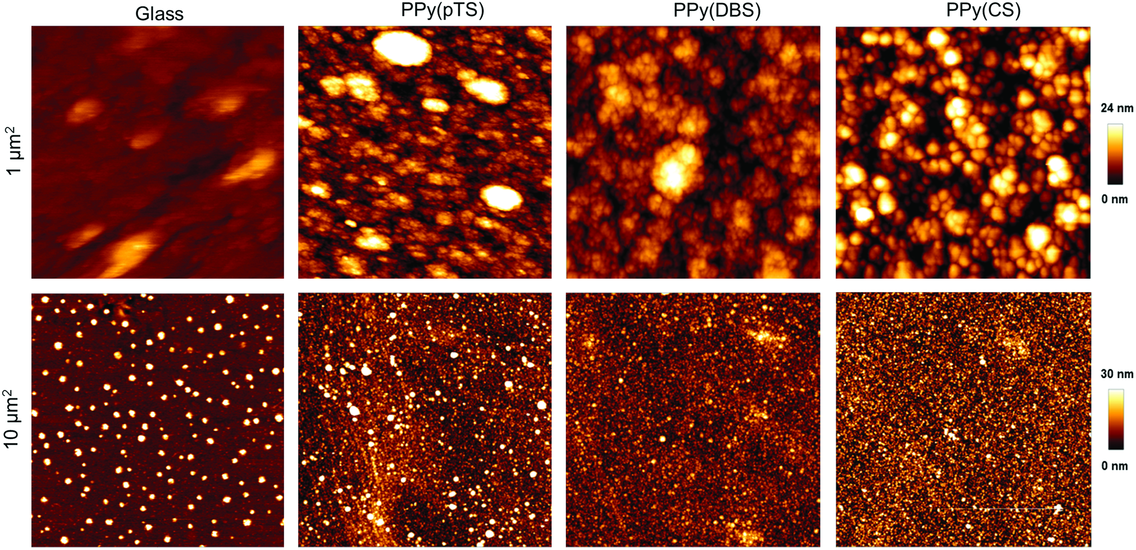
Characterization of material properties. Atomic force microscopy images of the topography of substrates tested. CS, chondroitin sulfate; DBS, dodecylbenzenesulfonate; PPy, polypyrrole; pTS, para-toluene sufonate. Color images available online at
Surface roughness was determined using AFM. Goniometry was performed to measure the contact angle for each substrate surface.
CS, chondroitin sulfate; DBS, dodecylbenzenesulfonate; PPy, polypyrrole; pTS, para-toluene sufonate; RMS, root mean square.
The surface wettability of each substrate was determined by CAG. Similar measures of contact angle were determined for all substrates, with each being moderately hydrophilic (i.e., less than 90°; Table 1).
24
Finally, CV demonstrated all PPy(doped) films had well-defined electrochemical responses with clear oxidation and reduction peaks indicating the presence of a redox-active polymer (Supplementary Fig. S1; Supplementary Data are available online at
Assessment of hNSC differentiation on unstimulated PPy(doped) films
Before differentiation on PPy films, hNSCs were maintained under conventional culture conditions as self-renewing cells able to form neurospheres and stably expressing cell proliferation marker Ki-67 and neural stem/progenitor markers vimentin, nestin, and sox-2 (Fig. 2a, b). 25 Subsequent transfer of hNSCs from standard laminin-coated glass substrates resulted in a differential effect on neuronal induction of hNSCs between PPy films (Fig. 2c, d). Specifically, qualitative assessment of differentiated hNSCs indicated that PPy doped with DBS provided the best support of MAP2 positive neurons, with networks of neurites being comparable to standard culture conditions, while CS and pTS doped films provided poor support with cultures comprising relatively few to negligible neurites, respectively (Fig. 2c, d). PPy(DBS) was chosen for subsequent experiments involving electrical stimulation.

Immunochemical characterization of hNSCs and substrate support of differentiation.
Differentiation of hNSCs on electroactive PPy(DBS) film
The scheme for culturing and stimulating hNSCs on doped PPy is illustrated in Figure 3. The impedances of the PPy(DBS) polymer were monitored for the duration of the hNSC electrical stimulation period. Three impedance values were derived from the resulting voltage waveforms (Fig. 3a), including total impedance (Zt=Vt/i), access resistance (Ra=Va/i; relating to changes in electrolyte), and polarization impedance (Zp=Vp/i; relating to changes at the electrode surface). 12 Notably, the Zp showed minimal change during the stimulation period, ranging from 5 ohms at day 1 to 4 ohms at day 3, indicating electrochemical stability of the PPy(DBS) film during cell stimulation.

Scheme for culturing and stimulating hNSCs on electroactive PPy(dopant) substrate.
Electrical stimulation of PPy(DBS)-based hNSCs altered cell differentiation, as illustrated by immunocytochemistry of neuronal marker Tuj1 and astrocyte marker GFAP, as well as cell distribution and neurite formation (Fig. 4). More specifically, in contrast to unstimulated glass-based and PPy(DBS)-based culture, electrical stimulation of PPy(DBS)-induced hNSCs to predominantly Tuj1-expressing neurons, with lower induction of GFAP expressing glial cells (Fig. 4a, b). Second, unlike unstimulated cultures, stimulated PPy(DBS)-based cultures comprised nodes or clusters of neurons joined by neurite networks (Fig. 4a). Third, electrically stimulated PPy(DBS) neurons exhibited a greater total neurite length per cell, mean neurite length, and maximum neurite length compared to other treatments (Fig. 4c). Although neurite number and branching were decreased for hNSCs differentiated on PPy(DBS) compared to standard glass-based differentiation, electrical stimulation of PPy(DBS) film resulted in a greater number of neurites and increased neurite branching compared to unstimulated cells on film (Fig 4d). Finally, qualitative assessment of SEM images supported the presence of fewer astrocytes for elecroactive PPy(DBS)-based differentiation. For all conditions tested, cells exhibited neurite outgrowths, with neurites connecting adjacent cells (Fig. 5). Micrographs also confirm cell attachment to all substrates (Fig. 5).
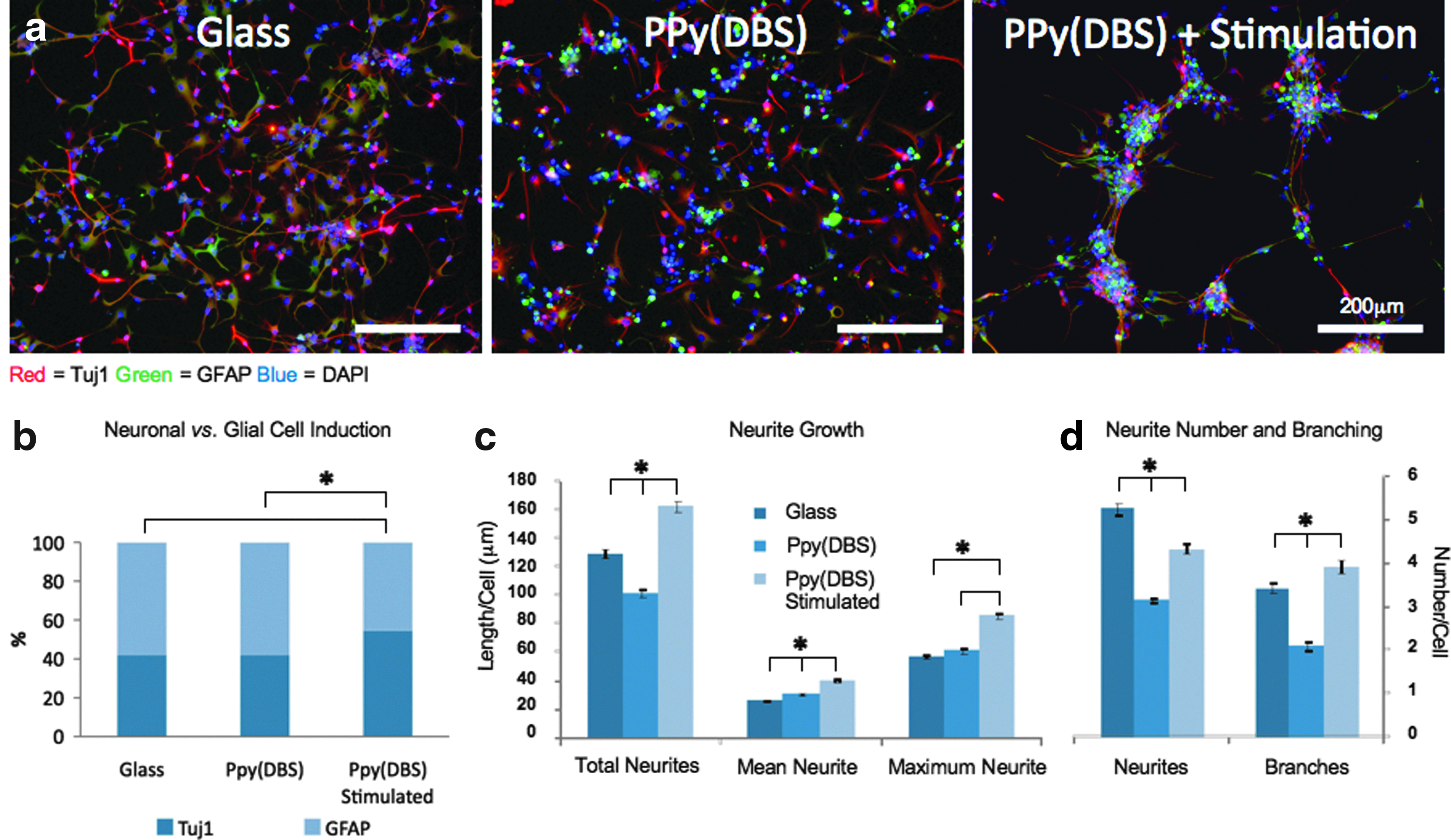
Effect of the electrical stimulation of PPy(DBS) substrate on hNSC differentiation.
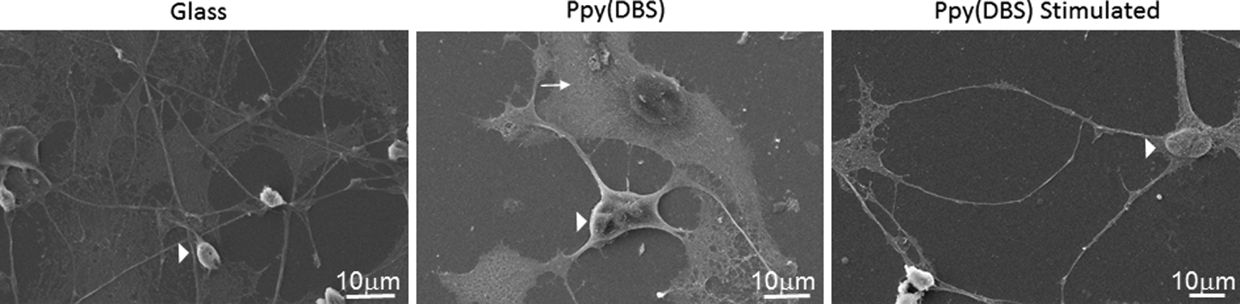
Scanning electron microscopy micrographs showing morphology of neurons (arrow heads) and glia (arrows) differentiated from hNSCs over 7 days on substrates tested.
Assessment of surface conductivity of PPy(DBS) film
Following the observation of cell clustering on electrically stimulated PPy(DBS) film, we postulated that it may reflect heterogeneous conductivity across the film surface with areas of higher and lower conductivity potentially underlying high and no cell clustering, respectively. We subsequently used SECM to assess conductivity across the surface of the PPy(DBS) film (Fig. 6). The SECM images show a surface that has an inherent conductivity represented by extensive green shading (Fig. 6), with mean±SD current values of 4.76±0.07 nA (Fig. 6a) and 4.81±0.05 nA (Fig. 6b), interposed by regions of higher and lower current representing regions of higher and lower conductivity, respectively. The distance between the different regions ranged from 10–20 μm (Fig. 6), corresponding to the distance between various cell clusters previously observed and shown in Figure 4a.
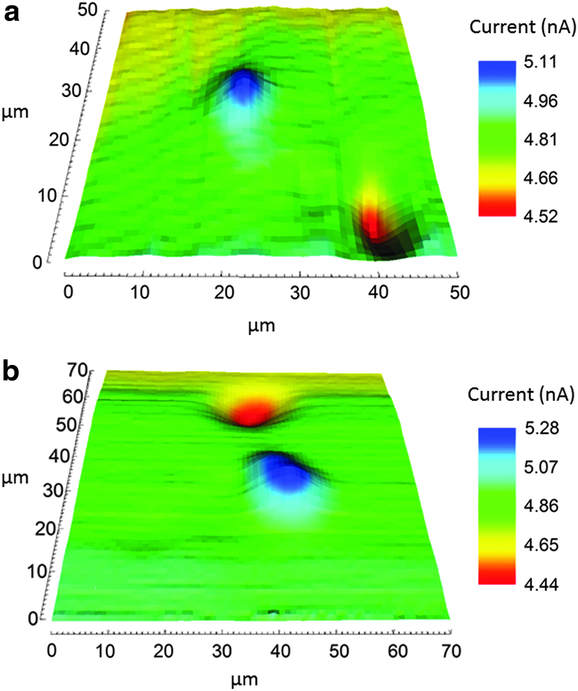
Scanning electrochemical microscopy images depicting surface conductivity at two different locations on PPy(DBS) film. In each case, inherent conductivity is represented by extensive green shading interposed by dark blue and red shading depicting areas with higher and lower conductivity, respectively.
Discussion
This study adds to the growing body of evidence for using electrical stimulation and CPs with bioactive dopants to influence the culture and differentiation of stem cells. We specifically report the differentiation of hNSCs to neurons and glia on laminin-coated PPy(DBS), with a higher proportion of neurons and lower glial cell numbers following electrical stimulation. Neurons exhibited clustering and increased neurite growth, with the latter manifesting as longer neurites with greater branching. Alternatively, standard laminin-coated glass-based differentiation of unstimulated hNSCs had greater numbers of shorter neurites with less branching.
Since AFM and CAG studies showed negligible differences between the surface roughness and contact angles of different substrates, it is unlikely that surface topography and surface energy influenced neuronal induction. Notwithstanding the potential effects of other properties (e.g., interacting surface chemistry) on the biocompatibility of a specific polymer/dopant system,8,26,27 our initial observation of unstimulated PPy(DBS) supporting neuronal induction affirmed further studies with electrical stimulation.
The observed tendency toward neuronal induction using electroactive PPy(DBS) extends the findings of Yamada et al. who report that electrical stimulation causes murine EBs to differentiate, somewhat specifically, into neuronal cells. 2 Importantly, while the overall composition of neurons and glial cells in the human brain remains contentious, with conventional opinion holding that glial cells are more numerous than neurons by a factor of 10, and more recent reports suggesting a smaller glia/neuron ratio of ∼1:1, since the glia/neuron ratio varies between different areas of the brain, there is a need for methods to modulate the proportion of glial to neuronal cells for region-specific modeling in vitro and tissue engineering for transplantation therapy. 28 To this end, based on our findings, electroactive PPy(DBS) might be useful to influence the tissue content of neurons and glia derived from hNSCs and potentially other stem cell types of human origin.
The pattern of cell clustering associated with electrical stimulation was unexpected, although a similar phenomenon has been reported for primary rat hippocampal neurons cultured on microelectrode arrays. 29 In addition, we have previously reported variable conductivity across the surfaces of PPy films, where nodular regions of polymer correlated with high current and extracellular matrix protein adhesion. 30 Our earlier findings, however, relate to nanoscale variation with phase separation in conductivity measured across 0.3–0.4 μm, whereas the present cell clustering occurred with micron resolution. Nonetheless, it is conceivable that areas of high conductivity presently measured are also associated with increased cell adhesion and/or galvanotaxis. While for now we can only surmise the cause of the heterogeneous conductivity, with one possibility being a heterogenous doping of the polymer, our findings suggest that modulation of conductivity and electroactivity of PPy(DBS) film and more broadly cell culture substrates can potentially be used to regulate maturation and organization of neural networks in vitro or in vivo.
In summary, the results presented here illustrate the utility of electrical stimulation and the biocompatibility of electroactive PPy(DBS) for differentiating clinically relevant human brain NSCs. The hNSCs described are self-renewing and multipotent, able to differentiate into multiple neural cell types, and have the potential to be used for both research and as a therapy for neurological diseases.25,31 Similarly, as a cytocompatible CP, PPy can be used for research and as a clinical device. Therefore, by interfacing hNSCs with electroactive PPy we have demonstrated a biologically relevant method for enhanced delivery of human neuronal tissues for research and clinical application.
Footnotes
Acknowledgments
The authors wish to acknowledge the use of facilities at the University of Wollongong Electron Microscopy Centre, ongoing financial support of the Australian Research Council (ARC) and the ARC Centre of Excellence for Electromaterials Science (ACES), support and assistance by the Australian National Fabrication Facility (ANFF)—Materials Node, and Mr Hongrui Zhang for assisting with AFM. Professor Gordon Wallace acknowledges the support of the ARC through an ARC Laureate Fellowship—the primary funding source for this work. The authors would like to dedicate this article to Dr. Nao Kobayashi, a remarkable colleague and cherished friend. Nao's passion and dedication to her scholarship was unparalleled and her friendship and caring spirit will be missed.
Disclosure Statement
No competing financial interests exist.
References
Supplementary Material
Please find the following supplemental material available below.
For Open Access articles published under a Creative Commons License, all supplemental material carries the same license as the article it is associated with.
For non-Open Access articles published, all supplemental material carries a non-exclusive license, and permission requests for re-use of supplemental material or any part of supplemental material shall be sent directly to the copyright owner as specified in the copyright notice associated with the article.