
Abstract
Rapid and controlled vascularization within biomaterials is essential for many applications in regenerative medicine. The extent of vascularization is influenced by a number of factors, including scaffold architecture. While properties such as pore size and total porosity have been studied extensively, the importance of controlling the interconnectivity of pores has received less attention. A sintering method was used to generate hydrogel scaffolds with controlled pore interconnectivity. Poly(methyl methacrylate) microspheres were used as a sacrificial agent to generate porous poly(ethylene glycol) diacrylate hydrogels with interconnectivity varying based on microsphere sintering conditions. Interconnectivity levels increased with sintering time and temperature with resultant hydrogel structure showing agreement with template structure. Porous hydrogels with a narrow pore size distribution (130–150 μm) and varying interconnectivity were investigated for their ability to influence vascularization in response to gradients of platelet-derived growth factor-BB (PDGF-BB). A rodent subcutaneous model was used to evaluate vascularized tissue formation in the hydrogels in vivo. Vascularized tissue invasion varied with interconnectivity. At week 3, higher interconnectivity hydrogels had completely vascularized with twice as much invasion. Interconnectivity also influenced PDGF-BB transport within the scaffolds. An agent-based model was used to explore the relative roles of steric and transport effects on the observed results. In conclusion, a technique for the preparation of hydrogels with controlled pore interconnectivity has been developed and evaluated. This method has been used to show that pore interconnectivity can independently influence vascularization of biomaterials.
Introduction
T
The physical properties of scaffolds that have drawn the most attention from researchers are mean pore size and total pore volume (porosity). Studies have shown that the rate and depth of tissue invasion depend on pore size. 4 A study conducted using chitosan found that pore sizes ranging between 70 and 120 μm allowed tissue invasion deeper than scaffolds with pore sizes less than 70 μm. 5 In a study with poly(lactic-co-glycolic acid) (PLGA) scaffolds, a broad range of pore sizes were investigated. The scaffolds had spherical pores, with mean pore diameter ranging between 79 and 312 μm. Histological analysis revealed scaffolds with pore sizes less than 200 μm favored high vessel densities, but poor penetration depths. Pore sizes greater than 200 μm had deeper tissue invasion with larger blood vessels. 6 Similar results were obtained with 80 μm pore size, which permitted only tissue growth near the surface. 7 A study conducted with pore sizes ranging from 300 to 700 μm showed that above 400 μm there was no difference in vascularization. 2 While there is some disagreement with the exact pore size that yield the best results, the general concept is that larger pore sizes seem to allow deeper vascularized tissue growth within biomaterials. However, these results may be further confounded by differences in interconnectivity of the pores in the scaffolds investigated.
Scaffold interconnectivity is commonly described in a binary way, either the pores are connected or they are not. However, the size of the connections is likely important. Interconnectivity may be more accurately described as the mean size of the opening between two neighboring pores.8,9 This characteristic feature of the scaffold influences movement within the scaffold whether it be diffusion of soluble factors, oxygen, and nutrients or cell migration, and tissue invasion. There is little information about the effects of interconnectivity of scaffolds on vascularized tissue ingrowth. Previously, an agent-based model of engineered tissue was used to study the significance of scaffold architecture on vascularization. Agent-based models are computational frameworks that study the interaction of autonomous agents with other agents and environmental factors. Within the vascularization model, endothelial cells are the agents that interact with each other and the microenvironment in the formation of blood vessels within the scaffold. A rule base that is driven by experimental findings and literature data govern the actions of each individual agent and its response to soluble and insoluble factors. 10 Using an agent-based model of engineered tissues, we have shown that interconnectivity may have a significant influence on vascularization of the scaffolds. Invasion depth and blood vessel density of scaffolds increased with interconnectivity at a constant pore size. 11 In some cases, small pore sizes allowed deeper invasion depths compared with larger pore sizes due to differences in interconnectivity. With this in mind, control over pore interconnectivity may influence scaffold vascularization.
There are many methods for creating porous hydrogels. Techniques include electrospinning, porogen leaching, and three-dimensional printing/patterning. 3 However, the distribution of shapes and sizes of pores varies with the fabrication process. 12 In addition, it may be difficult to control the interconnectivity of the pores. Sphere templating has been used to introduce monodisperse and uniform pores into a network.12–14 In this method, densely packed sintered microspheres are used to create a template for the pores. Following polymerization of the biomaterial around the template, the microsphere template is extracted resulting in an interconnected porous structure. This technique allows control over the pore structure and, more importantly, pore interconnectivity based on the time and temperature of microsphere sintering. While interconnectivity can be controlled, in theory, a systematic investigation of interconnectivity with this technique has not been performed.
Based on experimental and computational studies the interconnected pore structure of scaffolds assist in cell ingrowth and distribution within the scaffolds and thus may influence vascularized tissue formation. 15 The formation of vascular networks within a biomaterial is essential for the survival of engineered tissues. In this research, we sought to optimize the properties of porous poly(ethylene glycol) (PEG) hydrogels for accelerating vascularization based on the generation of hydrogels with controlled interconnectivity. Hydrogels were fabricated using microsphere templating and assessed for structure and mechanical integrity. Computational and animal models were used to examine the influence of pore interconnectivity on vascularization.
Materials and Methods
Materials
Poly(methyl methacrylate) (PMMA) microspheres (106–125 μm) were purchased from Cospheric (Santa Barbara, CA). PEG (Mn 8000), 2-hydroxy-2-methylpropiophenone (Irgacure 1173), acryloyl chloride (98%), thriethylamine (TEA, 99.5%), poly(lactic-co-glycolic acid) (PLGA 50:50, Mw=7000–17,000), poly(vinyl alcohol) (PVA, Mw=13,000–23,000), bovine serum albumin (BSA), sucrose (>99.5%), magnesium hydroxide (95%), PEG (Mn 3400), 3,6-dimethyl-1,4-dioxane-2,5-dione, and acrylic acid were obtained from Sigma-Aldrich (St. Louis, MO). Dichloromethane (99.9%), ethyl ether (anhydrous), and phosphate buffered saline (PBS) were obtained from Fisher Scientific (Hampton, NH). Recombinant rat platelet-derived growth factor-BB (PDGF-BB) with carrier protein was obtained from R&D Systems (Minneapolis, MN). Fluorescein isothiocyanate (FITC)-conjugated bovine serum albumin (FITC-BSA) was purchased from Invitrogen (Pittsburgh, PA).
Template and hydrogel preparation
A sphere templating technique developed based on previous publications was used to prepare porous scaffolds with varying levels of interconnectivity.13,16 A template was created by placing PMMA microspheres in a mold between two glass slides held together with a clamp. Individual microspheres were imaged using brightfield microscopy to determine the mean microsphere size (112.01±6.47 μm, n=300). The microspheres were placed in an 8-mm diameter Teflon mold with a thickness of 4 mm. The mold was sonicated for 20 min to improve packing of the microspheres. The mold was then heated using a vacuum oven to a desired specific temperature (160°C or 175°C) and the microspheres sintered for various times (0–30 h).
PEG-DA (875 mg, average Mn≈8000) was dissolved in 3.5 mL of deionized water. Irgacure 1173 (0.5% w/v) was added to the precursor as the photoinitiator. The hydrogel precursor was poured over the sintered microspheres while inside the mold and allowed to fill the void space in the mold. The sintered microspheres with the hydrogel precursor were polymerized under UV light (365 nm) for 5 min. The hydrogels were then washed with acetone several times and incubated overnight in acetone to dissolve the PMMA microspheres. The gels were removed from the acetone and washed with deionized water several times. The hydrogels were incubated in deionized water for 48 h to reach equilibrium. Figure 1 provides an illustration of the procedure for template and hydrogel preparation.

Fabrication of porous poly(ethylene glycol) (PEG) hydrogels based on sphere templating with poly(methyl methacrylate) (PMMA) microspheres. Packed microspheres are sintered at two different conditions (T1 and T2) to vary the level of microsphere fusion. PEG hydrogels are then polymerized around the template, which is subsequently leached out in acetone leaving a porous architecture within the hydrogels that was set by the microsphere template. T1 and T2 could represent different sintering times or temperatures.
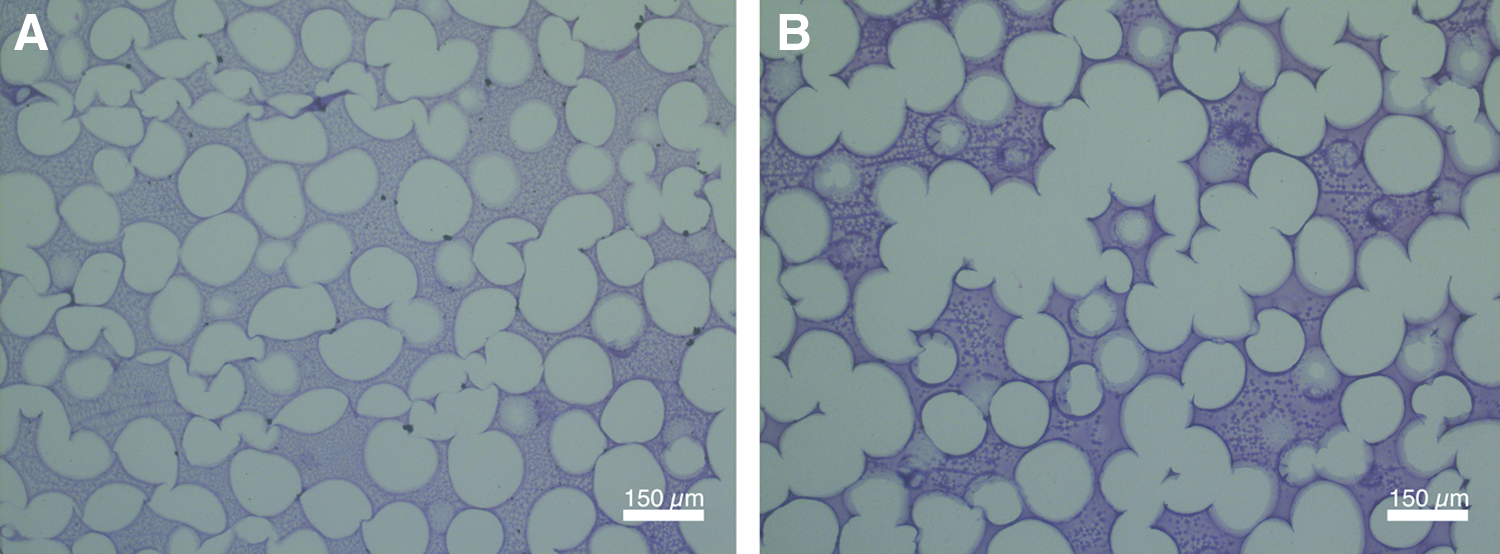
The PMMA templates were imaged using brightfield microscopy to determine the size of the interconnections and microsphere diameters. The resultant hydrogel pore size and interconnectivity was assessed based on selective partitioning of fluorescent protein within the pores imaged by confocal microscopy. 17 Briefly, hydrogels were incubated in a 0.5% (w/v) solution of FITC-BSA overnight. The following day a cross section of the hydrogels were cut and imaged using a PASCAL laser scanner microscopy system. The hydrogels were imaged using a 505–530 nm band pass filter with a 488 nm laser with a 10× objective. The resolution of the x and y directions was 1.75 μm/pixel. Images were obtained for a ∼60 μm thick section of hydrogel. For both the PMMA template and hydrogels, the interconnectivity values were divided by the mean pore size to obtain the normalized pore connectivity (NPC).
Mechanical testing
Hydrogels were generated using the sphere templating technique to generate three different interconnectivities: 0.24, 0.29, and 0.42 NPC (sintering at 175°C for times of 0, 45, and 90 min), for mechanical analysis (n=6). Hydrogels were prepared in a cylindrical shape with a 10 mm diameter and 4 mm height. An Instron 4465 compression tester (Instron Corp., Grove City, PA) was used to compress the hydrogels at a constant rate of 0.5 mm/min. A stress (σ) versus strain (ɛ) curve was generated for each of the hydrogels. The linear region of each plot is where the material followed Hooke's Law governed by the equation σ=Eɛ, where σ is stress (F/A) and ɛ is the strain (ΔL/LO). The slope was calculated using a linear regression of 0–10% of the strain range to obtain the Young's Modulus (E), which was averaged for six samples.
Multilayer gradient hydrogels
A hydrogel layer consisting of poly(lactic-co-glycolic acid) (PLGA) microspheres and poly(ethylene glycol)-co-(

Schematic representation of porous hydrogel with PLGA layer containing growth factor. Total thickness of the hydrogel and PLGA layers is 4 mm with a diameter of 10 mm. PLGA, poly(lactic-co-glycolic acid). Color images available online at
BSA, bovine serum albumin; DI, deionized; PDGF-BB, platelet-derived growth factor-BB; PEG, poly(ethylene glycol); PLGA, poly(lactic-co-glycolic acid); PVA, poly(vinyl alcohol).
Synthesis of PEG-PLLA-DA was performed using the procedure developed previously.19–21
Briefly, 20 g of PEG (Mn 3350) was mixed with 4.24 g of 3,6-dimethyl-1,4-dioxane-2,5-dione (
PDGF-BB was radiolabeled with 125I then loaded to quantify the amount of growth factor released from the hydrogel. 22 Briefly, PDGF-BB was radiolabeled with 125I using Iodo-beads (Pierce Chemical Co., Rockford, IL). The beads were washed with reaction buffer, placed into a solution containing 125I, followed by the addition of PDGF-BB into the solution for 15 min. The beads were removed, and the solution diluted to a final volume of 3 mL and dialyzed against deionized water for 48 h to remove excess 125I. Fifteen milligrams of PLGA microspheres containing radiolabeled PDGF-BB was combined with 50 μL of PEG-PLLA-DA (10%, w/v). Two different hydrogel conditions, low and high interconnectivity (NPC of 0.24 and 0.42), hydrogels (n=3) were prepared as described above. Each hydrogel was placed in 2 mL of PBS in a transwell plate incubated at 37°C. Release samples were collected daily from each well and replaced with fresh PBS. The release of PDGF-BB from free microspheres and microspheres incorporated in the hydrogel were measured with a γ counter (Packard Instrument, Meriden, CT). Summation of the previous day's data is used to calculate the cumulative release. Radioactive decay of the Iodine was adjusted for using the decay half-life equation. Percent release of growth factor was calculated from an average of three parallel studies.
Animal model
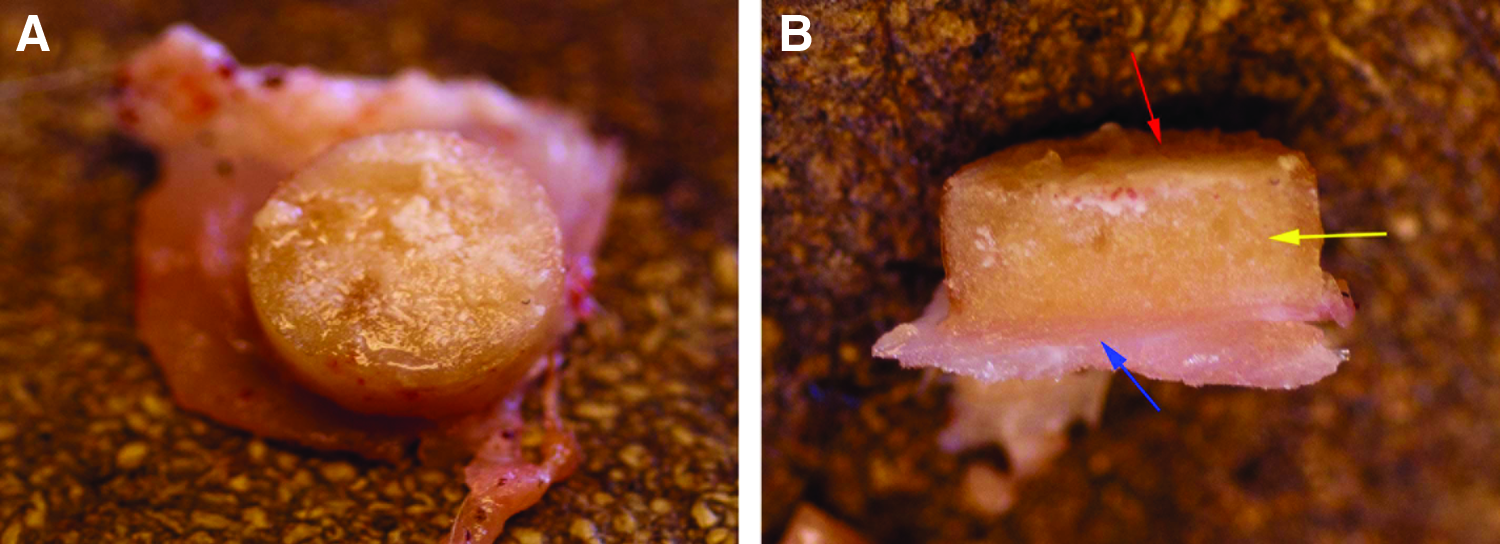
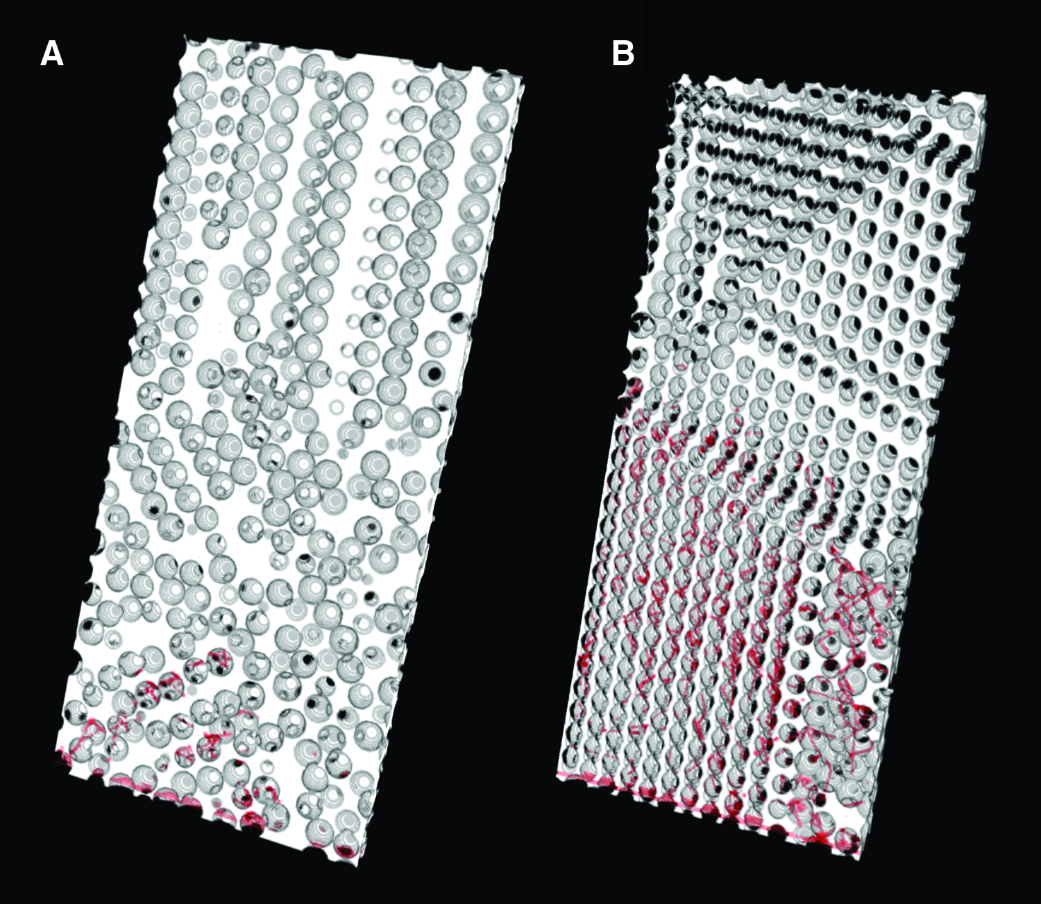
All animal experiments were carried out at Edwards Hines, Jr. VA Hospital using procedures approved by the Institutional Animal Care and Use Committee. A rodent subcutaneous implantation model was used for evaluating the hydrogels in vivo. 23 Hydrogels with a mean pore size of 129 μm and NPC values of 0.24 and 0.42 (sintered at 175°C for 0 and 90 min), n=5, were synthesized and prepared under sterile conditions, Figure 3. Polypropylene shells were fabricated in the shape of a top hat, with a height of 4 mm, a diameter of 10 mm, and an integral sewing ring extending from the open end of the hat. The shells were autoclaved for sterilization before animal surgery and all hydrogels were made to fit in the shells. The multilayer hydrogel was prepared to fit into the top hat in an orientation in which the PLGA microspheres were distal to the tissue bed allowing generation of a PDGF-BB gradient within the hydrogel.
Cross-sectional slices of hydrogels with a mean pore size of 129 μm sintered at 175°C for
Male Lewis rats (300–400 g, n=5; Charles River, Wilmington, MA) were anesthetized initially with 5% isofluorane through a nose cone and maintained at 2–4% isofluorane/35% oxygen mixture during the procedure. Their backs were shaved and skin scrubbed with isopropyl alcohol followed by a povidone–iodine antiseptic solution. A longitudinal incision was made along the spine and the skin separated using blunt dissection. Polypropylene shells containing the hydrogels were implanted subcutaneously and secured with four evenly spaced sutures through the sewing ring. Each rat received two implants with the implant location determined randomly. The skin incision was closed using 4-0 nylon suture.
At 3 and 6 weeks after implantation, 100 μg AlexaFluor 647-conjugated Isolectin GS-IB4 from Griffonia simplicifolia (Invitrogen) was injected into animals through the tail vein under general anesthesia. The animals were then perfusion fixed with 4% paraformaldehyde and the implanted samples harvested. The harvested tissue, including the entire hydrogel and underlying muscle, were cut into two symmetric pieces Figure 4. One half was paraffin embedded and sectioned (5 μm thickness) for histological staining. The other half was embedded in Optimal Cutting Temperature compound, frozen, and sectioned (50 μm thickness) for fluorescence imaging. Sections were cut through the center of the sample so they contained cross sections of the gel with the interface of the underlying muscle.
Histological and vascular analysis
Paraffin-embedded tissue sections were stained with Hematoxylin and Eosin (H&E) and Masson's Trichrome. For quantification of tissue invasion, the H&E-stained sections were digitally imaged (5× objective, 1.26 μm/pixel) using an Axiovert 200 inverted microscope. The depth of tissue invasion was defined as the maximum depth of tissue ingrowth within the pores from the underlying muscle. Due to tissue shrinkage during processing, the invasion depth was divided by the height of the imaged sample to obtain a normalized invasion depth. Masson's Trichrome-stained sections were used to evaluate the overall tissue structure.
Frozen tissue sections were imaged with confocal microscopy (Carl Zeiss AG, Jena, Germany) with dual fluorescence for AlexaFluor 647-conjugated isolectin-labeled endothelial cells (633 nm excitation, 650 nm longpass filter, far red) and tissue/hydrogel structure (488 nm excitation, 505–530 nm bandpass filter, green) with a 20× objective. The vasculature was imaged and vascular density calculated based on the equation: Vascular Density (%)=(Area of Lectin-Positive Staining/Area of Tissue)*100.
Modeling scaffold vascularization
Computer models of scaffolds were combined with an agent-based model of angiogenesis to further study the effect of biomaterial pore interconnectivity on vascularization. Scaffolds were modeled as having spherical pores with a constant pore size and interconnectivity. The interconnectivity of the scaffolds was defined as the diameter of the opening between adjacent spheres. 8 The scaffolds were generated by specifying a random pore location for the first pore. Subsequent pore locations were generated by searching all locations within the scaffold that would satisfy the condition of the specified interconnectivity. Once a location met the criteria a pore was placed followed by repeated searching for locations from added pores that would satisfy the interconnectivity criterion. Each pore has a minimum of three different pores adjacent to it. This process was repeated until no locations within the scaffold could satisfy the specified interconnectivity.
Scaffolds were generated with a constant spherical pore size (150 μm) and interconnectivity. The NPC was used to quantify connectivity and is defined as the ratio of the pore throat diameter to the pore size. Two NPC scaffold conditions were selected for study based on the in vivo results, 0.37–0.47 (interconnectivity 55.5–70.5 μm) and 0.20–0.27 (interconnectivity 30–40.5 μm). Ten unique scaffolds were created for each condition. The bulk porosity and porosity at the surface, where the host blood vessels come into contact, was calculated for each of the scaffolds to compare the two types of scaffolds.
A gradient is formed due to the transport of PDGF-BB within the hydrogel. The gradient was modeled using Fick's second law. The assumption for this model is that diffusion is the only mechanism of transport and the proteins do not bind to the hydrogel structure. Boundary condition at the distal end was defined based on the release kinetics of PDGF-BB from the PLGA, and the tissue interface was treated as an infinite sink. Diffusion coefficients within the hydrogels were determined by fitting data to the experimental results with radio-labeled protein. The concentration profiles were obtained by solving Fick's second law using MATLAB. The predicted temporal and spatial variations in concentration were combined with the model scaffolds. Simulation of angiogenesis within these scaffolds was performed using the agent-based model previously described 10 and average invasion depths quantified for 1–6 weeks.
Statistical analysis
All statistical data are expressed as means±standard deviation. In vitro data were analyzed using one-way ANOVA followed by a Tukey's post test for normally distributed data or Dunns method for data that failed normality criteria. Growth factor release in vitro studies were analyzed with one-way repeated measures ANOVA using SigmaStat (San Jose, CA). Paired Student's t-test was used for comparison of in vivo data due to each animal receiving one implant of the two different NPC scaffolds. Values of p<0.05 were considered statistically significant.
Results
Hydrogel preparation
PMMA microspheres were sintered at two different temperatures, 160°C and 175°C, to develop templates for generating hydrogels with varying interconnectivity. The time at which the templates were sintered ranged between 0 and 30 h, 0 h signifying the time at which the oven achieved the desired temperature. The interconnectivity of the spheres in the template and the pores in the resultant hydrogels increased with longer sintering times and higher temperatures (Fig. 5). The pores in the hydrogels made from the templates agreed well with the structure of the PMMA template. The ranges of interconnectivities obtained for 160°C and 175°C were 33–55 μm (NPC 0.23–0.42) and 33–54 μm (NPC 0.23–0.42), respectively. There was statistical difference in NPC (p<0.05) between the hydrogel and PMMA template sintered at 160°C at each time point (Fig. 5A) with the hydrogels exhibiting a slightly higher value. For the 175°C sintered condition, all had statistical differences (p<0.05), except for the 0, 0.25 and 1.25 h time points. Pore size did not exhibit a significant correlation with sintering time. The range of pore sizes obtained when sintering at 160°C (0–30 h sintering time) was 143–154 μm, with a mean pore size of 150±2.60 μm. Similarly for the 175°C sintering condition (0–1.5 h sintering time), the range of pore size obtained was 122–139 μm, with a mean pore size of 129±6.77 μm. While time did not appear to influence pore size, there was a statistical difference (p<0.001) between mean pore sizes for the two sintering temperatures.
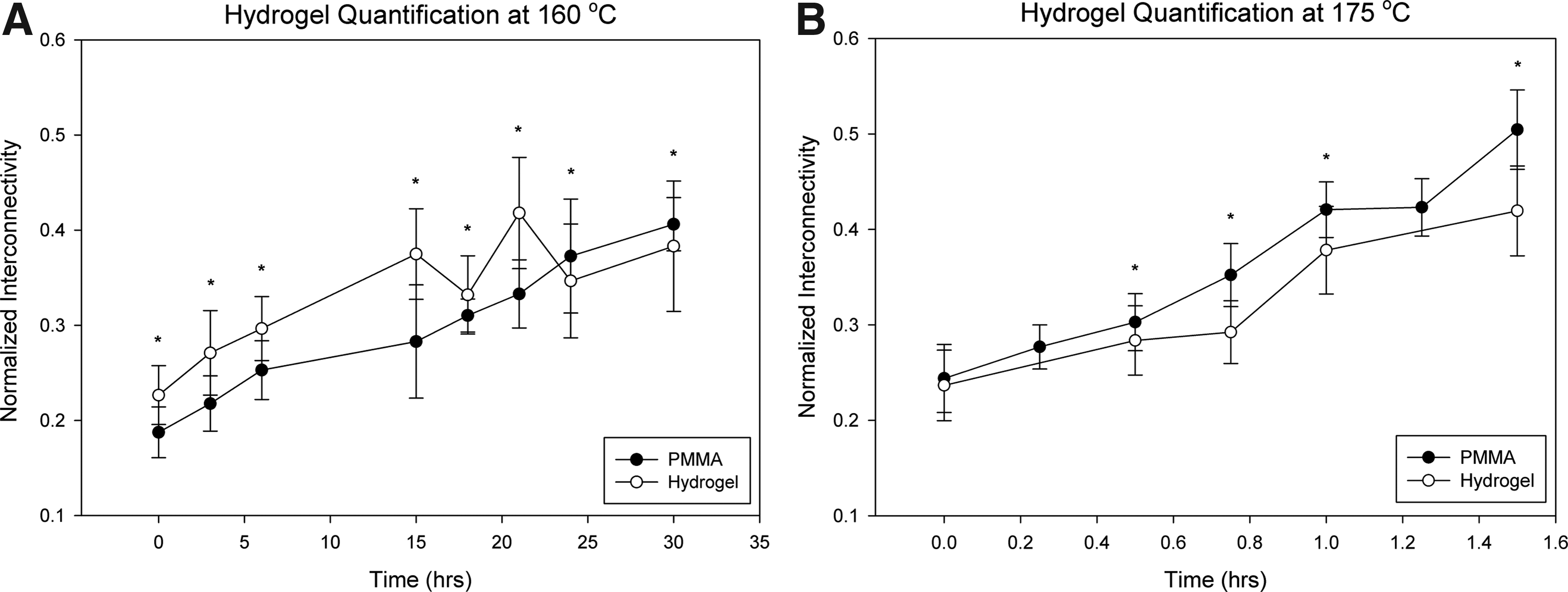
Plots of normalized pore interconnectivity versus incubation time plots for PMMA templates and resultant hydrogels sintered at
Compression testing was conducted on the hydrogels to determine the influence of hydrogel pore interconnectivity on mechanical properties. The elastic modulus of the hydrogels decreased as the interconnectivity of the hydrogels increased with a significant difference between the lower (1284±182 kPa) and higher (852±232 kPa) hydrogel interconnectivities (NPC values of 0.24 and 0.42), (Fig. 6).

Hydrogels exhibit decreasing compressive modulus with interconnectivity. Low, medium, and high NPC values were 0.24, 0.29, and 0.42, respectively. *p<0.05 (n=6).
Growth factor release in vitro
Multilayer hydrogels were prepared by placing a PEG-PLLA layer with PLGA microspheres containing PDGF-BB on the distal surface of the hydrogels. The release of PDGF-BB from the hydrogels was observed by measuring the release of 125I proteins in the incubation solution. The percent release of PDGF-BB varied with hydrogel interconnectivity (Fig. 7). There was a statistical difference (p<0.001) in release between the lower and higher interconnectivity release profiles. There was an initial burst of release for both conditions the first 4 days followed by a sustained release. The growth factor is released more rapidly from gels with higher interconnectivity.
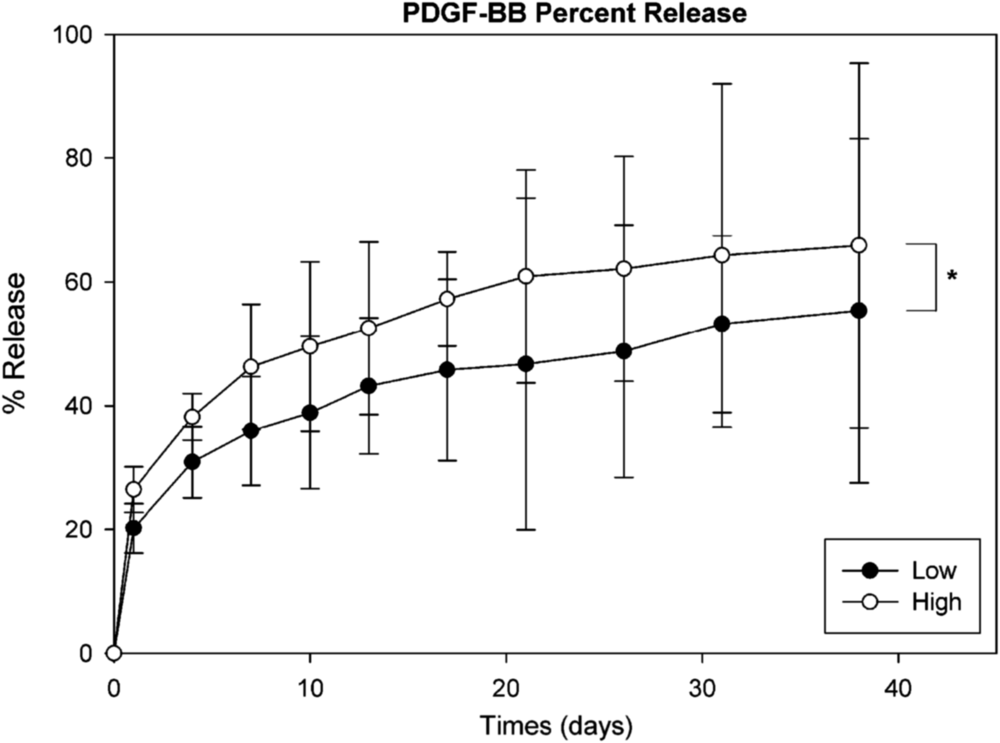
Percent release of platelet-derived growth factor-BB (PDGF-BB) from low (NPC 0.24) and high (NPC 0.42) interconnectivity hydrogels. *p<0.05.
Vascular tissue invasion in vivo
PEG hydrogels with two different interconnectivities (NPC 0.24 and 0.42) were implanted subcutaneously in rats. At weeks 3 and 6, the hydrogels were harvested and processed for histological analysis. Tissue invasion was observed within the hydrogel pores for both conditions (Fig. 8). Qualitative inspection of the H&E stains at 3 weeks suggests that the depth of tissue invasion within the hydrogel is larger for the higher interconnectivity scaffolds compared with the lower. At 3 weeks, tissue was present in the entire hydrogel volume for the high interconnectivity hydrogels compared with nearly half for the lower. For both conditions, the tissue invaded can be seen within the channels connecting the pores (Fig. 8B, D).
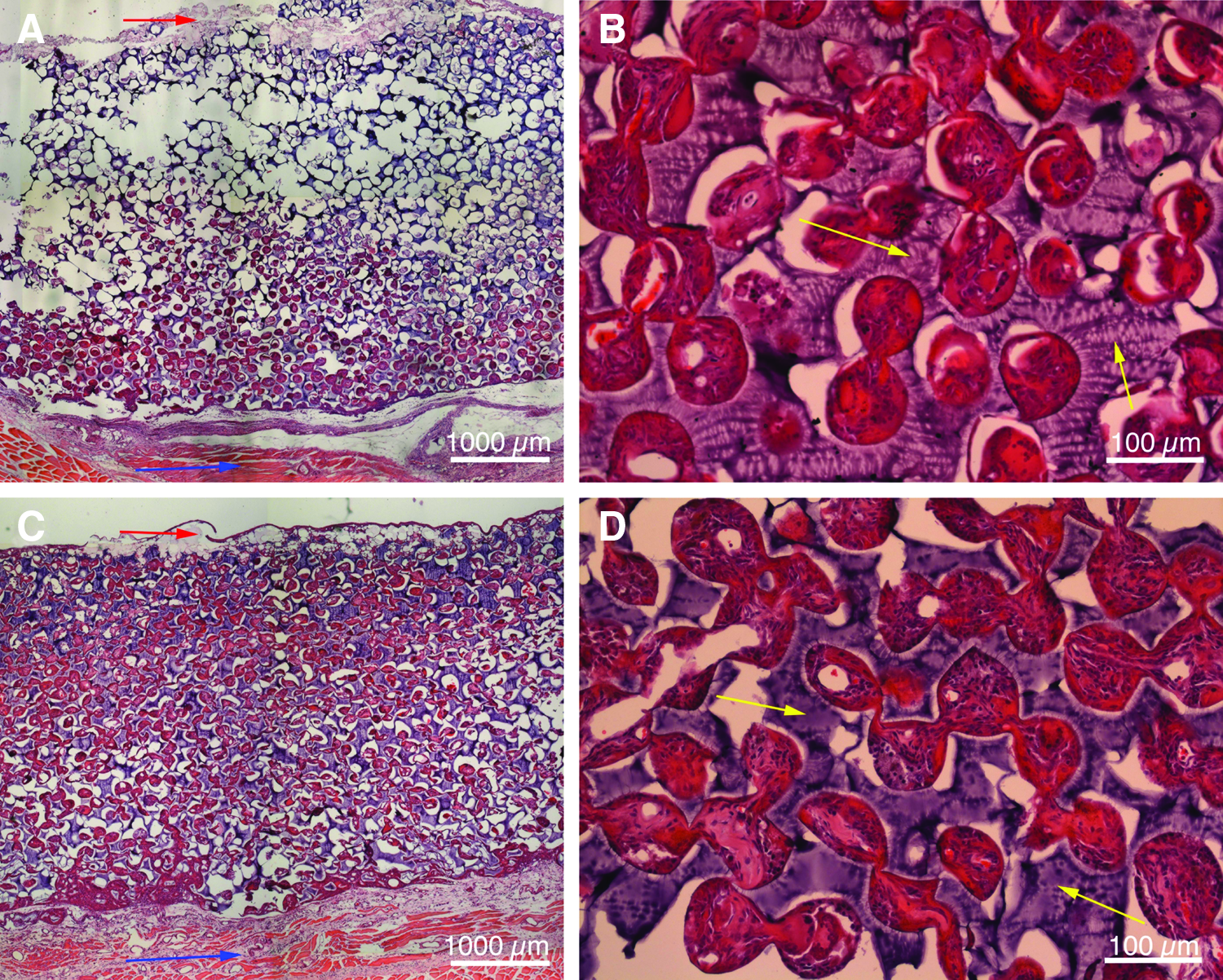
Hematoxylin and Eosin staining of
Histological staining shows that some regions appear to be progressing to granulation tissue as collagen can be seen within the pores of the hydrogel scaffolds (Fig. 9). In many areas, a robust foreign body response can be seen within the tissue with multinucleated foreign body cells present within the pores. A layer of inflammatory tissue (red) was present at the tissue–polymer interface, while collagen (blue) is present within the center of the pores. Collagen is observed for both conditions at 3 and 6 weeks.
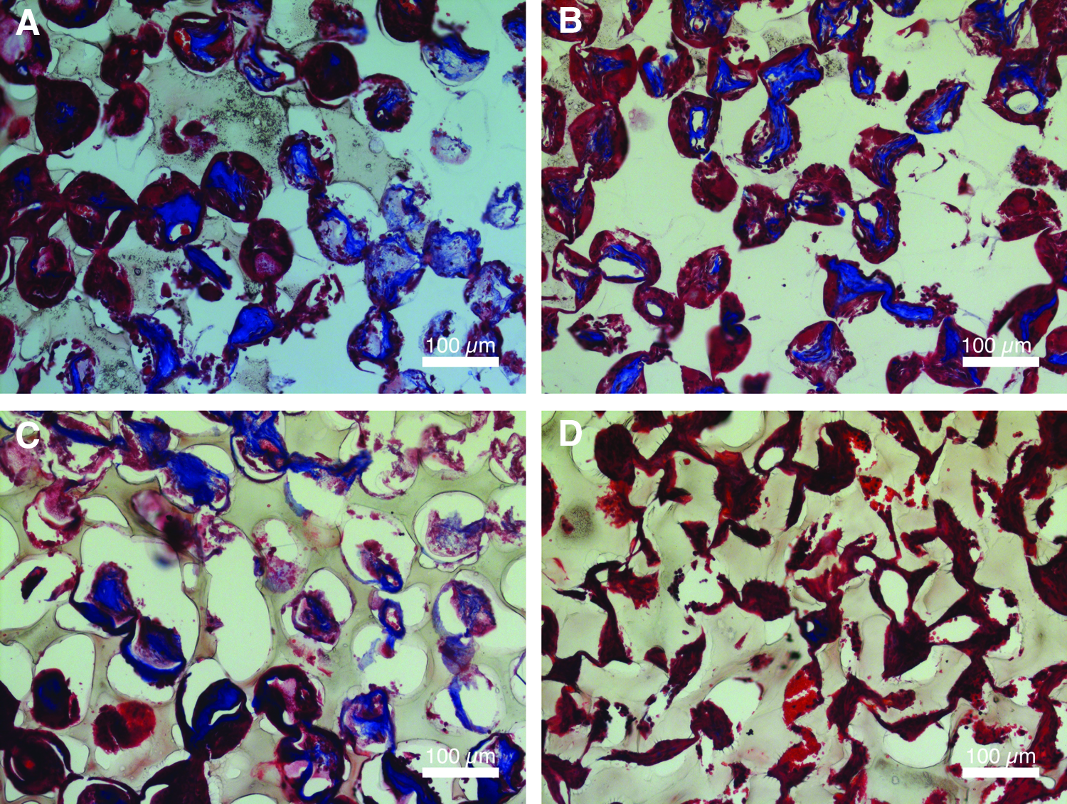
Masson's Trichrome staining for
Absolute and normalized depths of tissue invasion into the porous PEG hydrogels were quantified (Fig. 10). Significant tissue invasion was observed for all hydrogels at 3 and 6 weeks. There was a significant difference (p=0.004) in absolute depth of tissue invasion at week 3 between the lower (1334.62±191.70 μm) and higher (2463±404.81 μm) interconnectivity hydrogels. When comparing the same groups between time points there were no statistical differences (Fig. 10A). However, when examining normalized values, there was statistical difference between time points and also between the two conditions (Fig. 10B). For the lower interconnectivity hydrogels there was an increase in invasion depth from 3 to 6 weeks, which was opposite for the higher interconnectivity hydrogels, where there was a decrease in invasion depth.

Vascular analysis
To evaluate vascularization within pores of hydrogels, animals were injected with fluorescently labeled isolectin (AlexaFluor 647) and perfusion fixed with 4% paraformaldehyde before tissue harvest. Vessels were observed in the tissue present in the pores of the hydrogels throughout the scaffolds (Fig. 11). Blood vessel density was measured for all conditions (Fig. 12). At 3 weeks, blood vessel density was higher for the higher interconnectivity (NPC 0.42) compared to the lower interconnectivity (NPC 0.24), 9.14%±2.03% and 4.38%±0.54%, respectively. The blood vessel density decreased for both conditions from week 3 to 6 (1.44%±0.18% for higher and 1.19%±0.15% for lower interconnectivity hydrogels). There was no significant difference in vessel density between the higher and lower interconnectivity hydrogels at week 6. The diameter of the vessels was not different between the two groups.

Vascularization within porous PEG hydrogels. Confocal microscopy images of isolectin (red)-labeled blood vessels in
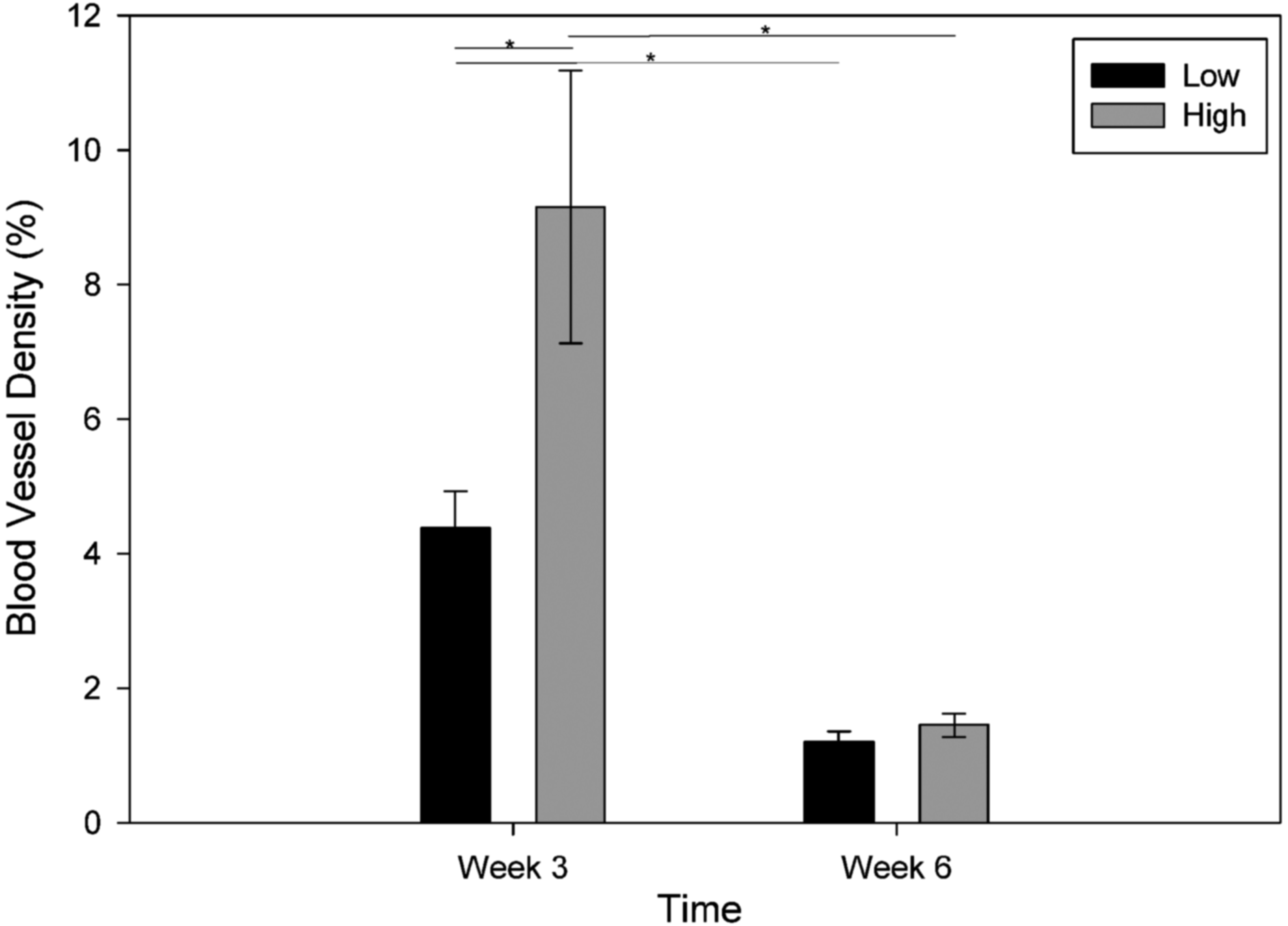
Blood vessel density versus time for low (NPC 0.24) and high (NPC 0.42) hydrogels. Blood vessel density decreased from week 3 to 6 for both conditions. *p<0.05.
Simulated scaffolds
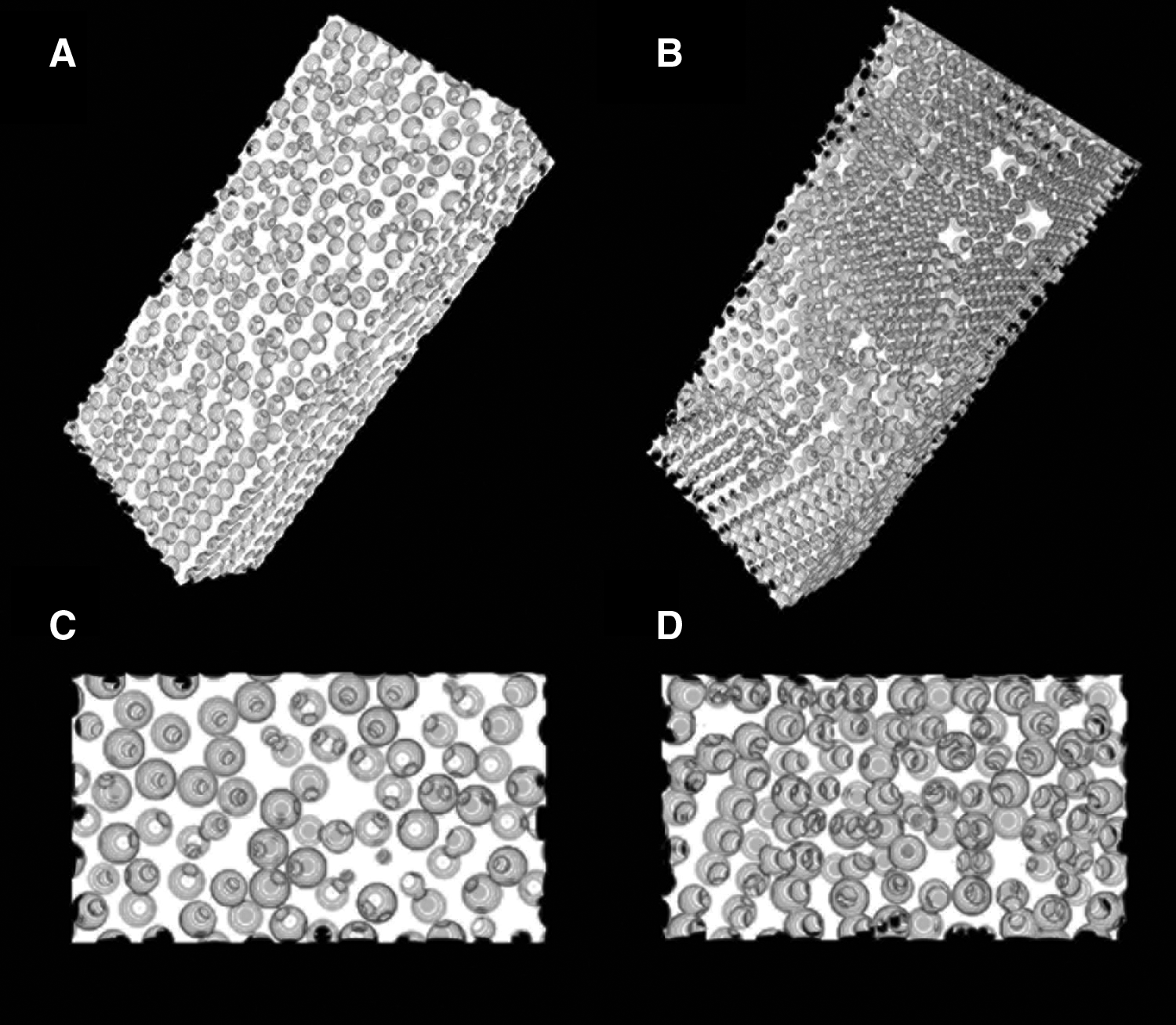
Interconnectivity influences a number of processes with regard to vascularization. Blood vessel formation could be altered due to steric hindrance of vessel growth or alterations in growth factor transport as suggested by the release studies. An agent-based model of angiogenesis in porous polymer scaffolds was used to examine the effect of interconnectivity on vascularization with greater control over the polymer properties. Vascularization was modeled within the porous structure using a previously described agent-based model. 11 Computer modeled scaffolds were generated with NPCs similar to those used in experimental studies. For these scaffolds, porosity is dependent on the pore interconnectivity (Table 2). The lower pore throat diameter (0.20–0.27 NPC) produced scaffolds with a porosity of 63.25%±0.10% whereas the higher (0.37–0.47 NPC) had a mean bulk porosity of 77.63%±0.59%. The interface porosity had mean porosities of 64.04%±2.16% and 78.91%±1.43%, respectively. Three-dimensional renderings of scaffolds with corresponding interfaces are shown in Figure 13.
NPC, normalized pore connectivity.
The gradient that formed due to PDGF-BB transport within the hydrogel was modeled using Fick's second law. Diffusion coefficients within the scaffold were determined by fitting experimental radio-labeled protein release (Fig. 7) to the model. Figure 14 illustrates the predicted concentration gradients within the two scaffold conditions (0.20–0.27 and 0.37–0.47 NPC) at 3 and 6 weeks. The difference between the gradients for the same time point is ∼20%. At 3 weeks, the lower interconnectivity scaffolds have a higher concentration gradient compared with the higher interconnectivity due to slower diffusion within the scaffold. Similar trends are observed for the 6 week time point, however with a lower bulk concentration. This difference in concentration within the scaffolds may contribute to the observed differences in vascularized tissue invasion.

Modeled concentration of PDGF-BB within scaffolds at 3 and 6 weeks for low (0.20–0.27 NPC) and high (0.37–0.47 NPC) interconnectivity scaffolds.
To compare the relative contributions of steric hindrance and altered concentration profiles, vascularization was modeled within both scaffold NPC conditions and both concentration profiles. This means that high NPC scaffolds were modeled within both its predicted concentration profile and the concentration profile of the low NPC. These are referred to as HIHC (high interconnectivity/high concentration) and HILC (high interconnectivity/low concentration), respectively. Analogous conditions were examined for low NPC (i.e., low interconnectivity/high concentration (LIHC) and low interconnectivity/low concentration (LILC)). Figure 15 illustrates model simulations of vascularization of the scaffolds with constant pore size and varied interconnectivities after 6 weeks. Scaffolds with a greater pore throat diameter allows for deeper invasion depths within the scaffolds. The depth of vessel invasion is greater in the higher interconnectivity groups for all time points (Fig. 16). The different concentration gradients (low and high) did not alter vascularization significantly within the scaffolds. Maximum tissue invasion is observed at week 4 for all conditions, followed by a regression for weeks 5 and 6.
Three-dimensional renderings of scaffold vascularization for

Quantitative analysis of average invasion depth of scaffolds for model results generated for low (0.20–0.27 NPC) and high (0.37–0.47 NPC) interconnectivity hydrogels with low and high concentration profiles. High interconnectivity/high concentration, high interconnectivity/low concentration, low interconnectivity/high concentration, and low interconnectivity/low concentration are represented as HIHC, HILC, LIHC, and LILC, respectively.
Discussion
In this research, the relationship between vascularization and pore interconnectivity within porous PEG hydrogel scaffolds was investigated. A sphere template technique was used to create hydrogels with a constant pore size and controlled interconnectivity to isolate the effect of interconnectivity on vascularization13,16 Using PMMA microspheres as a template, hydrogels were created with varying interconnectivity, but a relatively uniform pore size (130–150 μm). The temperature and time at which the microspheres were sintered allowed for hydrogels with varying interconnectivities to be created based on the structure of the microsphere template. The sintering process may have resulted in a smaller effective pore size in the high interconnectivity gels due to greater pore overlap. The pore size varied little for the same sintering temperature at different times. There was no clear trend observed, as an increase in sintering time did not decrease the effective pore size. Because of these observations, the mean pore size was calculated for the 160°C and 175°C sintering temperature and used in further discussion. However, there was a statistical difference (p<0.001) between mean pore sizes for the two different sintering temperatures. The interconnectivity ranged from 33 to 55 μm depending on the sintering time and temperature. Mechanical testing conducted on the hydrogels show that increasing interconnectivity also resulted in a decreasing elastic modulus, likely due to differences in bulk porosity.
A source of the growth factor (PLGA microspheres) was placed on one end of the gels to create a gradient stimulus for tissue and vessel invasion. In this system, there was an initial burst release of PDGF-BB from the hydrogels followed by a sustained release profile for ∼40 days. The release of the entrapped growth factor was controlled primarily by the microsphere degradation rate. However, pore structure also influenced release. PDGF-BB was released more rapidly from the higher interconnectivity hydrogel. The larger pore throat allows a more rapid diffusion through the gel and release into the surrounding solution. 24 The gradient hydrogels were then evaluated in a rodent subcutaneous implantation model. There was significant tissue invasion for both interconnectivity conditions at week 3. Surprisingly, tissue invasion for the higher interconnectivity had completely vascularized nearly all hydrogels by 3 weeks with tissue and blood vessels present throughout the volume of the hydrogel. Tissue invasion was observed at the lower interconnectivity; however, a smaller portion of the hydrogel contained tissue. The presence of larger interconnectivities contributed to significantly greater tissue invasion within the hydrogels. Although the difference in interconnectivities between the hydrogels is 20 μm, it is large enough to allow a significantly greater tissue invasion within the hydrogel.
The sphere templating technique was used so gels with a constant pore size and controlled interconnectivity could be generated. Other pore structures could be used. However, the spherical geometry allows for control over the interconnectivity that is difficult to achieve. It is possible that other geometries, such as cylindrical pores, may facilitate directed invasion of vascularized tissue, but this would require investigation of the influence of the nature of pore connections on this response. These interactions are more complicated due to the multiple interfaces that would exist in these systems. The pore structure can also influence curvature of the pore, which can influence cell behavior. Interface roughness is well known to influence cell and tissue response to a biomaterial. The introduction of rougher surfaces may result in deviation from the results described in this study. In all studies, a gradient of growth factors were included to stimulate tissue growth into the porous structure. PEG invokes a minimal inflammatory response when implanted, and previous work has shown that subcutaneous hydrogel implants in the absence of growth factors resulted in invasion depths <1000 μm.25,26 For this reason, gradients of PDGF-BB were used to increase tissue and vessel invasion into the complex porous structure to explore the influence of the pore geometry.
In our system PLGA microspheres are still present and releasing significant levels of growth factor at week 3. This stimulus likely stimulates cell proliferation and migration into the scaffold. At this relatively early stage, the blood vessel density is high. At week 3 the blood vessel density is higher within the higher interconnectivity hydrogel. While differences in the depth of tissue invasion was expected, the increased density was not anticipated as the values are normalized by tissue area. In general, granulation tissue forms within the pores, which is expected to have a relatively constant density. The difference in blood vessel densities may result from the more rapid vascular invasion into the gels with higher interconnectivity. The vessels present in the pores had sufficient time to proliferate to generate more vessels. In the lower interconnectivity case, where the rate of tissue invasion was slower, data at 3 weeks may still be showing the initial sprouting process. Both groups exhibit a decrease in blood vessel density at week 6 likely due to remodeling of the networks. In addition, by week 6, a significant amount of growth factor has been released so the concentration within the scaffolds was low.
Previous studies have looked upon scaffold pore size as the main contributor to promoting vascularization within a biomaterial. Although there is not agreement on the ideal pore size, the general consensus is that larger pores promote rapid and deeper vascularized tissue growth. Similar results were obtained from this study compared with others, where increased pore size allows for vascularization deep within the hydrogel. However, the results obtained in this study agree with what others have found.2,27 In studies with PLGA scaffolds, it was observed that larger pores (>200 μm) allowed deep vascularization within the biomaterial. In a study with scaffolds seeded with cells, the optimal pore size for vascularization was determined to be 40 μm.13,28 Bezuidenhout et al. investigated scaffolds with pore sizes ranging between 63 and 180 μm with a uniform NPC of ∼0.50. Their study determined no clear relationship between vascularization and pore size. 29 In the present study pores were smaller in size, 129 μm, however the amount of tissue growth was significantly greater. This pore size allowed for vascularization deep within the hydrogel, ∼60% vascularized for low interconnectivity by 6 weeks, and ∼90% vascularization by 3 weeks for the higher interconnectivity case. It is clear that pore size may have an effect on tissue growth, however, its contributions to vascularization of a biomaterial is also dependent on the interconnectivity of the scaffolds that was observed for this study where a uniform pore size and varied NPC was investigated.
Tissue invasion presented a more complex behavior with temporal differences between low and high interconnectivity gels at 3 and 6 weeks. Tissue invasion increased from 3 to 6 weeks for the lower interconnectivity hydrogel and decreased for the higher interconnectivity. A number of possible mechanisms exist for this behavior. First, at week 6 significant amount of PLGA microspheres have degraded and nearly 60% of the PDGF-BB is released. At this point there is a much lower concentration gradient in the scaffolds supporting tissue ingrowth. Second, the blood vessels that are assembled at the end of the hydrogel may be immature. This may result in insufficient oxygen and nutrient transport to support the new tissue resulting in tissue regression. This is not observed for the lower interconnectivity case due to the slower diffusion of growth factor within the hydrogel, which results in varying gradients within the gels. Another possibility is that the maximum amount of tissue ingrowth for the lower interconnectivity was not observed due to limitations with the invasive analysis procedure used, presenting only a single time point of the dynamic procedure. Tissue invasion may have peaked at 4 or 5 weeks so this 6 week time point could also represent regression in the lower interconnectivity.
A computational model was used to evaluate vascularization within scaffolds with different pore sizes and varying levels of interconnectivity. 11 Results were based on experimental findings with porous scaffolds that were not accurately modeled, as they had neither spherical pores, narrow size distributions, nor controlled interconnectivity. In this study, model scaffolds were generated to more accurately mimic the structure of hydrogels produced using the sphere templating technique. From these studies, the interconnectivity of the scaffolds was shown to have a significant impact in porosities obtained, 63.25% and 77.63%. The porosity values obtained for higher interconnectivity scaffolds are similar to what is experimentally obtained from sphere templating technique, 73.7%, with a constant pore size and a NPC value of 0.50. 16 This suggests the method of generating scaffolds that have similar properties as actual scaffolds created by the sphere templating technique for further study in computational modeling of vascularization. Predictions of PDGF-BB concentrations were combined with the model scaffolds to provide insight into the relative roles of steric hindrance and altered transport on vascularization. When high NPC scaffolds were modeled with its predicted concentration and the concentration of the low NPC scaffolds, the results obtained showed no difference in tissue invasion (Fig. 16). Similar trends are observed with the low NPC scaffolds. The two different concentration profiles for the same NPC scaffolds produced almost identical tissue growth for all time points observed. The pore interconnectivity appears to alter vascularization through steric hindrance of ingrowth rather than altered transport of growth factors.
Comparison of experimental findings to the agent-based model provides insight into the potential roles of steric hindrance and altered growth factor distribution on vascularization within in vivo scaffolds. In addition, the model allows insight into vascularization at intermittent time points to help explain the experimental findings. From the in vivo experiments invasion results could only be quantified at distinct time points, 3 and 6 weeks. As stated above, there was regression of invaded tissue for the higher interconnectivity hydrogel from week 3 to 6. The agent-based model of tissue growth allowed identification of a potential maximum invasion point between those two times. This trend was not observed for the lower interconnectivity hydrogel. However, the model suggests that this results from the maximum invasion being lower in this condition and that regression may have indeed occurred in the system. Overall, the model allows insight that is not possible in experimental studies.
Conclusions
In this study, we investigated the role of interconnectivity of hydrogels on vascularization using both computational and animal models. Hydrogels with controlled pore size and interconnectivity were fabricated using a sphere templating technique. The hydrogels with larger interconnectivity allowed for extensive vascularization and formation of blood vessel networks within the pores in vivo. Pore interconnectivity is an important feature for optimization in the fields of tissue engineering and regenerative medicine.
Footnotes
Acknowledgments
This work was supported, in part, by funding from the Veterans Administration and National Science Foundation (IIS 1125412).
Disclosure Statement
No competing financial interests exist.