
Abstract
To increase the epithelial proliferation of an auto-tissue-engineered lamellar cornea, 3.0×10⁶ corneal epithelial cells (CECs) were combined with 3×10⁵ mouse embryonic stem cells (ESCs) pretransfected with the HSV-tk gene (CECs+ESCs-TK group), and 3.3×10⁶ corneal epithelial cells (CECs group) were seeded between the acellular porcine corneal stroma and the amniotic membrane using the centrifugal cell seeding method. After 4 days of perfusion culture (treatment with ganciclovir starting on day 2), a thicker corneal epithelium (four to five layers) formed in the CECs+ESCs-TK group compared with that observed in the CECs group (two to three layers). More stem/progenitor cell (K3 −, p63+, ABCG2+, and integrin-β1+) and proliferation phenotypes (Ki67+) were measured in the CECs+ESCs-TK group compared with the CECs group using immunofluorescence staining, real-time quantitative reverse transcription polymerase chain reaction, and flow cytometry. Consistent with these findings, the colony-forming efficiency and cellular doubling time were significantly different between the CECs+ESCs-TK group (16.18%±3.98%, 28.45±2.03 h) and CECs group (11.96%±2.60%, 36.3±1.15 h). In a rabbit lamellar transplantation model, the CECs+ESCs-TK group had better epithelial barrier functions and wound healing abilities compared with the CECs group. Furthermore, ESCs-TK could be completely and safely removed by ganciclovir. Thus, the ESCS-TK coculture system could serve as a potential strategy for corneal tissue engineering.
Introduction
T
For the maintenance of epithelial viability in corneal reconstruction, the seeding cells, bioengineered scaffolds, and proper strategies were evaluated. 2 First, to acquire the highly proliferative properties of corneal epithelial cells (CECs) for seeding, various methods have been applied, including different feeder cells, conditioned media, cytokines or growth factors, 3 as well as the immortalization of CECs. 4 In our laboratory, an immortalized permanent cell line has been established from a normal human cornea using serial culture, 5 and we also demonstrated that the functional properties of stem-like CECs could be markedly enhanced by the microenvironment of embryonic stem cells (ESCs).6–9
The second major problem associated with corneal reconstruction is the absence of a suitable scaffold for the stem cells to grow and be transferred to the eye. Currently, several natural or synthetic biomaterials, including amniotic membranes (AMs), 10 fibrin gels, 11 silk, 12 and collagen, 13 have been applied for the reconstruction of corneal substitutes. Furthermore, from the perspective of the natural extracellular matrix composition and regional-specific cues for cellular proliferation and adhesion, the acellular porcine corneal stroma (APCS) may provide a promising alternative scaffold. 14 As we previously reported, the APCS 15 was developed by our group and has been successfully applied in corneal limbus 16 and stroma17,18 reconstruction.
Finally, an appropriate reconstruction strategy also plays a key role in epithelial growth. In recent years, after 4–6 weeks of construction, two to six layers of the epithelium had formed on different scaffolds using air–liquid interface culture.12,13,19 However, to further decrease the culture time for clinical application and to mimic the native ocular environment of the epithelium, a centrifugal cell seeding method 20 and dynamic culture system 21 were used, respectively.
On the basis of our previous studies, the viability of an auto-tissue-engineered lamellar cornea (ATELC) was increased using the ESC microenvironment in a controlled and safe manner. Briefly, these strategies were simultaneously applied in the reconstruction process. First, using coculture with ESCs, multilayers of rabbit autologous passage 1 (P1) epithelial cells were rapidly centrifugally seeded between the APCS and AM. After an increase in cell adhesion induced by the sheer force of the perfused culture, the ATELC was transplanted into a rabbit model. To avoid undesirable differentiation and tumorigenesis of ESCs, 22 ESCs were pretransfected with the herpes simplex virus thymidine kinase (HSV-TK), a suicide gene (ESCs-TK). When the viability of the CECs was enhanced, all of the ESCs-TK were induced to undergo apoptosis gradually by treating the culture with ganciclovir. 6
Materials and Methods
Animals
Female and male New Zealand white rabbits (aged 10 weeks and weighing 2–3 kg) were purchased from the Animal Laboratory of Sun Yat-sen University (Guangzhou, China). All of the animal experiments were performed with permission obtained from the Medicine Ethics Committee in Zhongshan Ophthalmic Center, Sun Yat-sen University, and complied with the Association for Research in Vision and Ophthalmology (ARVO) statement on the use of animals in ophthalmic and visual research.
Preparation of APCS and AM
APCS preparation: The APCS was prepared as previously described. 15 Briefly, native porcine corneas were immersed in a bicarbonate-mixed salt solution containing phospholipase A2 (PLA2) (200 U/mL; Sigma, P0861) and 0.5% (w/v) sodium deoxycholate (SD) (Sigma, D6750) for 6 h at 37°C. Next, the samples were immersed in a bicarbonate-mixed salt solution containing only PLA2 for 2 h at 37°C followed by six washes of 30 min each in a bicarbonate-mixed salt solution at 10°C to remove the residual PLA2 and SD. All of the steps were performed with continuous shaking in a thermostat-controlled water bath.
AM preparation: Proper informed consent was obtained from all AM donors in accordance with the principles of the Declaration of Helsinki on Biomedical Research. For the ATELC construction, the AM was prepared as previously described and sectioned into 2.3×2.3-cm pieces. 20
Cell preparation and culture
Primary CECs, which were obtained from a single 1×1-mm2 auto-rabbit corneal limbal tissue explant (100 μm thickness, left eye), were prepared as previously described and cultured in corneal epithelial medium. 6
Mouse ES-E14 cells, which had been transfected with the suicide gene of herpes simplex virus thymidine kinase (ESC-TK) and selected with 1 μg/mL puromycin (Sigma), were plated at a density of 400/cm2 in 1% gelatin (Sigma)-coated tissue culture dishes containing mouse ESC culture medium. 6
Control and experimental groups
A total of 3.3×10⁶ cells were seeded for each of the two groups according to the different types of cells. In the control group (CECs group), 3.3×10⁶ passage 1 (P1) auto-rabbit CECs were seeded. In the experimental group (CECs+ESCs-TK group), 3×10⁶ P1 auto-rabbit corneal CECs and 3×10⁵ ESCs-TK were mixed uniformly before the reconstruction process.
Culture strategies of ATELC
First, as we previously reported, three layers of cells were centrifuged onto the AM in both groups (14 mm2 in diameter) (1800 bpm, 4 min). 20 Next, after the membrane was covered with APCS (upside-down), it was fixed in the chamber (Ismatec pump; Ismatec, Minucells) with the AM (facing up for 4 days with continuous perfusion culture. 21 After 24 h of perfusion culture (20 mL/h) on both sides of the ATELC, 20 μM ganciclovir was added into the medium. 6 On day 4 postconstruction, each ATELC specimen was collected for observation of Hematoxylin and Eosin (H&E) staining, transmission electron microscopy (TEM, H600; Hitachi), and immunofluorescence staining. After all of the cells were isolated from the ATELC using the previously reported methods, 21 the DNA content and proliferative properties were observed using ELISA, real-time quantitative reverse transcription polymerase chain reaction (real-time qRT-PCR), flow cytometry, colony-forming efficiency (CFE), and the methylthiazol tetrazolium cell proliferation assay (MTT). 8
Removal of ESCs-TK by ganciclovir treatment
To track the ESCs-TK in a coculture system, all of the ESCs-TK were labeled with DiO (5 μg/mL; Invitrogen) for 30 min at 37°C before the reconstruction process. The expression of DiO was identified using fluorescence microscopy and flow cytometry analysis using the CellQuest Pro software (n=10; FACSCalibur flow cytometry, BD Biosciences). 6
HE and immunofluorescence staining
To measure the morphology and differentiation of the cells of ATELC, specimens from both groups were analyzed using H&E staining (n=4), TEM (n=4), quantitative DNA analysis (n=10; Sigma), flow cytometry (n=10), and immunofluorescence staining (n=4). The primary antibodies were K3 (1:100; Millipore), P63 (1:50, Millipore), ABCG2 (1:50, Millipore), Ki67 (Abcam ab8191; 1:100), integrin-β1 (Abcam 78502; 1:100), desmocollin 2 (Abcam 72792; 1:100), and integrin-β4 (Abcam ab29042; 1:100). The secondary antibodies included Alexa Fluor 594 IgG (Invitrogen, A11012; 1:100) and Alexa Fluor 488 IgG (Invitrogen A-11001; 1:100); the nuclei were counterstained with Hoechst 33342 (blue color; Invitrogen). The examinations were performed using a laser scanning confocal microscope (LSM 510 META; Carl Zeiss).
Evaluation of differentiated phenotypes and growth capacity of epithelial cells in ATELC using a quantitative assay
Real-time qRT-PCR was performed to detect the mRNA expression levels of K3, P63, and ABCG2. As previously described, 8 the total RNA from each group (n=10) was isolated using TRIzol (Invitrogen) according to the manufacturer's instructions and quantified by absorption at 260 nm. A total of 0.5 μg of RNA was reversely transcribed into cDNA in a 20-μL volume reaction system using the SYBR Prime Script™ 25 RT-PCR Kit (DRR063S; Takara). Samples of synthesized cDNA were divided into aliquots and stored at−80°C. Real-time qRT-PCR was performed and analyzed using an ABI PRISM 7000 sequence detection system (Applied Biosystems, Inc.). The PCR was performed in a 20-μL volume reaction system containing 10 μL of 2×SYBR Green reaction mix (SYBR Green system [DRR063S; Takara]), 0.4 mM paired primers, and 1 μL of cDNA. The sequences of the PCR primer pairs are listed in Table 1. The thermal cycling consisted of denaturation for 3 min at 95°C followed by 40 cycles of 15 s at 95°C and 30 s at 60°C. The PCR amplification efficiency of the primer sets was determined to be essentially 100% before qPCR. The comparative Ct (▵▵CT) method was used to compare the mRNA expression levels of the genes of interest. GAPDH was selected as an internal control gene.
As previously described, flow cytometry (n=10) was performed to quantitatively evaluate the differentiation and proliferation markers of CECs in both groups. The primary antibodies were K3 (1:100; Millipore), P63 (1:50; Millipore), ABCG2 (1:50; Millipore), and Ki67 (Abcam ab8191; 1:100). The secondary antibody was Alexa Fluor 488 IgG (Invitrogen A-11001; 1:100); all of the results were analyzed using the CellQuest Pro software (BD Biosciences).
The proliferative capacities of epithelial cells were also measured using the MTT cell proliferation assay (n=10) and CFE (n=10) assays as previously reported.5,8
Rabbit lamellar keratoplasty
ESCs-TK were labeled using DiO (5 μg/mL; Invitrogen) for 30 min at 37°C before construction, and the ATELC of both groups were implanted into the right rabbit corneas by routine lamellar keratoplasty (n=14, 100-μm thickness, 6.25-mm diameter). Each of the grafts was sutured into the recipient bed with eight interrupted 10–0 nylon sutures. Tobramycin–dexamethasone eye ointment was applied four times a day for 7 days after LKP. On postoperative day 4, the AM was removed, and sodium fluorescein staining was performed to assess the epithelial integrity. In addition, slit lamp examination was performed to assess the optical clarity of the cornea, corneal neovascularization, and degradation of corneal grafts. On postoperative days 4, four rabbits from each group were killed, and their corneas were observed using confocal microscopy in vivo, H&E, and immunofluorescence staining. On postoperative day 20, 10 rabbits were killed, and corneal specimens from these rabbits were evaluated using light transmittance assays and immunofluorescence staining for collagen III (1:50; Acris). 21
Statistical analysis
The values are expressed as the means±standard deviation. The SPSS 11.0 software was used for the statistical analyses, and the differences between two groups were compared using the independent samples t-test. The statistical significance was defined as p<0.05.
Results
Physiological morphology of ATELC
The morphology of primary epithelial cells was mosaic and homogeneous. Most of the cells were K3+ (CEC-specific marker), P63−, ABCG2− (corneal stem/progenitor cell markers), and Ki67- (proliferation marker) (Fig. 1A). The ESCs-TK stably exhibited a clonal appearance and showed Oct-4 expression (ESC marker) (Fig. 1B). After 4 days of continuing perfusion culture, multilayers of the epithelium were formed on the ATELC in both groups. In the CECs group, there were only two to three layers of squamous cells with large nuclei on the ATELC, and the DNA content of the samples was 3960±579.66 ng. In addition, the TEM image showed that a few microvilli, desmosomes, and hemidesmosomes in the cell–cell and cell–scaffold junctions. Similarly, immunofluorescence staining also revealed a few markers of cell–cell junctions (desmocollin-2) and cell–scaffold junctions (integrin-β4). However, in the CECs+ESCs-TK group, four to five layers of flattened cells with a small size were formed more compactly on the ATELC; the DNA content was 6440±1298.89 ng (n=10, p<0.05), and more microvilli, desmosomes, and hemidesmosomes were observed using TEM (n=4). Moreover, immunofluorescence staining demonstrated higher expression levels of desmocollin-2 and integrin-β4 compared with those observed in the CECs group (Fig. 2).
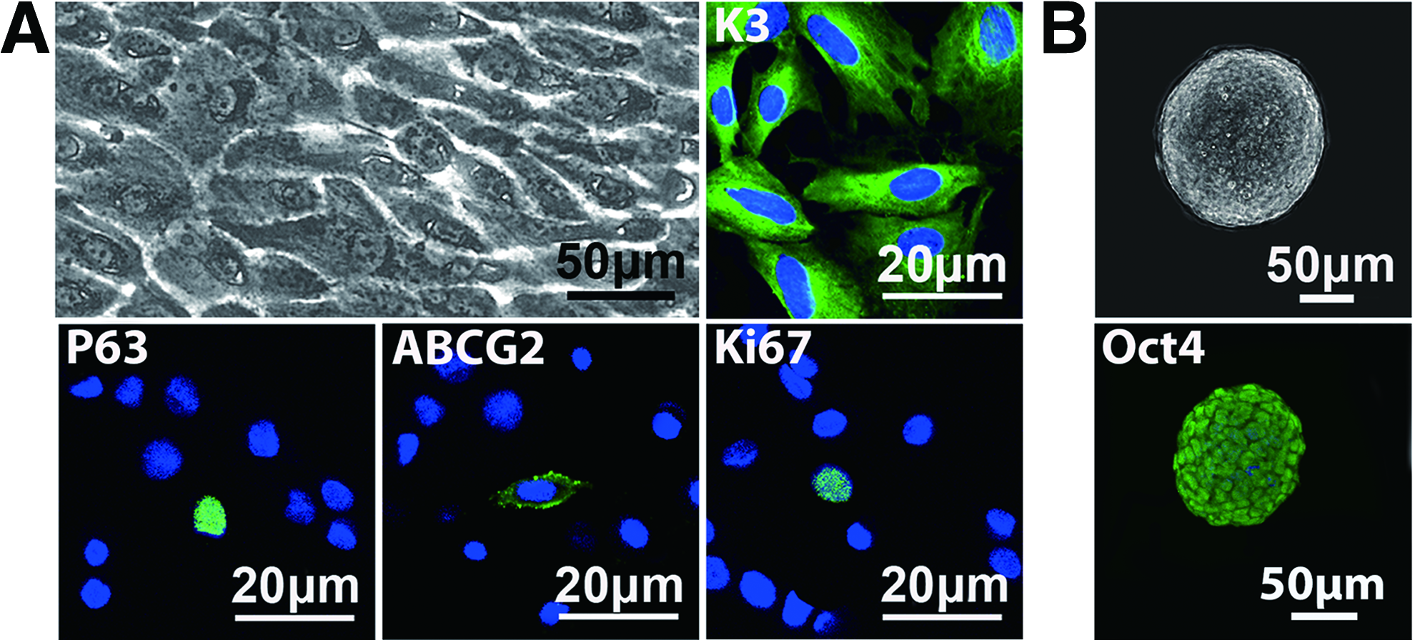
Phenotypes of auto-rabbit corneal epithelial cells (CECs) and mouse embryonic stem cells (ESCs) that had been pretransfected with the HSV-TK gene.
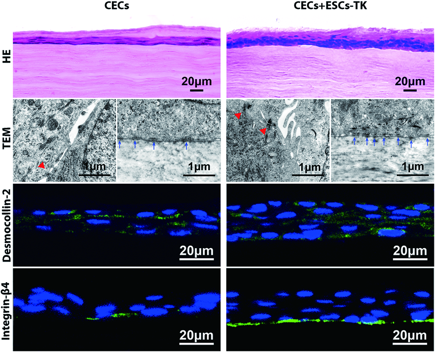
Histological evaluation of the morphological and adhesive properties of the auto-tissue-engineered lamellar cornea (ATELC) after 4 days of perfusion culture in vitro. (left column: CECs group, right column: CECs+ESCs-TK group). (from top to bottom) Hematoxylin and eosin (H&E) staining shows that two to three layers and four to five layers of the corneal epithelium formed between the amniotic membrane and the acellular porcine corneal stroma (APCS) in the CECs group and CECs+ESCs-TK group, respectively. In contrast to the CECs group, the nuclei of the epithelial cells in the CECs+ESCs-TK group were smaller, and the arrangement of the cells was more compact. More microvilli, desmosome (desmocollin-2), and hemidesmosome (integrin-β4) were also observed in the cell–cell and cell–APCS junctions using TEM and immunofluorescence staining, respectively, in the CECs+ESCs-TK group (green). ▵ desmosome in the cell–cell junction;↑hemidesmosome in the cell–APCS junction. Color images available online at
Removal of ESCs-TK from ATELC
Using flow cytometry analysis, 98.77%±0.38% of ESCs-TK were labeled by DiO before the reconstruction process. After the cells were cocultured with CECs for 24 h, approximately 12.4%±0.78% of the cells were DiO positive. After the medium was treated with 20 μM ganciclovir for 3 days, the expression of DiO markedly decreased from 3.4%±0.78% (2 days) to 1.33%±0.68% (3 days) and to 0.13%±0. 15% (4 days) (n=10, p<0.05) (Fig. 3).
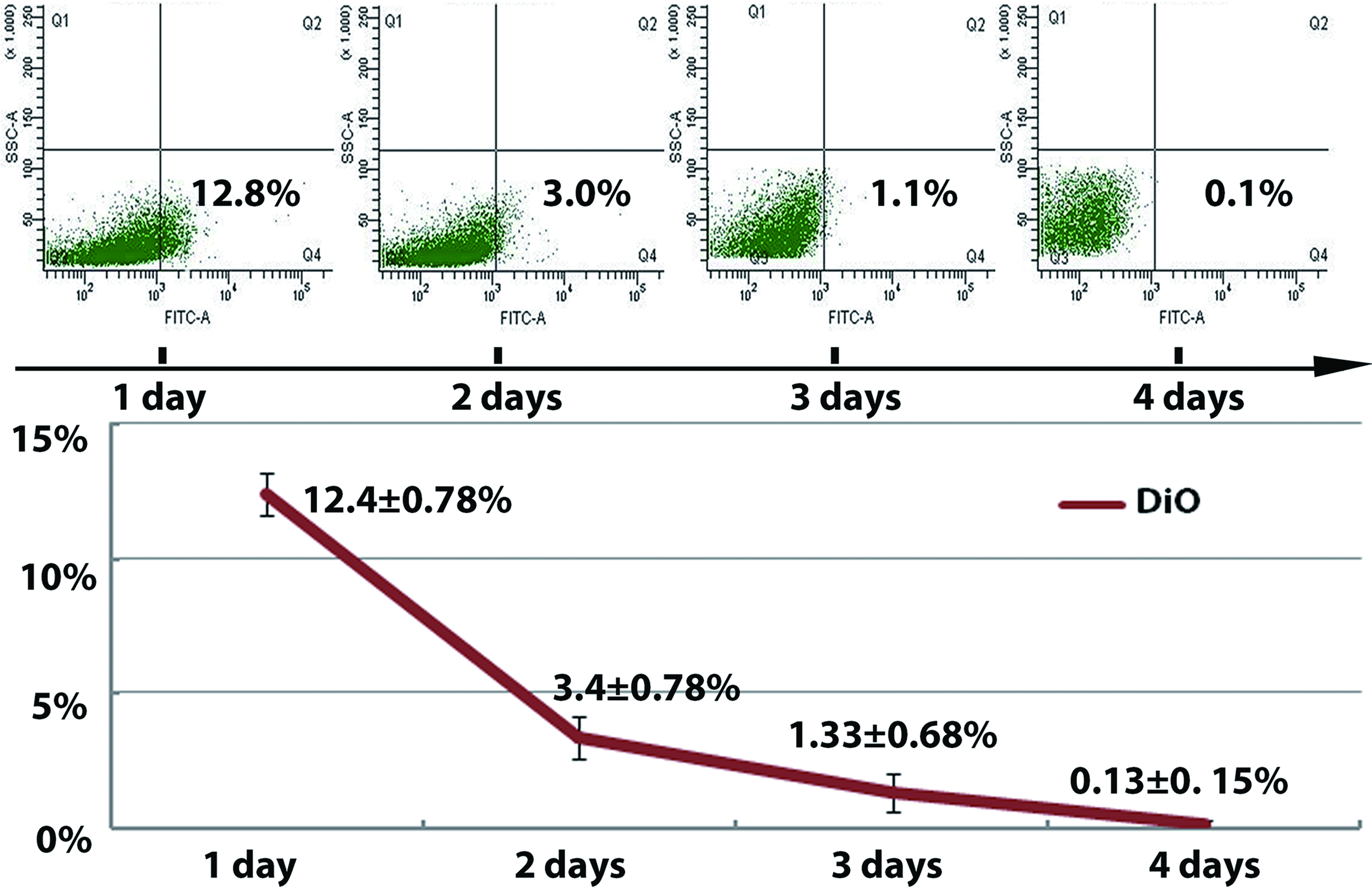
CECs were efficiently isolated from cocultured ESCs-TK through ganciclovir treatment in culture. It was observed that 98.77%±0.38% of ESCs-TK could be labeled using DiO. After the cells were seeded onto APCS using the centrifugal cell seeding method and cocultured for 24 h with CECs in vitro, 20 μM ganciclovir was added to the media to remove the ESCs-TK, and this effect was confirmed by DiO detection. The expression of DiO decreased from 12.4%±0.78% (1 day) to 3.4%±0.78% (2 days), to 1.33%±0.68% (3 days), and to 0.13%±0.15% (4 days), indicating that 3 days of treatment with ganciclovir resulted in the depletion of nearly all of the ESCs-TK. The data are expressed as the means±SD (n=10, p<0.05). Color images available online at
Cocultured ESCs-TK can enhance stem/progenitor cell phenotypes of CECs in ATELC
It has been shown that more basal cells of the ATELC express stem/progenitor cell phenotypes in the CECs+ESCs group (K3−, P63+, ABCG2+, and integrin-β1+) (Fig. 4A). Real-time qRT-PCR analysis demonstrated that the expression of the corneal specific marker K3 mRNA (1.58±0.12) in the CECs+ESCs-TK group was significantly lower compared with that observed in the CECs group (1.03±0.23). In addition, the expression of P63 mRNA and ABCG2 mRNA (1.52±0.25) in the CECs+ESCs-TK group was increased from 1.08±0.15 to 1.69±0.26 and from 0.99±0.18 to 1.52±0.25, respectively (n=10, p<0.05) (Fig. 4B). Consistent with the real-time qRT-PCR results, quantitative flow cytometry analyses also showed that the percentage of Ki67 (15.47%±2.20%), P63 (14.63%±2.23%), and ABCG2 (13.77%±1.91%) expression in the ATELC in the CECs+ESCs-TK group was significantly higher than that found in the CECs group (Ki67 [10.83%±1.53%], P63 [8.50%±0.96%], ABCG2 [8.13%±0.85%]) (Fig. 4C, n=10, p<0.05). Furthermore, the cell-cloning efficiency of the cells in the ATELC of the CECs+ESCs-TK group (9.72%±3.5%) was also significantly higher compared with that of the CECs group (6.29%±1.96%) (Fig. 4E, F, n=10, p<0.05). MTT proliferation assays also showed that the doubling time of the CECs+ESCs-TK group (28.45±2.03 h) was significantly shorter than that of the CECs group (36.3±1.15 h) (Fig. 4G, n=10, p<0.05).

The cocultured ESCs-TK promoted the expression of stem/progenitor phenotypes and the proliferative properties of epithelial cells in ATELC.
Rabbit lamellar keratoplasty
As shown in Figure 5A, slight edema and diffused excoriation were observed in the ATELC of the CECs group after the AM was removed on postoperative day 4 using slit lamp biomicroscopy and sodium fluorescein staining. In contrast, the ATELC of the CECs+ESCs-TK group displayed higher transparency after the AM was removed on postoperative day 4, and details of the pupil boundary and iris texture could be clearly observed. In addition, the sodium fluorescein staining of the animals in this group was negative (n=4). None of the DiO-labeled ESCs-TK could be observed on postoperative day 4 after the cells were treated with 20 μM ganciclovir four times daily (Fig. 5B top left). By using confocal microscopy in vivo, it was clear that all of the CECs were compactly and intactly formed on the ATELC (Fig. 5B top right). The H&E staining showed one to two layers of epithelial cells on the ATELC and abundant active stromal keratocytes (myofibroblasts) in the recipient bed were observed in the CECs group. Compared with these findings, four to five layers of epithelium on the ATELC and a few keratocytes in the recipient bed were observed in the CECs+ESCs-TK group (Fig. 5B, top-middle). Similar to the diffusion of keratocytes in the recipient bed, collagen III, which is secreted by active keratocytes, was higher in the CECs+ESCs-TK group, as assessed using immunofluorescence staining (Fig. 5B, middle-botttom). Moreover, the edematous grafts of the CECs group were gradually alleviated during re-epithelialization, and the light transmittance of the grafts in the CECs group was lower compared to that of the CECs+ESCs-TK group on postoperative day 20 over a range of wavelengths from 300 to 800 nm (Fig. 5B, bottom, n=10, p<0.05).

The physiological function, the morphology, light transmittance of ATELC in the auto-rabbit lamellar corneal transplantation model.
Discussion
Similar to many milestones that have been reached by tissue engineering in heart, lung, liver and, vascular regeneration in recent years, increased attention has been focused to the integration of cells and scaffolds to restore or to establish normal tissue function in vitro. 20 The identification of the appropriate cell types, scaffold material, seeding strategies and culture conditions are the next challenges in tissue engineering. In the future, one of the challenging aims is the treatment of corneal failure with functional corneal equivalents using corneal epithelial stem/progenitor cells. The key focus of this study was to promote the viability of CECs in corneal equivalent reconstruction.
The viability of CECs could be remarkably improved using the ESCs-TK coculture system
When the cornea is seriously injured, corneal transplantation is imperative. However, healthy tissue for culture in this disease is often difficult to obtain. Thus, in this study, to avoid excessive wounding to the healthy cornea and the immunological rejection of a corneal transplant, only a 1-mm2 single corneal explant was trephined from the rabbit auto-corneal limbus. To acquire a sufficient number of epithelial cells for reconstruction after 14 days of culture, we found that only a few of the P1 rabbit CECs expressed the stem/progenitor cell phenotypes and highly proliferative properties before the construction process (Fig. 1).
Recent studies have shown that cytokines or growth modulators secreted by ESCs can promote the viability of somatic cells in vitro.23,24 Our previous studies also demonstrated that the proliferation of CECs and endothelial cells could be improved by the conditional medium from ESC cultures.7,8,25 Furthermore, somatic cells may be reprogrammed into progenitor cells using the coculture system by direct ESC-cell contact.26,27 We also reported that ESC-cell direct contact culture may rapidly and remarkably enhance the stem/progenitor properties of rabbit CECs through the integrin beta-FAK-P13K/Akt pathway in vitro. 6
In this study, we also showed that the seeding of multiple layers of cells between the APCS and AM by the centrifugal cell seeding method and culturing in dynamic media for 4 days maintained and significantly enhanced the proliferation of the epithelial cells by the favorable extracellular microenvironment of the ESCs. As evaluated by real-time qRT-PCR, flow cytometry, colony-forming efficiency, and MTT assay, the ATELC of the CECs+ESCs-TK group clearly expressed the undifferentiated phenotypes and demonstrated highly proliferative properties compared with the CECs group, which may facilitate corneal re-epithelialization in vivo. 1
As the immunofluorescence staining images showing, more of the basal cells expressed progenitor/stem phenotypes (K3−, P63+, ABCG2+, and integrin-β1+) and proliferation marker (Ki67+) in the ATELC of the CECs+ESCs-TK group compared with the CECs group (Fig. 4). By maintaining the native basement membrane components of the cornea, the substrate properties of APCS 21 under basal cells may play a critical role in the maintenance of cocultured epithelial cells in their undifferentiated state, 28 which is required for corneal wound healing.
Multiple layers of the corneal epithelium can be rapidly constructed on APCS by centrifugal cell seeding and AM covering
The intact corneal epithelium on the surface can defend against external mechanical irritation, protect against infection and inflammation, and prevent excessive scar formation. In corneal reconstruction engineering, the adhesive properties of the epithelium also play a vital role in withstanding the operation of corneal transplantation and the friction of eye blinking in vivo. In this study, during the reconstruction process, all of the cells were cultured between the AM and APCS and fixed onto a chamber of a perfusion culture system. By the protection and limitation of AM, cell loss may be avoided during construction. Thereby, the density of the cells was increased, which could promote the proliferative and adhesive properties of cells vice versa. 29 Moreover, using the perfusion culture system, the fluid shearing force, supply of sufficient oxygen and nutrition, and the proliferation and adhesion properties could also been improved. 30 As shown in the Figure 2, more ultrastructures of desmosomes and hemidesmosomes were formed between the cell–cell and cell–matrix junctions in the ATELC of the CECs+ESCs-TK group compared with the CECs group. Consistent with these results, more desmocollin-2 (desmosome marker) and integrin-β4 (hemidesmosome marker) were detected using the immunofluorescence staining in the CECs+ESCs-TK group than the CECs group (Fig. 2). Furthermore, after additional layers and a higher density of CECs with higher proliferative and adhesive functions were constructed in the ATELC of the CECs+ESCs-TK group in vitro, the corneal transplantation was enhanced compared with that obtained in the CECs group.
Intact multiple layers of the epithelium could increase the restoration of transparency and decrease the formation of scar after corneal transplantation
Covering of the AM could also protect the corneal epithelium from transplantation and the formation of cell junctions in the ATELC in vivo. After the AM was removed on postoperative day 4, the corneal barrier functional properties were intact, and the corneal transparency was completely restored in the ATELC of the CECs+ESCs-TK group (Fig. 5A). After 20 days of observation, four to five layers of the epithelium were maintained on the ATELC; a few differentiated myofibroblasts and some collagen III were formed on the receiving bed, and the ATELC showed high optical transparency and favorable epithelial barrier function in the CECs-ESCs+TK group throughout the observational period (Fig. 5B). In contrast, on postoperative day 4, the barrier function of the ATELC had not completely formed in the CECs group. Even after 20 days of observation, the ATELC of the CECs group restored its transparency, and the light transmittance of the cornea was always lower compared with that of the CECs-ESCs+TK group. The cornea consisted of highly organized collagen I and microstructures. If the cornea was deeply wounded, it would be substituted by newly synthesized type III collagen and lose its spatial organization. However, only a slight alteration in the cornea resulted in a marked decrease in visual acuity. Thus, the intact corneal epithelium of the ATELC in the CECs-ESCs+TK group plays an important role in limiting the formation of the ECM and decreases the activity of stromal keratocytes (myofibroblasts) in wound healing, both of which are closely correlated to the opacity of the corneal stroma.1,2,31
ESCs could be completely removed by ganciclovir treatment
ESCs are undifferentiated resident cells with high proliferative capacities and pluripotent potential and may promote the proliferation of cocultured somatic cells by serving as the stem cell microenvironment; however, this method still remains limited by the tumorigenicity or impurity of the induced differentiation of the cells. 22 Thus, in this study, the ESCs were pretransfected with the HSV-TK gene (a suicide gene) and were sensitive to ganciclovir. Moreover, after the stem/progenitor phenotypes of CECs were significantly enhanced using this coculture system and treatment with ganciclovir, nearly all of the DiO-labeled ESCs-TK could be depleted in vitro, with none of the cells detectable 4 days after transplantation (Figs. 3 and 5B, top). These results were consistent with and supported by our previous reports that the ESCs-TK could be completely removed by adding ganciclovir in vitro as evaluated by flow cytometry analysis of apoptosis experiments (Annexin V and PI staining), and observation of undetected Dil track in ESCs-TK after 3 days of ganciclovir treatment 6 ; as well as in vivo by tracking the ESCs-TK with transfected GFP fluorescence. 32
During the entire observation period, there was an absence of any tumor formation, which was consistent with our previous studies in cell coculture and leukemia mouse treatment.6,32 These results also showed that, the gradual removal of ESCs from surrounding cells by treatment with ganciclovir in vitro and in vivo, did not destroy the intact barrier function of the corneal epithelium and maintained the corneal transparency consistently, which is also consistent with previous studies. 33
Although the ESC-TK coculture system has obvious advantages in promoting the proliferation of CECs in the rabbit corneal transplantation model, there is still improvement in ATELC reconstruction, as assessed using different parameters, including the percentage or time of the ESCs-TK coculture. Furthermore, whether the ATELC is appropriate for other transplantation models, such as corneal limbus transplantation procedures, requires further studies.
Conclusion
In conclusion, this work reconstructed a viable ATELC on APCS using ESCs-TK coculture, the centrifugal seeding method, and AM covering. The ATELC could be transplanted into the rabbit model to rapidly restore corneal transparency. Moreover, treatment with ganciclovir completely removed the ESCs-TK without forming any teratoma. This controllable and safe method using the ESC microenvironment to construct a more viable ATELC demonstrates its expansive potential clinical applications.
Footnotes
Acknowledgments
This work was supported by the National High Technology Research and Development Program (863 Program) of China (No.2012AA020507) (W.Z.C.), Natural Science Foundation of China (No. 81270971) (W.Z.C.), Guangdong Natural Science Foundation (No. S2012010009113) (W.Z.C.), the Fundamental Research Funds of State Key Laboratory of Ophthalmology of China (2012PI05) (W.Z.C.). The authors thank Professor De-Quan Li from the Baylor College of Medicine for his helpful comments and revision.
Disclosure Statement
No competing financial interests exist.