
Abstract
Polycationic nanocomplexes are a robust means for achieving nucleic acid condensation and efficient intracellular gene deliveries. To enhance delivery, a multilayered nanoparticle consisting of a core of electrostatically bound elements was used. These included a histone-mimetic peptides, poly-l-arginine and poly-d-glutamic acid was coated with silicate before surface functionalization with poly-l-arginine. Transfection efficiencies and duration of expression were similar when using green fluorescent protein (GFP) plasmid DNA (pDNA) or GFP mRNA. These nanoparticles demonstrated significantly higher (>100%) and significantly longer (15 vs. 4 days) transfection efficiencies in comparison to a commercial transfection agent (Lipofectamine 2000). Reprogramming of human foreskin fibroblasts using mRNA to the Sox2 transcription factor resulted in three-fold higher neurosphere formation in comparison to the commercial reagent. These results demonstrate the potential of these nanoparticles as ideal vectors for gene delivery.
Introduction
N
Polyplexes prepared from a combination of cationic and anionic polymer-based derivatives are reported to yield efficient gene delivery and subsequent expression through facilitated endosomal escape into the cytoplasm, based on the proton sponge effect, as well as some putative retrograde trafficking and compartment-specific unpackaging elements within the nucleus.11,14 However, several hurdles still need to be overcome for clinical applications using polyplexes. Polyplex systems often have low complex stability in a biological milieu such as serum, thereby leading to undesirable complex disassociation or aggregation and decreased transfection activity concomitant with increased adverse effects.15–17 Therefore, polyplexes that form highly stable complexes under extracellular conditions are required, especially for systemic administration. However, a drawback of polyplexes that exhibit higher stability is that intracellular release of an enclosed nucleic acid may be hindered.
Results and Discussion
To determine particle uptake and transfection efficiency due to the presence or absence of the different layers and components, fluorescein isothiocyanate (FITC) labeled poly-l-arginine (PLR) containing particles encapsulating pDNA for mCherry (Addgene: pcDNA3.3_mCherry) were transfected into MC3T3-E1 cell lines. Cationic PLR was conjugated with pDNA in the presence of histone h3 tail peptide (HTP) and/or poly-d-glutamic acid (PDGA) followed with or without encapsulation in a silicate layer and a final layer of PLR. Histone tail peptides were used to enable loosening of pDNA from the cationic polymer (PLR) by acetylation. Silicate coating was used to stabilize and condense the quarternary core composed of pDNA/mRNA, PLR, HTP, and PDGA. An outermost layer of PLR was used to provide a positive charge to the particle so that it can effectively adhere to the negatively charged cell membrane. Confocal imaging for plasmid expression indicated that the nanoparticle with all components, that is, poly-l-arginine (PLR) coating of silicate encapsulated quarternary core, demonstrated good transfection efficiency (as observed by greater intensity of green in Fig. 1A) while showing intensive expression (as observed by greater intensity of red mCherry in Fig. 1A). Particles made using PLR complexed with pDNA demonstrated minimal expression, which was increased by layering with silicate followed by another layer of PLR. Complexation with PDGA demonstrated effective release of pDNA from the cationic polyplex, resulting in greater expression of transfected pDNA. Confocal microscopy also revealed that silicate-coated nanoparticles are extremely resilient to aggregation and dissociation in serum (no extracellular green/yellow clusters) in comparison to their binary, ternary, and quaternary core counterparts without silicate stabilization. Qualitative observation demonstrated PDGA inclusion to greatly increase the number of particles taken up by cells in both silicate and nonsilicate-coated complex transfections. The particle with the most efficient expression was used to study the duration of plasmid expression. We transfected these nanoparticles encapsulating pDNA to green fluorescent protein (GFP) into human foreskin fibroblasts (HFFs). Results indicated high expression on day 3 that was reduced by day 14 with minimal expression by day 21 (Fig. 1B).

Morphologic analysis of silicate-polyplexed nanoparticles by transmission electron microscope clearly showed the core-shell structure (Fig. 2A). The average nanoparticle diameters are in the range from 120 to 170 nm with a narrow particle size distribution, which was consistent with dynamic light scattering analysis. The prepared nanoparticles were characterized by Fourier transform infrared (FTIR) spectra in (Fig. 2B). The characteristic absorbance peaks at 1637 and 3310 cm−1 corresponds to the amide group and N-H group stretching, respectively. 18 In Figure 2B(b), the FTIR spectra of the core-shell particles show absorption bands at 1090 and 795 cm−1, which can be attributed to asymmetric vibration and symmetric vibration of Si-O bonds. 19 The absorption peaks of the amide group and the N-H group shifted to 3369 and 1644 cm−1, which indicated their interaction with silicate shells. 18 Therefore, the FTIR spectra conclusively indicated the formation of silicate coating.
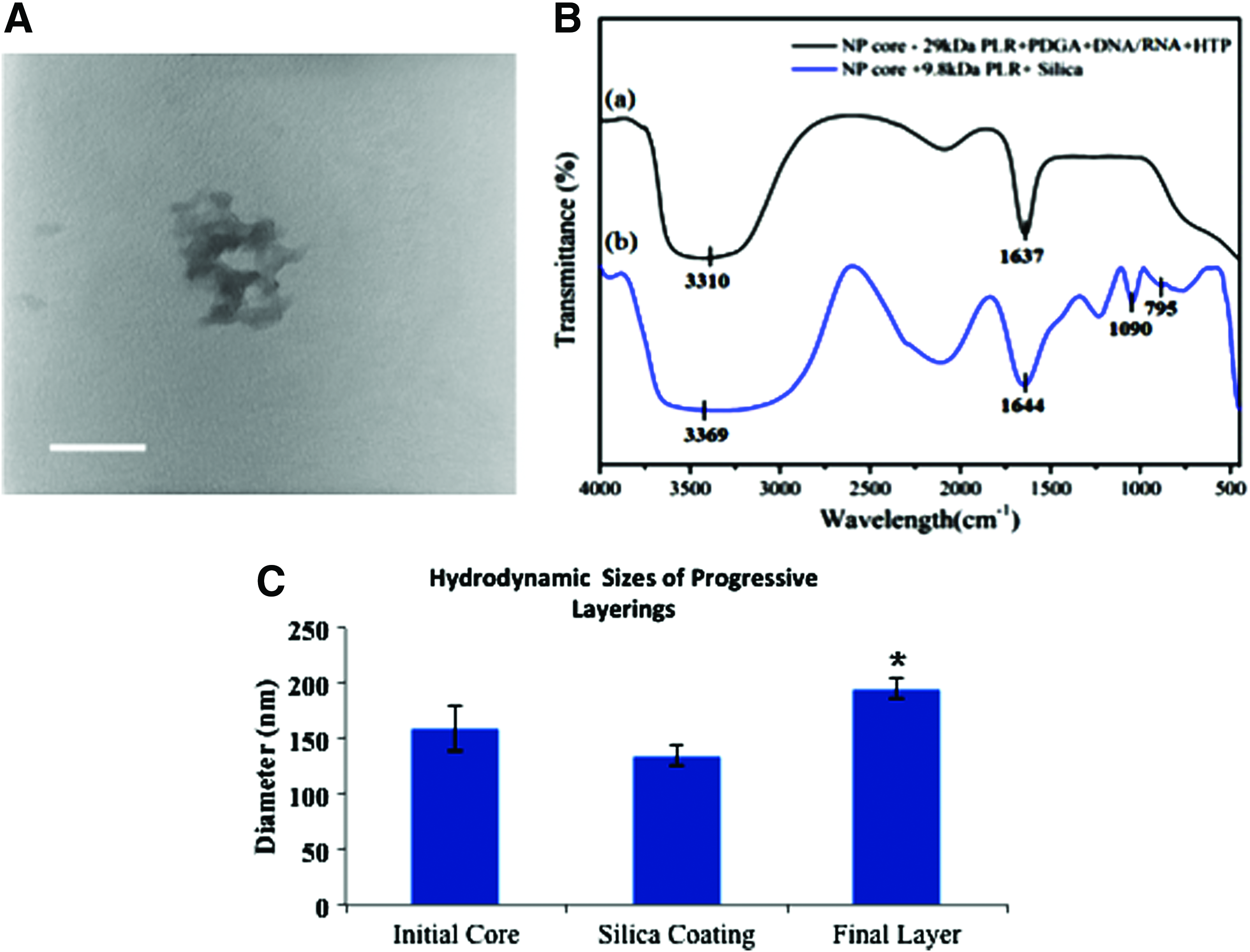
Silicate coating of ternary complexes was observed to condense nanoparticles (Fig. 2C). This could be explained by a number of factors, most likely due to the documented instability of nanocomplexes formed with polypeptides with heterogeneous charge distributions.20,21 Since formation of cores (152.6±8.5 nm) occurs within an acidic solution (pH ∼6), it is possible that silicate coating (140.0±5.1 nm) within an alkaline solution (pH=7.4) causes anionic components (PDGA and DNA) to increase their negative charge densities. Within the presence of anionic monomeric silicate species, the anionic nanoparticle constituents may seek to repulse themselves inward, exposing more cationic species for silicate condensation and oligomeric network formation and effectively sealing a “semi-stable” core within a shell of silicate. This phenomenon has recently been documented in the form of “dual-responsive” nanoparticles.15,22 Not unexpectedly, further layering on this silicate shell yielded a final particle with a hydrodynamic diameter increase of 15–30 nm (171.2±10.2 nm). The ostensibly loose polyplex morphology of these quaternary complexes also lends support to the idea that silation gels the polyplexes as a whole rather than merely at the surface.
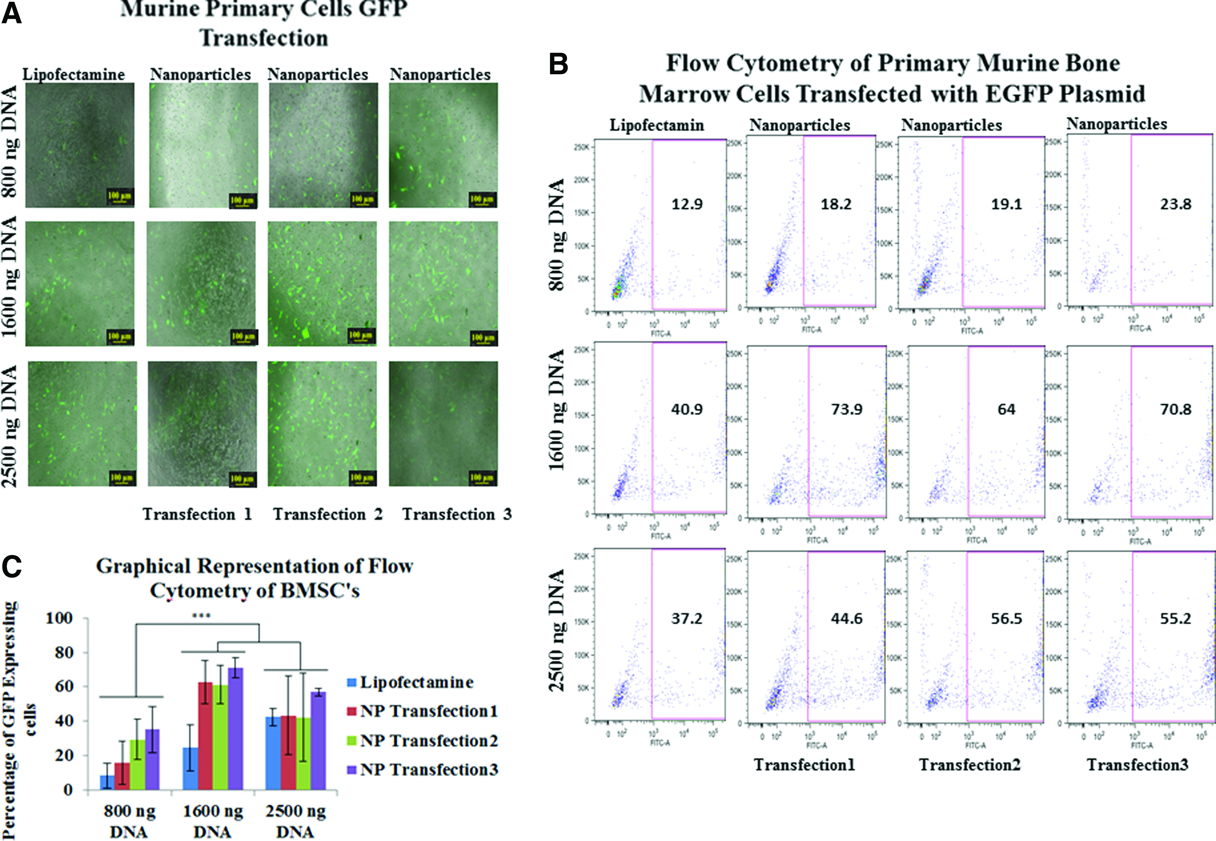
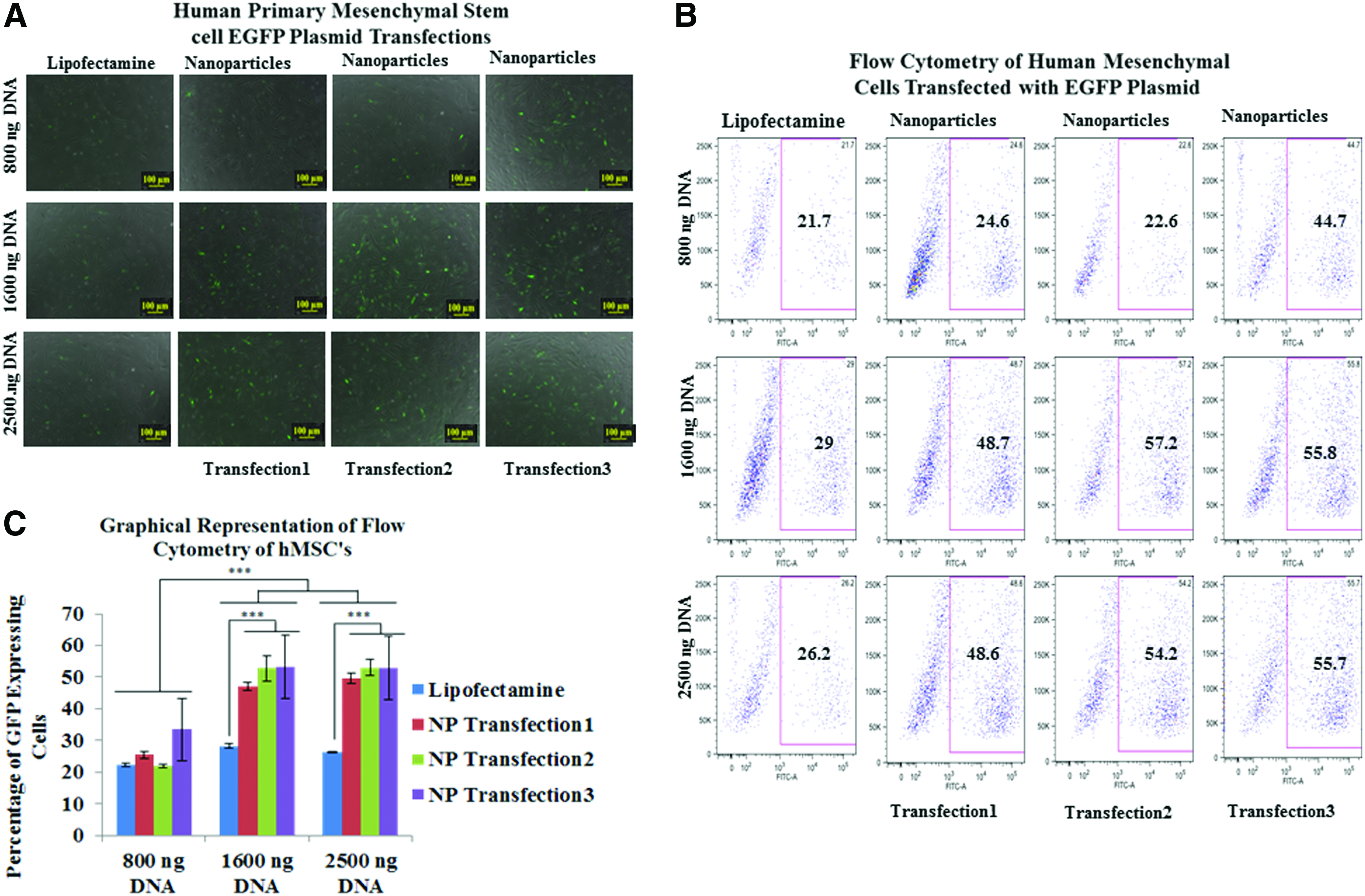
These multilayered nanoparticles demonstrated higher transfection efficiencies when compared with lipofectamine transfections in both primary bone marrow stromal cells (BMSCs) and primary human mesenchymal stem cells (HMSCs) (Figs. 3 and 4). Transfection efficiencies of pDNA in BMSCs were 1.41, 1.48, and 1.84 times higher for 800 ng; 1.8, 1.56, and 1.73 for 1600 ng; and 1.19, 1.15, and 1.48 for 2500 ng of transfected pDNA for single-, double-, and triple nanoparticle-mediated transfections (Fig. 3). Transfection efficiencies of pDNA in HMSCs were 1.14, 1.04, and 2.05 times higher for 800 ng; 1.67, 1.97, and 1.92 times for 1600 ng; and 1.85, 2.06, and 2.12 for 2500 ng (Fig. 4), compared with Lipofectamine for single, double, and triple transfections, respectively. Efficient expression of GFP in both cell types was obtained when using 1600 ng of pDNA, where expression was 130% higher in BMSCs and 70% higher in hMSCs relative to lipofectamine. There was a statistically significant difference between the percentage of GFP expressing cells transfected with pDNA using lipofectamine and nanoparticles for the single transfections whereas for repeated second and third transfection of nanoparticles, there was no statistically significant difference between the nanoparticle transfected groups for both BMSC's and HMSCs, at any of the concentrations of pDNA except for the transfection in HMSCs at 800 ng of pDNA concentration using lipofectamine compared with thrice nanoparticle transfected cell group. The pDNA and mRNA to GFP was transfected at 1600 ng concentration into the HFFs using nanoparticles and lipofectamine, and it was observed that the nanoparticle transfected cell groups showed greater expression compared with Lipofectamine over 21 days. However, due to its cytosolic burst release feature, Lipofectamine transfected cell group showed comparable transfection and expression efficacy at day 3. The duration of mRNA to GFP expression in HFFs was slightly longer as compared with duration of pDNA expression. This can be attributed to the fact that the half life of mRNA is longer than pDNA for expression of gene of interest. The GFP expression via mRNA as in case with pDNA was almost equal for lipofectamine and nanoparticle transfected cells at day 3 due to burst release profile of lipofectamine. However, again as in the case with pDNA expression, the cells transfected with nanoparticles showed a longer duration of expression (Fig. 5).
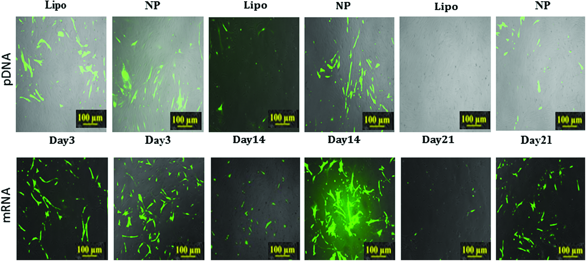
(Upper Panel) Fluorescent imaging of human foreskin fibroblasts (HFF) transfected with GFP pDNA—expression is assessed for more than 21 days (Lower Panel). Fluorescent imaging of HFFs transfected with GFP mRNA synthesized using in-vitro mRNA synthesis kit—expression is assessed for more than 21 days. Color images available online at
The cytotoxicity of these nanoparticles was assessed using Promokine Apoptotic/Necrotic/Healthy Cell Detection kit via flow cytometry in human dermal fibroblasts and HMSCs. There was a statistically significantly difference between the percentage of healthy cells in the cell groups transfected with nanoparticles as compared with the percentage of healthy cells in the cell groups transfected with Lipofectamine2000 with the cell group transfected with nanoparticles showing a fold higher viability as compared with Lipofectamine2000-transfected groups. This shows that these multilayered polymeric nanoparticles can be safe for delivery of functional nucleic acids in vitro as well as in vivo (Supplementary Fig. S3; Supplementary Data are available online at
Neurospheres are free-floating spherical condensations of cells with neural stem/progenitor cell characteristics that can be derived by reprogramming of HFFs using Sox2 transcription factor. 4 The efficiency of generating neurospheres on delivery ofSox2 mRNA to fibroblasts was compared using the two delivery vectors (naonoparticles and Lipofectamine). The number of neurospheres in cultures transfected with nanoparticles was found to be three-fold higher at 12 days post-transfection (Fig. 6). Nanoparticle transfection of Sox2 mRNA had altered the gene expression profile of cells in the affected cultures. Fibroblasts such as HFFs can be characterized by a gene expression profile that includes high levels of expression of the Col1a1, Col2a1, and Fap genes, but on transfection, many neural progenitor and pluripotency genes are upregulated, such as Nanog, Oct4, Sox2, Sox1, Nestin, and Musashi. Primers and Ct values for qPCR are given in Supplementary Tables S2 and S3, respectively. Expression patterns of these genes were examined through quantitative reverse transcription polymerase chain reaction analysis for days 6, 8, and 12 post-transfection. Figure 6A and B shows the expression levels of each gene after nanoparticle transfections with reference to Lipofectamine-induced transfection. Most of the neural progenitor and pluripotency genes were expressed at levels two-fold or greater in nanoparticle-transfected cultures compared with Lipofectamine-transfected cultures at all time points. All of the genes analyzed increased in expression through day 12 except Oct4, in which no significant change in expression occurred between the three time points. Sox2 expression pattern was included in the analysis to determine differential delivery patterns between Lipofectamine and nanoparticles. The steady increase in Sox2 expression levels till four-fold over Lipofectamine delivery shows that nanoparticles continue to provide Sox2 delivery through day 12, which subsequently increases expression of other marker genes, indicative of a neuroprogenitor. Sox1 is a particularly strong indicator of neural induction, as it is one of the earliest transcription factors expressed in ectodermal cells committed to a neural lineage and upregulation of Sox1 is directly correlated to neural determination and differentiation.23,24 As shown in Figure 6C, fibroblast reprogramming using nanoparticles to deliver Sox2 led to an eight-fold increase in Sox1 expression over untreated cells and a 6-fold increase over Lipofectamine by day 12. This indicated increased levels of neural differentiation in the cells treated with Sox2 pDNA via this silica-stabilized and multistage polyplex delivery system.

In this work, we demonstrate that a coating of a silicate gel core with PLR yields a cationic nanoparticle with potentially useful therapeutic nucleic acid delivery properties that include high pDNA/mRNA binding ability as well as high rates of cellular uptake and extended release into the nucleus. We have shown that pDNA/mRNA complexed with PDGA, PLR, and HTP and stabilized by silicate before a further layering by PLR can induce effective delivery of nucleic acid payloads into the cell to initiate protein translation of a pDNA sequence, as well as reprogramming of differentiated cells into a desired phenotype using time-released mRNA. In early qualitative studies, quaternary core complexes composed of (pDNA/RNA+PDGA+PLR+HTP) also demonstrated significantly enhanced transfection into the murine osteoblast cell lines (MC3T3-E1) (Fig. 1A) along with greater gene expression of mCherry pDNA.
Cationic polymers such as poly(ethylenimine), chitosan, poly(L-arginine), poly(L-ornithine), and poly(L-lysine) form relatively stable complexes that are capable of mediating effective gene transfer at various amines to phosphate ratios (n/p), where the “n” to “p” ratio determines the quantity of DNA that can be complexed with the polymer. Using improvements to mere electrostatic binding between such polymers, pDNA delivery was shown to be much more efficient compared with the commercially available transfection reagent Lipofectamine. We have successfully demonstrated the increased efficacy conferred by multilayered complexes designed for nuclear-specific unpackaging and biocompatibility.
The positive charge of PLR in the outermost coating of nanoparticles leads to a strong electrostatic interaction with the negatively charged cell surface membrane, leading to facilitated particle wrapping and uptake (Fig. 1A). This is in agreement with the recent demonstration that poly-ethyleneimine (PEI) nanoparticles, which are similarly negatively charged, are taken up into the cells with high efficiency. 25 However, the latter study did not look at nucleic acid delivery but showed that the attachment of ligand such as folic acid further enhances uptake in cancer cells. 26 Previous research has been done to study PEI delivery with silica as well as silica delivery of DNA to find the optimal nanoparticle composition to get enhanced transfection efficiencies.26–28 Cationic polymers bind to sulfated proteoglycans, which act as cellular receptors on cholesterol-rich lipid rafts—this characteristic makes them excellent for ubiquitous targeting in vitro. 29 Carboxylate-containing polymers have been shown to further influence DNA-polymer complexation and release kinetics, where±ratios remain similar but additional negative moieties serve to promote electrostatic unpackaging during cellular uptake while increased surface area due to increased number of polyplexes facilitates transfection.30–33 This complexation is frequently observed to be reversed after exposure to the multitude of proteins, salts, and other molecules present within serum, so standard transfections frequently utilize serum-free medium; indeed, heparin, dextran sulfate, and alginate are well documented as destabilizing cationic polyplexes.
A paradox is that, with DNA constructs, the disassociation of DNA from polymer is desired within the nucleus. To achieve this compartment-specific unpackaging and efficient gene transfer to the nucleus, numerous studies have utilized cationic histone-mimetic peptides, which endogenously are nuclear localized and subjected to various modifications, including deprotonation by acetyl-CoA and histone acetyltransferase.34–36 It has been previously shown that histone H3 tail peptides (HTP) formed polyplexes that interacted with the transcriptionally activating HAT complex HBO1, and that polyplexes containing H3K4Me3 achieved fast pDNA transcription after microinjection into cellular nuclei.35,37 Building on this work, this study aimed at exploiting this very property of trimethymated histone tail peptides to gain enhanced gene transcription on delivery, holding off exposure of the moieties until endosomal internalization leads to acidifying conditions that dissolve the transiently stabilizing silicate. In addition, due to the intrinsic nature of histones and their exclusive existence within the nucleus, domains for histone winding are similar to domains for nuclear-specific targeting mediated by karyopherin and importin binding to highly conserved lysine- and arginine-rich regions.38–42 Therefore, the quaternary core complexes composed of (pDNA/mRNA+PDGA+PLR+HTP) demonstrated significantly enhanced transfection into the murine osteoblast cell line (MC3T3-E1) (Fig. 1A) along with greater gene expression of mCherry pDNA. However, it has been previously observed that trimethylated HTPs enhance the rate of gene expression but do not greatly enhance the rate of transfection as compared with nonmethylated HTPs. This might be because of the presence of histone methyltransferases in the nucleus of the cell. These methyltransferases might be interacting with and methylating the nonmethylated HTP, although at a slower rate. Therefore, this effect can potentially outweigh any initial advantages in transcriptional activation by the pretrimethylatedpolyplexes. Since the deprotonations induced by HTP's occur at key arginine and lysine residues, it is possible that poly(L-arginine) may also act as a substrate for such enzymatic activity by methyl and acetyltransferases, among others.43–45 One unfortunate effect of cationic polymer transfections is that the majority of the payload is dissociated during initial phases of uptake (perhaps through interaction with the anionic cell membrane), 46 and that the remaining payload frequently remains bound to its cationic carrier during nuclear dispersion. In seeking to mediate many of the outlined inefficiencies, recent studies have demonstrated oligomeric silicate coating of bare polyplexes to effectively seal the payload during initial phases of uptake, additionally preventing destabilization by polyanions and contributing to buffering during endosomal uptake (via the proton sponge effect, a phenomenon whereby lysosomes are prematurely ruptured due to osmotic influx).15,47–49 Further layering on this anionic silicate layer presents a highly functionalized nanoparticle surface with a stable core capable of controlled nucleic acid release. Our results suggest that silicate is capable of reversibly stabilizing highly complex and otherwise readily destabilizable quaternary cores of nanoparticles, which subsequently are released into the cell and take advantage of many of the outlined benefits of histone tail peptides, cationic polymers, and anionic polymers. We posit that use of D-isomers of poly (glutamic acid) as an anionic polymer presents a time-release element to our polymeric delivery system, due to the fact that D-isomers are unlikely to be proteolytically degraded and have been documented to have limited immunogenicity.50–52 If a nanoparticle's charge becomes increasingly anionic during its residence within the cell, it will be more likely to repulse anionic nucleic acids as has been previously documented with poly(glutamic acid)'s inclusion within vectors.30,31 We observed no visible polyplex internalization 30 h after transfection with silicate-coated poly (L-arginine)-DNA complexes (despite gene expression), while similar complexes containing poly(D-glutamic acid) are capable of sustaining prolonged release over the course of roughly a week and maintaining gene expression in 55–70% transfected cells (Fig. 4). In addition, our multilayered polyplexes are stable in serum for more than 1 week, while their cores rapidly destabilize in the presence of anionic polymers alone.
Using the nanotechnology here, we demonstrate superior expression of pDNA in primary murine adherent bone marrow cells and primary human mesenchymal cells. We also demonstrate reprogramming of primary HFF cells using nonviral nanocarriers. These carriers, which consist of biomimetic polypeptides and biocompatible materials, condense genetic payloads (pDNA and mRNA) with high efficacy and help achieve an optimum extracellular stability and intracellular unpackaging and release. This technology is based on a few simple tenets: If a “semi-stable” particle on the threshold of its stability can be released into the cytosol, functionalization will greatly increase payload delivery. In addition, the trigger for this loss of stability can be anything ranging from hydrolytic and enzymatic degradation to conformational change.
Supplementary Data
Supplementary Data contains detailed methods involved in formation of the dual layered core-shell structure nanoparticle for the optimization of nanoparticle design for transfection into murine osteoblast cell line and primary bone marrow cells along with detailed calculations on the optimized concentrations of each of the components and their interactions with each other.
Footnotes
Acknowledgments
The authors would like to acknowledge the funding from NIH/NIA grant R01 AG030637. They are grateful for the guidance rendered by Dr. Deanna Thompson at RPI for her help with examination of neurospheres. They are also thankful to Aniket Tolpadi for all the help with the design of figures and artwork for the nanoparticle schematic used in this article.
Disclosure Statement
No competing financial interests exist.
References
Supplementary Material
Please find the following supplemental material available below.
For Open Access articles published under a Creative Commons License, all supplemental material carries the same license as the article it is associated with.
For non-Open Access articles published, all supplemental material carries a non-exclusive license, and permission requests for re-use of supplemental material or any part of supplemental material shall be sent directly to the copyright owner as specified in the copyright notice associated with the article.