
Abstract
Skeletal myoblasts have been extensively used to study muscle growth and differentiation, and were recently tested for their application as cell therapy and as a gene delivery system to treat muscle and nonmuscle diseases. However, contamination of fibroblasts in isolated cells from skeletal muscle is one of the long-standing problems for routine expansion. This study aimed to establish a simple one-step process to purify myoblasts and maintain their purity during expansion. Mixed cells were preplated serially on laminin- and collagen type I-coated surfaces in a different array for 5, 10, and 15 min. Immunocytochemical staining with antibodies specific to myoblasts was performed to evaluate myoblast attachment efficiency, purity, and yield. It was found that laminin-coated surface favors the attachment of myoblasts. Highest myoblast purity of 78.9%±6.8% was achieved by 5 min of preplating only on the laminin-coated surface with a yield of 56.9%±3.3%. Primary cells, isolated from skeletal muscle (n=4), confirm the enhancement of purity through preplating on laminin-coated surface for 5 min. Subsequent expansion after preplating enhanced myoblast purity due to an increase in myoblast growth than fibroblasts. Myoblast purity of ∼98% was achieved when another preplating was performed during passaging. In conclusion, myoblasts can be purified and efficiently expanded in one step by preplating on laminin-coated surface, which is a simple and robust technique.
Introduction
S
Despite multiple applications of myoblasts in both basic research and clinics, one of the long-standing challenges for the routine culture of myoblasts in vitro is the contamination of fibroblasts during the isolation of cells from skeletal muscle. This fibroblast population increases dramatically during expansion as they proliferate faster than myoblasts. To avoid this problem, basic research on myoblasts has commonly used stable cell lines, although these cells lack typical myoblast morphological and functional properties. 11 However, for clinical application, primary myoblasts from an autologous and allologous origin are used, and they need to be expanded in vitro to achieve adequate amounts of cells for transplantation. Fibroblast overgrowth during expansion significantly reduces myoblast purity and requires purification before clinical application.
Several myoblast purification procedures have been reported in the literature, including serial preplating,12,13 selective adhesion of myoblasts and fibroblasts, 14 percoll density centrifugation, 15 cell sorting by size, 16 antibody tagging,17,18 and supplementation of fibroblast growth inhibitor.18,19 Cell sorting techniques have been shown to increase myoblast purity but with relatively poor yield. In addition, these procedures are complicated and require specialized equipment. The usage of growth inhibitor, such as mitomycin C and irradiation, has also been tested to remove fibroblasts and has shown effective purification of myoblasts. However, these techniques reduce the proliferative activity of myoblasts and are not suitable for expansion. In contrast, the preplating technique is simple and is most commonly used to purify myoblasts. Preplating techniques traditionally utilize collagen type I-coated surfaces, which enable fibroblasts to attach faster than myoblasts, and the myoblast population is obtained after multiple preplatings.12,13,18 However, the yield of myoblasts is low in conventional preplating techniques and these techniques fail to completely remove fibroblasts.
However, these techniques are not viable for use in cell expansion either due to an incapability of myoblast proliferation or a failure to completely remove fibroblasts. The presence of a small fraction of fibroblasts after purification may significantly reduce myoblast purity during expansion due to fibroblast overgrowth. This study aimed to develop a simple and effective preplating technique that not only purifies myoblasts but also maintains their purity during expansion. For this purpose, laminin-coated surfaces, which are known to facilitate myoblast attachment and proliferation,20,21 were used alone or in combination with conventional collagen type-I-coated surfaces. The yield and purity of myoblasts were evaluated using immunocytochemical staining with myoblast-specific markers to optimize the preplating technique. Purification of several primary cells isolated from human skeletal muscle tissue was also tested to evaluate the effectivity of preplating technique. Moreover, the optimized preplating technique was applied for serial passages to evaluate the maintenance of purity during expansion.
Materials and Methods
Muscle cell harvesting and culture
This research was approved by the Universiti Kebangsaan Malaysia Research and Ethics Committee (UKMREC) with approval code of FF-037-2013 and FF-313-2010. Cryopreserved primary human skeletal muscle myoblast cells (HSMMs; Lonza) and cells harvested from human skeletal muscle tissue were used in this study.
HSMMs of passage 2 were thawed and seeded into a 75-cm2 tissue culture flask with F10:DMEM (1:1; Sigma) containing 10% fetal bovine serum (FBS; PAA Laboratories), before incubation. All of the cell incubations in this study were performed at 37°C with an atmospheric condition of 5% CO2. Waste medium was replaced every 48 h with a fresh culture medium. After reaching 80% confluence, cells were treated with trypsin-EDTA (Mediatech) for 5 min to detach cells from the culture surface. Cell suspension was then centrifuged and resuspended in fresh culture medium and used for preplating experiments (at passage 4).
Human skeletal muscle samples were collected as redundant tissue from four consented patients undergoing lower limb surgery. Samples were processed within 24 h after surgery. Muscle tissues were clean from fat, connective tissue and blood vessel. Tissue samples were minced into small pieces and digested with 0.25% trypsin (Sigma) in a 37°C incubator shaker for 10 min. Undigested tissue was separated by centrifugation at 500 rpm for 5 min. The supernatant was collected and neutralized with the same volume of trypsin inhibitor. Digestion steps were performed for undigested tissue an additional two to three times. Finally, all the supernatants were centrifuged at 1000 rpm for 10 min. Pellets were resuspended with F10:DMEM containing 10% FBS. Cells were seeded on a 75-cm2 culture flask (Greiner) and incubated at 37°C with an atmospheric condition of 5% CO2. The first medium change was performed 72–96 h of seeding and subsequent changes were performed every 48 h. After reaching 80% confluence, the cells were treated with trypsin-EDTA for 5 min. The cell suspension was then centrifuged and resuspended in the fresh culture medium and used for preplating experiments (at passage 1).
Immunofluorescence staining
Immunofluorescence (IF) staining was performed with the anti-desmin and/or anti-fibroblast antibodies to identify myoblasts and fibroblasts, respectively. This technique was also used to evaluate myoblast and fibroblast attachment efficiency, as well as myoblast purity and yield. Briefly, adherent cells were fixed with 4% paraformaldehyde (Sigma) and permeabilized with 0.05% triton X-100 (Sigma). After masking the nonspecific proteins with 10% goat serum (Gibco), cells were incubated for 1 h at room temperature with a mixture of rabbit anti-human desmin (1:250; Novus Biologicals) and mouse anti-fibroblast clone TE-7 (1:100; Millipore). Cells were then immunolabeled with a mixture of Alexa Fluor 594 goat anti-rabbit and Alexa Fluor 488 goat anti-mouse (1:250; Molecular Probes), followed by counterstaining with dye 4′-6-diamidino-2-phenylindole (DAPI; Molecular Probes). HSMMs and primary cells isolated from skeletal muscle tissue were also stained only for anti-human desmin antibody using a similar protocol. At least five images were captured randomly from each plate using a Nikon A1 confocal microscope. Cells were manually quantified for total myoblasts (desmin positive), total fibroblasts (fibroblast marker positive), and total cells (DAPI positive). In the case of single staining with desmin, the total number of fibroblasts was calculated by subtracting the number of total myoblasts from the total number of cells.
Surface coating with laminin and collagen type I
Surface coating with laminin (Sigma) has been described elsewhere. 21 Briefly, 50 μg/mL of laminin solution was added to the culture surface and incubated for 1–2 h. Culture surfaces were washed with prewarmed phosphate-buffered saline (PBS; Sigma) before cell culture use.
Rat tail collagen type I (Sigma) was used to coat the culture surface according to the manufacturer's protocol. Briefly, the culture surface was incubated with 250 μg/mL collagen type I for 2–3 h. Excess fluid was then removed, and the culture surface was dried overnight at ambient temperature. The culture surface was washed with prewarmed PBS before cell culture use.
Preplating
Figure 1 demonstrates the preplating experiment on different orders of laminin- and collagen type I-coated surfaces. For the serial plating experiment, 2.5×10³ cells/cm2 were seeded on the first plate (collagen type I- or laminin-coated surface) and incubated for 5, 10, or 15 min. Unattached cells were then transferred to the second plate (collagen type I- or laminin-coated surface) and similarly incubated either for 5, 10, or 15 min. Finally, the rest of the unattached cells were transferred onto the third plate (plain surface). The attached cells after serial preplating on different surfaces were incubated for 24 h before IF staining. To evaluate the myoblast purity of cells that were used for preplating, cells were seeded onto a plain surface without serial plating and incubated for 24 h before IF staining (control). The preplating experiment with primary cells was performed only for the best condition to confirm the effectivity of the purification techniques. The purity and yield of myoblasts for each plate of preplating were evaluated using the following equations:
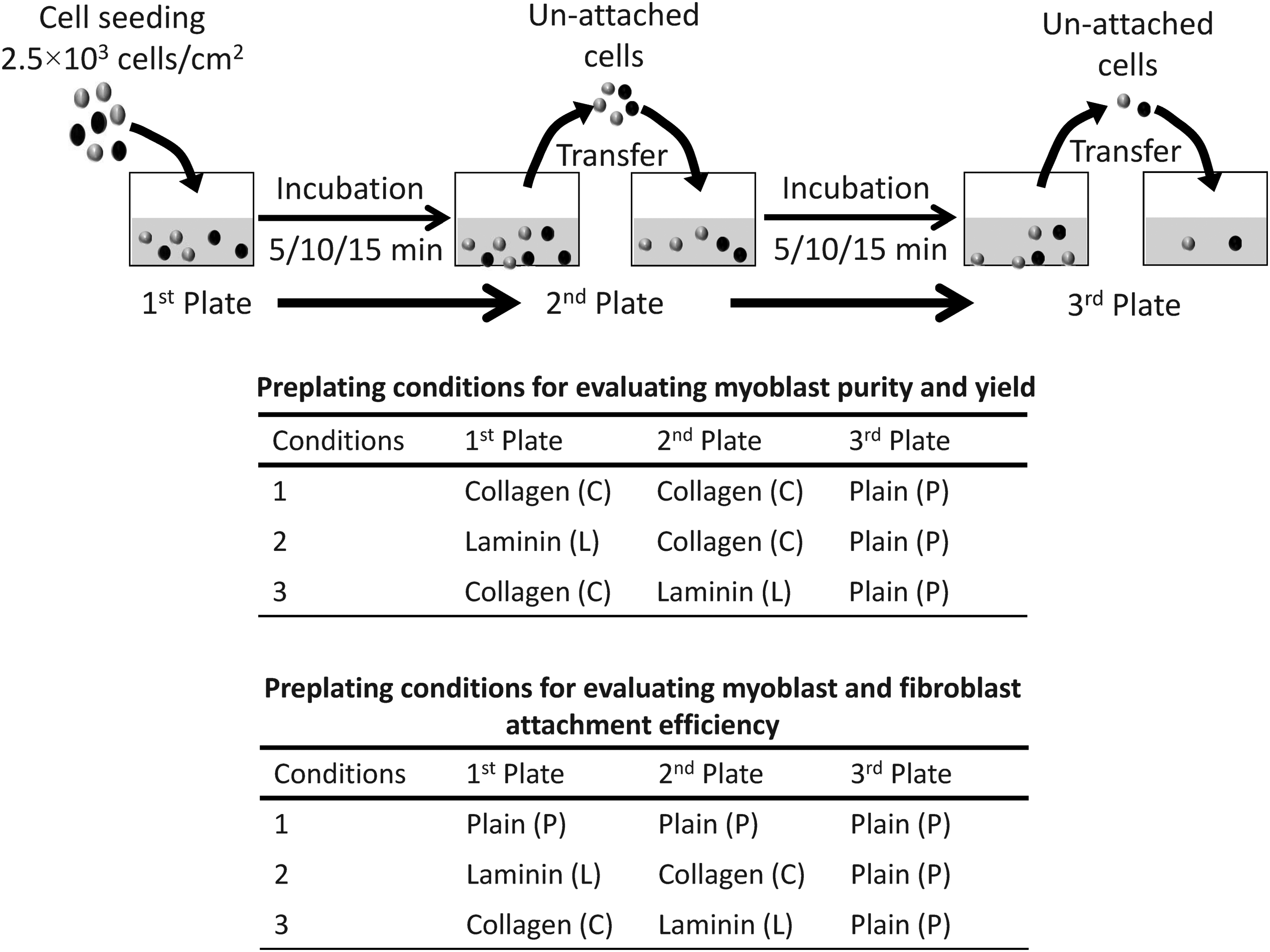
Schematic demonstration of the preplating experiment performed using various combinations of culture surfaces with an incubation time of 5, 10, and 15 min. Preplating conditions were also listed for evaluation of myoblast purity and yield, as well as myoblast and fibroblast attachment efficiency.
Attachment efficiency
The attachment efficiency of myoblasts and fibroblasts was evaluated on plain, collagen type I- and laminin-coated surface for the incubation period of 5, 10, or 15 min using the preplating experiment as described in the previous section. The conditions are listed in Figure 1. The efficiency of myoblast and fibroblast attachment was evaluated only at the first plate of serial preplating. Briefly, a mixed population of myoblasts and fibroblasts were seeded on a different culture surface (first plate) and incubated for 5, 10, or 15 min to facilitate the attachment of myoblasts and fibroblasts. Unattached cells were transferred to subsequent plates as shown in Figure 1. Finally, adherent cells were incubated for another 24 h, and IF staining was performed to evaluate the number of attached myoblasts and fibroblasts (as described in the Immunofluorescence Staining section). The efficiency of myoblast and fibroblast attachment for the 5-, 10-, or 15-min incubation on different culture surfaces was evaluated using the following equation:
Purification and subsequent cell expansion
To evaluate the maintenance of myoblast purity during cell expansion, purification and expansion of myoblasts were performed on laminin-coated surface for 2 subsequent passages. Purification was performed through preplating of mixed cells on laminin-coated surface for 5 min, and attached cells on laminin-coated surface were expanded for 144 h. Finally, cells were trypsinized and preplated onto laminin-coated surface for 5 min to perform another round of purification. Myoblast purity was evaluated at 0 h (initial sample without purification), 24 h (after first purification), 144 h (after trypsinization without purification), and 168 h (24 h after second purification), using the protocol described in earlier sections.
Moreover, the growth rate of myoblasts and fibroblasts was also evaluated on laminin-coated surface along with that on plain surface. For this purpose, a mixed population of cells were seeded on plain and laminin-coated surface at seeding density of 2.5×10³ cells/cm2 and cultured for 144 h. Adherent cells at 24 and 144 h were stained with anti-desmin and anti-fibroblasts (as described earlier) to determine the myoblast and fibroblast population, respectively. The growth rate of myoblasts and fibroblasts was evaluated using the following equation:
Growth rate (h−1)=Ln (cell concentration at 144 h/cell concentration at 24 h)/120 h
Statistical analysis
All the experiments were performed in triplicate. The values are shown as mean±standard deviation. Student's t-tests were performed to determine statistical significance. p-Value <0.05 was considered as statistically significant.
Results
Identification of myoblasts and fibroblasts
Myoblasts and fibroblasts in a mixed culture were identified by means of immunostaining as an invasive technique. Only two types of cells were identified in culture (Fig. 2). A group of cells were positive for only desmin and identified as myoblasts, whereas the other group was positive for only the fibroblast-specific marker and identified as fibroblasts. On the basis of this observation, for further experiments in this study, immunostaining was employed only with desmin, and desmin-positive cells were identified as myoblasts and desmin-negative cells were identified as fibroblasts.
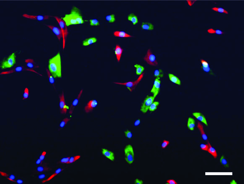
Immunostaining of a mixed population of primary muscle cells using a specific antibody for myoblasts (anti-desmin antibody; red) and fibroblasts (anti-fibroblast antibody; green). The cells were counterstained with DAPI (blue). Scale bar indicates 50 μm. DAPI, 4′-6-diamidino-2-phenylindole. Color images available online at
Attachment efficiency of myoblasts and fibroblasts
To evaluate the efficiency of myoblast and fibroblast attachment on plain, laminin- and collagen type I-coated surfaces in relation to time, a mixed population of myoblasts and fibroblasts were seeded onto designated culture surface and incubated for 5, 10, and 15 min. Representative images from each condition are shown in Figure 3A.
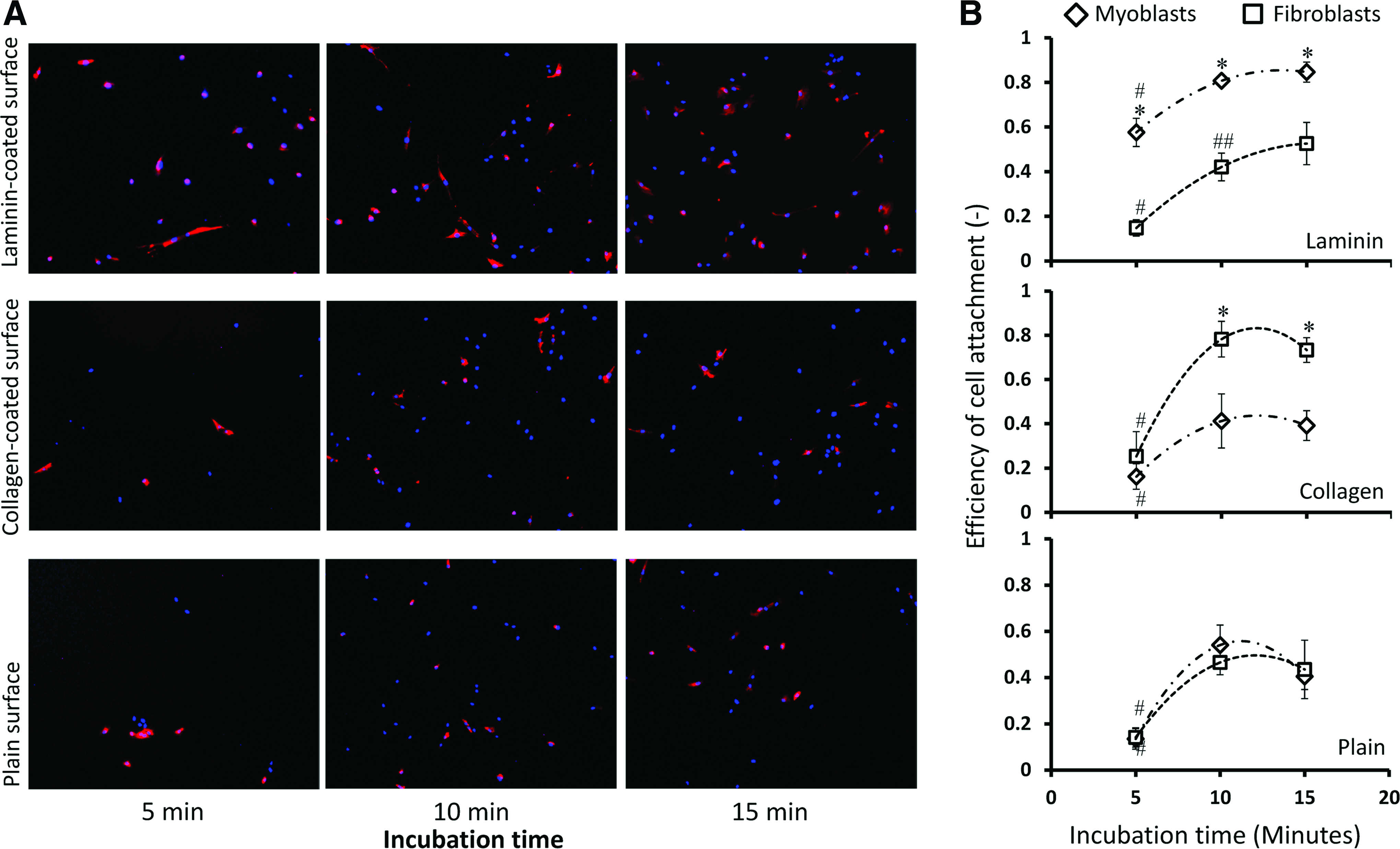
Preferential attachment of myoblasts and fibroblasts on plain and laminin- and collagen-coated surfaces.
It was found that myoblasts preferentially attached to the laminin-coated surface and that the efficiency of myoblast attachment (Am) on laminin-coated surfaces was significantly higher for an incubation period of 5, 10 and 15 min compared to fibroblasts (Fig. 3B). The attachment of myoblasts onto the laminin-coated surface was initiated immediately as the Am value for 5-min incubation was 0.58±0.06, which increased significantly to 0.81±0.02 for the 10-min incubation. The value of Am for the 15-min incubation showed a further increase (0.85±0.05) but was not significantly different from that of 10 min. It was also found that the efficiency of fibroblast attachment (Af) on laminin-coated surfaces increased significantly with an increase in incubation time and that the rate of the increase was comparatively higher compared to myoblasts. Fibroblast attachment on laminin-coated surface for the 10- and 15-min incubation increased 2.8- and 3.6-fold compared to the 5-min incubation, respectively, whereas for myoblasts it increased 1.4- and 1.5-fold, respectively.
In contrast to myoblasts, fibroblasts preferentially attached onto collagen type I-coated surfaces, and the Af on collagen type I-coated surfaces for 10- and 15-min incubations was significantly higher compared to Am, although no significant differences were observed for the 5-min incubation (Fig. 3). The Af value on the collagen type I-coated surface increased sharply from 0.25±0.11 at 5 min of incubation to 0.78±0.08 at 10 min of incubation. No further increase was detected for the 15-min incubation period.
On a plain surface, the efficiency of myoblast and fibroblast attachment also increased with an increase in incubation time; however, no significant difference was observed for myoblast and fibroblast attachment.
Efficiency of myoblast purification
To purify myoblasts, the serial plating technique was employed using HSMMs, as demonstrated schematically in Figure 1. The quantitative evaluation for purity is demonstrated in Figure 4A. The highest purity of myoblasts was achieved by incubating the mixed population of cells on the laminin-coated surface for 5 min (first plate of serial plating), and the purity was estimated to be ∼79%, which was 1.7 times higher compared to the initial population and was statistically significant. However, the purity of myoblasts significantly decreased after 10- and 15-min incubations on the laminin-coated surface (first plate of serial plating). High-purity myoblast populations were also obtained from two other conditions. In one case, two subsequent plating on collagen-coated surfaces for 10 min resulted in a myoblast purity of ∼71% on a subsequent plate, that is, on a plain surface. In another case, the cells incubated for 15 min first on the collagen type I-coated surface and then subsequently on the laminin-coated surface resulted in a purity of ∼74% on the laminin-coated surface.
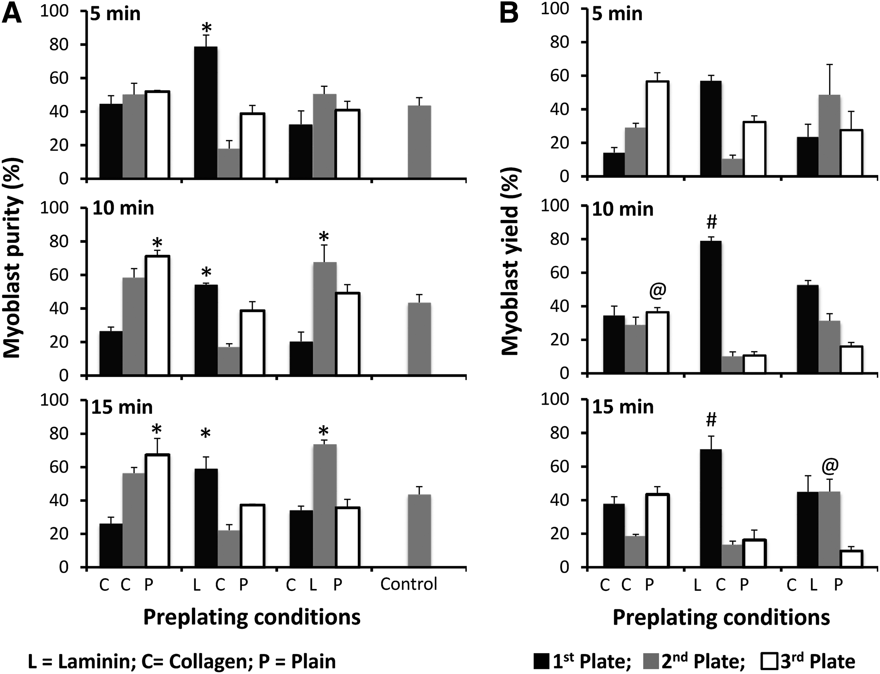
Myoblast purity
To understand the effectiveness of the purification process, the yield of myoblasts in each condition was also evaluated (Fig. 4B). The yield of myoblasts on the laminin-coated surface (first plate of serial plating) after the 5-min incubation was ∼57%. However, the yield of myoblasts on laminin-coated surface with 10- and 15-min incubations was 1.4 and 1.3 times higher compared to the 5-min incubation, respectively (p<0.05), although the purity was significantly lower. Two subsequent platings on collagen-coated surfaces for 10 min yielded ∼37% myoblasts on a plain surface. The incubation of cells for 15 min on the collagen type I-coated surface and subsequently on the laminin-coated surface resulted in a myoblast yield of ∼45% on the laminin-coated surface. These results indicated that myoblasts can be purified with a high efficiency in one step by plating a mixed population of cells on the laminin-coated surface for only 5 min.
The one-step purification process through 5 min of preplating on laminin-coated surface was tested for primary cells isolated from lower limb skeletal muscle tissues (n=4). The age of patients was ranging from 56 to 84 (Fig. 5). The myoblast population at the end of the primary culture varies between samples and it was ranging from 43.48%±2.39% to 63.76%±2.69%. In all samples, one-step purification significantly increases the purity of myoblasts. The purification index, which indicates the ratio of myoblast purity after preplating to the initial population, was ranging from 1.36 to 1.60. However, there were no significant differences observed for the purification index between samples. These results confirm the effectivity of one-step purification for primary cells isolated from skeletal muscle tissues.
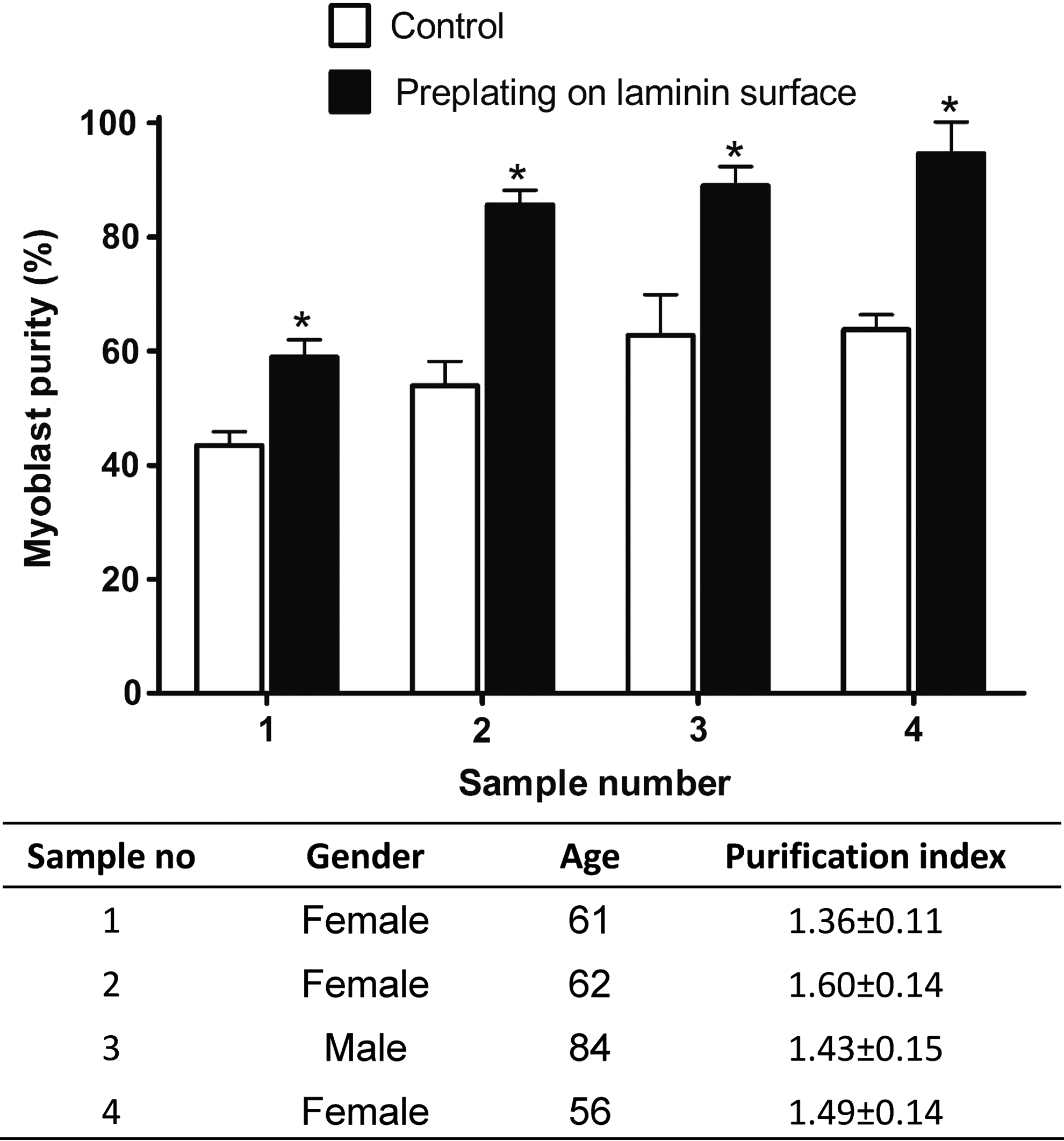
Purity of myoblasts for primary cells isolated from skeletal muscle tissue of lower limb. Purification was performed at the end of primary culture using 5 min of preplating on laminin-coated surface. *Significantly higher purity of myoblasts compared to control (p<0.05).
Maintenance of myoblast purity during expansion
To evaluate the maintenance of myoblast purity during expansion, one-step purification of myoblasts using a laminin-coated surface (5-min incubation after seeding) was incorporated with continuous culture for two subsequent passages. As shown in Figure 6a, the purity of the myoblasts at the start of the experiment was ∼52%, which reached ∼78% after the first purification. The purity of myoblasts during in vitro culture for 144 h was increased to ∼89%, which was further increased to 98% after the second purification step at subsequent passages. The increase in myoblast purity during in vitro culture on the laminin-coated surface was due to a significantly higher growth of myoblasts compared to fibroblasts (Fig. 6b). In contrast, the growth rate of myoblasts on plain surface was significantly low than that of fibroblasts. These results indicated that the one-step purification of myoblasts on laminin-coated surfaces will help to maintain the purity throughout the expansion process.
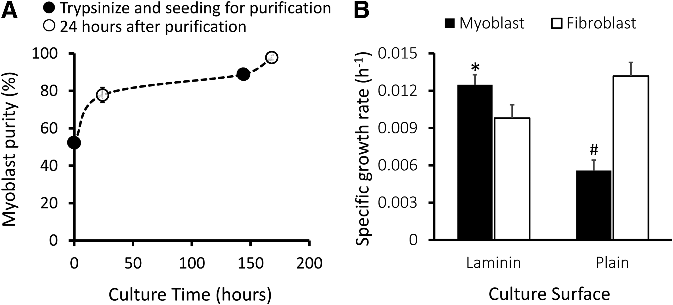
Purification of myoblasts and subsequent expansion.
Discussion
Isolation of cells from skeletal muscle produces mixed populations of myoblasts and fibroblasts. Generally, myoblasts are separated from fibroblasts during isolation of cells from muscle tissue12,13; however, absolute purity cannot be achieved.12,13,18,22 Even a small contamination of fibroblasts in the separated myoblasts could result in a continuous reduction of purity during cell expansion, as fibroblasts grow faster than myoblasts in culture using plain surface. For clinical application, it was previously estimated that at least 1 billion pure myoblasts are required for efficient treatment. 3 Baj et al. 23 previously reported that irrespective of patient age, an adequate number of cells can be produced within 1 month; however, only half of the population was identified as myoblasts. To permit rapid clinical application, the myoblast culturing process requires coordination between the purification and expansion processes. Thus, in this study, we aimed to develop a simple and robust one-step myoblast purification protocol that can be use concurrently with the myoblast expansion process.
Collagen, laminin, and fibronectin are major extracellular matrix (ECM) proteins found in tissues and play an important role in regulating cell function in vitro and in vivo. Generally, ECM proteins bind to cell surface receptors such as integrin and activate signaling pathways that regulate cell morphology, attachment, migration, proliferation, differentiation, and apoptosis. Primary human skeletal muscle cells contain two distinct cell types, that is, skeletal myoblasts and fibroblasts, which can be confirmed using immunostaining with specific markers. Immunocytochemical staining of myoblasts for desmin, an intermediate filament that is found only in myoblasts, was used to distinguish between myoblasts and fibroblasts. 24
Preferential attachment of myoblasts and fibroblasts on different ECM proteins was studied extensively, resulting in the development of preplating techniques to purify myoblasts. It was reported that myoblasts attach preferentially onto laminin- and collagen type IV-coated surfaces, whereas fibroblasts prefer fibronectin- and collagen type I-coated surfaces. 25 Previously, Kühl et al. 25 reported that plating a mixed population of myoblasts and fibroblasts from embryonic mouse thigh muscle on a laminin-coated surface for 10–20 min resulted in a faster attachment of myoblasts than fibroblasts, which yielded nearly 90% purity. Our results also confirmed the preferential attachment of myoblasts and fibroblasts on laminin- and collagen type I-coated surfaces, respectively. Moreover, we showed that the incubation time after seeding was very critical to achieve higher purity of myoblasts. Incubation of mixed cell populations for 5 min on a laminin-coated surface facilitated the attachment of ∼60% myoblasts and ∼15% fibroblasts, resulting in a high purity of myoblasts with high yield. However, at 10- and 15-min incubations on the laminin-coated surface, the rate of increase in fibroblast attachment was higher compared to myoblasts (Fig. 3), thus resulting in a significant decrease in myoblast purity. In addition, the myoblast yield increased as more myoblasts attached with increasing incubation time.
Traditionally, a gelatin/collagen type I-coated surface was used for serial plating to purify myoblasts. As fibroblasts attach faster on collagen type I-coated surfaces, the aim was to separate fibroblasts by serial preplating, leaving myoblasts in suspension, which were finally transferred to another culture surface to achieve pure myoblasts. Previous studies using serial preplating on a collagen type I-coated surface have been shown to achieve myoblast purity ranging between 78% and 95% of the total cell count.12,13,18,22 In this study, we demonstrated that two subsequent incubations of a mixed population for 10 min on a collagen type I-coated surface resulted in ∼71% purity. Moreover, 15-min incubations on the collagen type I-coated surface and then on the laminin-coated surface also resulted in high purity (79%). In both cases, the yield of myoblasts was significantly lower compared to the 5-min incubation on the laminin-coated surface, although the myoblast purity was nearly similar. These results suggested that instead of separating fibroblasts first using the collagen type I-coated surface, one-step purification using a short incubation (5 min) on the laminin-coated surface was more suitable to achieve a high purity and yield of myoblasts. These results were persistent for primary cells isolated from skeletal muscle myoblasts.
Laminin is one of the major adhesive glycoprotein components of skeletal muscle. It was reported that myoblasts attach onto the laminin-coated surface using the α7β1 integrin receptor, which is responsible for myoblast motility. 26 Enhancement of myoblast migration on laminin-coated surfaces has been shown to facilitate in vitro expansion by reducing the formation of myotubes, which is the terminally differentiated state of myoblasts.21,27 In addition, laminin-coated surfaces enhance myoblast growth by increasing cell division as well as the number of proliferative cells in culture.20,21,28 The results of the current study demonstrated that myoblasts grew at a higher rate compared to fibroblasts on the laminin-coated surface which, in turn, enhances the myoblast purity during culture. Myoblast purification using laminin-coated surfaces in subsequent passages gave rise to ∼98% purity of myoblasts from an initial 52% purity. Taken together, these results indicated that the incorporation of one-step purification of myoblasts using the laminin-coated surface with continuous culture not only helps to purify myoblasts but also maintains purity during expansion.
Conclusion
In conclusion, laminin-coated surfaces favor myoblast attachment. A short incubation of a mixed population of myoblasts and fibroblasts on a laminin-coated surface results in a high purity with moderate yield. The subsequent expansion of cells on the laminin-coated surface maintains the purity due to the increase in myoblast growth compared to fibroblasts. Taken together, these findings indicate that myoblasts can be purified and expanded efficiently in one step by preplating on the laminin-coated surface, which is a simple and robust technique.
Footnotes
Acknowledgments
This study was funded by the UKM Medical Centre Fundamental Fund (FF-037-2013 and FF-313-2010), the Tissue Engineering Centre grant (PPM-17VA001-01-0000000), and Arus Perdana (UKM-AP-TKP-10-2010). The authors also acknowledge Mr. Mohd Asyraf B. Mat Afandi and Ms Nor Kamalia Binti Zahari for their technical support.
Disclosure Statement
No competing financial interests exist.