
Abstract
Besides the advance in scientific knowledge and the production of different compounds, cell culture can now be used to obtain cells for regenerative medicine. To avoid microbial contamination, antibiotics were usually incorporated into culture media. However, these compounds affect cell biochemistry and may modify the differentiation potential of cultured cells. To check this possibility, we grew human adipose tissue-derived stem cells and differentiated them to adipocyte with or without antibiotics commonly used in these culture protocols, such as a penicillin–streptomycin–amphotericin mix or gentamicin. We show that these antibiotics affect cell differentiation. Therefore, antibiotics should not be used in cell culture because aseptic techniques make these compounds unnecessary.
Introduction
T
Despite their effectiveness to avoid bacterial and fungal growth, antibiotics can have a number of significant disadvantages. One of them is a potential influence on cultured cell biochemistry. We accomplished a very extensive literature review and, surprisingly, only found a very small number of reports on this topic, and none of them in human cells. Thus, in rat primary hepatocyte cultures, penicillin 360 μM (∼210 U/mL)–streptomycin 175 μM (250 μg/mL) inhibited protein synthesis. 5 In primary adipoblasts from young obese Zucker rats, the penicillin 1 mg/mL (∼1785 U/mL)–streptomycin 33 μg/mL mix modified the heparin-releasable lipoprotein lipase activity. 6 In mouse B16/F10 melanoma cells, penicillin 100 U/mL–streptomycin 100 μg/mL causes a moderate stimulation in dopa oxidase and tyrosine hydroxylase activities, but a slight inactivation in the dopachrome tautomerase activity. 7 The absence of studies on this issue maybe is the cause for the unawareness about the effect of antibiotics used in cell culture on many different aspects of cell biochemistry.
Since the influence of these antibiotics on cell differentiation, at concentrations found in cell culture, has not been usually analyzed, we have studied their effect on adipocyte differentiation of hASCs.
Materials and Methods
Biological samples, growth conditions, and cell differentiation
STEMPRO human adipose-derived stem cells (hASCs) (#R7788-115; Invitrogen™, Life Technologies™) are isolated from human adipose tissue collected during liposuction procedures and cryopreserved from primary cultures. Before cryopreservation, the hASCs are expanded for one passage in the MesenPRO RS™ Medium. Each lot of hASCs originates from a single donor of human lipoaspirate tissue.
Cells were grown, without antibiotics, in the MesenPRO RS Medium containing glucose (5 mM) and fetal bovine serum (FBS 2%) complemented with the MesenPRO RS Growth Supplement and
Determination of adipocyte markers
Intracellular droplets of lipids were stained with the hydrophilic stain Nile Red, which becomes fluorescent when partitioned in a hydrophobic environment. For quantitative determination, a NovoStar BMG Labtech microplate instrument (Ex: 485 nm/Em: 572 nm) was used. This determination was replicated, at least, six times.
The Adipogenesis Detection Kit (Abcam) was used to quantify triglyceride accumulation in cells, in triplicate in three independent experiments, following the manufacturer's conditions. In the assay, triglycerides are solubilized and hydrolyzed to glycerol, which is subsequently oxidized to convert the probe to generate color (λmax=570 nm). A NovoStar BMG Labtech microplate instrument was used for the measurements.
For quantitative determination of leptin in cell culture supernatants, the Leptin Human ELISA Kit (Abcam) was used. Media were centrifuged for 5 min at 1400 rpm (SX4250 rotor, Beckman Coulter Allegra X-22) and then supernatants were diluted for use in the quantification reaction. These experiments were performed in triplicate in three different experiments. Concentrations were determined according to the manufacturer's instructions.
Determination of mitochondrial enzyme activities and levels
The Complex IV Human Specific Activity Microplate Assay Kit from Mitosciences (Abcam) was used according to the manufacturer's instructions for the determination of respiratory complex IV (CIV)-specific activity and levels. CIV is immunocaptured within the wells and the activity is determined colorimetrically by the oxidation of reduced cytochrome c as an absorbance decrease at 550 nm. Subsequently, in the same wells, the quantity of enzyme is measured by adding a CIV-specific antibody conjugated with alkaline phosphatase. The phosphatase changes a substrate from colorless to yellow at 405 nm. The citrate synthase (CS)-specific activity was assayed following previously described protocols. 8 Protein concentration was determined by the Bradford protocol (#500-0006; Bio-Rad). 9 All enzyme determinations were performed in triplicate in at least three independent experiments using a NovoStar BMG Labtech microplate instrument.
Determination of hydrogen peroxide levels
The production of the hydrogen peroxide was measured in triplicate by using 2′,7′-dichlorofluorescin diacetate (Sigma) as described previously, 10 with slight modifications. A BD FACSAria sorter cytometer was used for measurements of intracellular fluorescence. Weasel software was used for flow cytometry data analysis.
Statistics analysis
The statistical package StatView 6.0 was used to perform all the statistics. Data for mean, standard deviation, and number of replicates or independent experiments are presented. The Kolmogorov–Smirnov test was used to check the normal distribution. For normal variables, the unpaired two-tailed t-test was used to compare parameters. The p-values lower than 0.05 were considered statistically significant.
Results
The hASCs cultured in the adipogenic differentiation medium slowed down proliferation and showed a morphological change with an important rise in cell volume, mainly due to a lipid accumulation, as demonstrated by the significant increase in Nile Red staining (p<0.0001), a vital stain for the detection of intracellular lipid droplets, 11 and cell triglycerides (p<0.0001) (Fig. 1A, B). Moreover, the levels of secreted leptin, another adipocyte marker, 12 were significantly higher after the differentiation protocol (p=0.0004) (Fig. 1C). All these results support the adipocyte differentiation of hASCs.
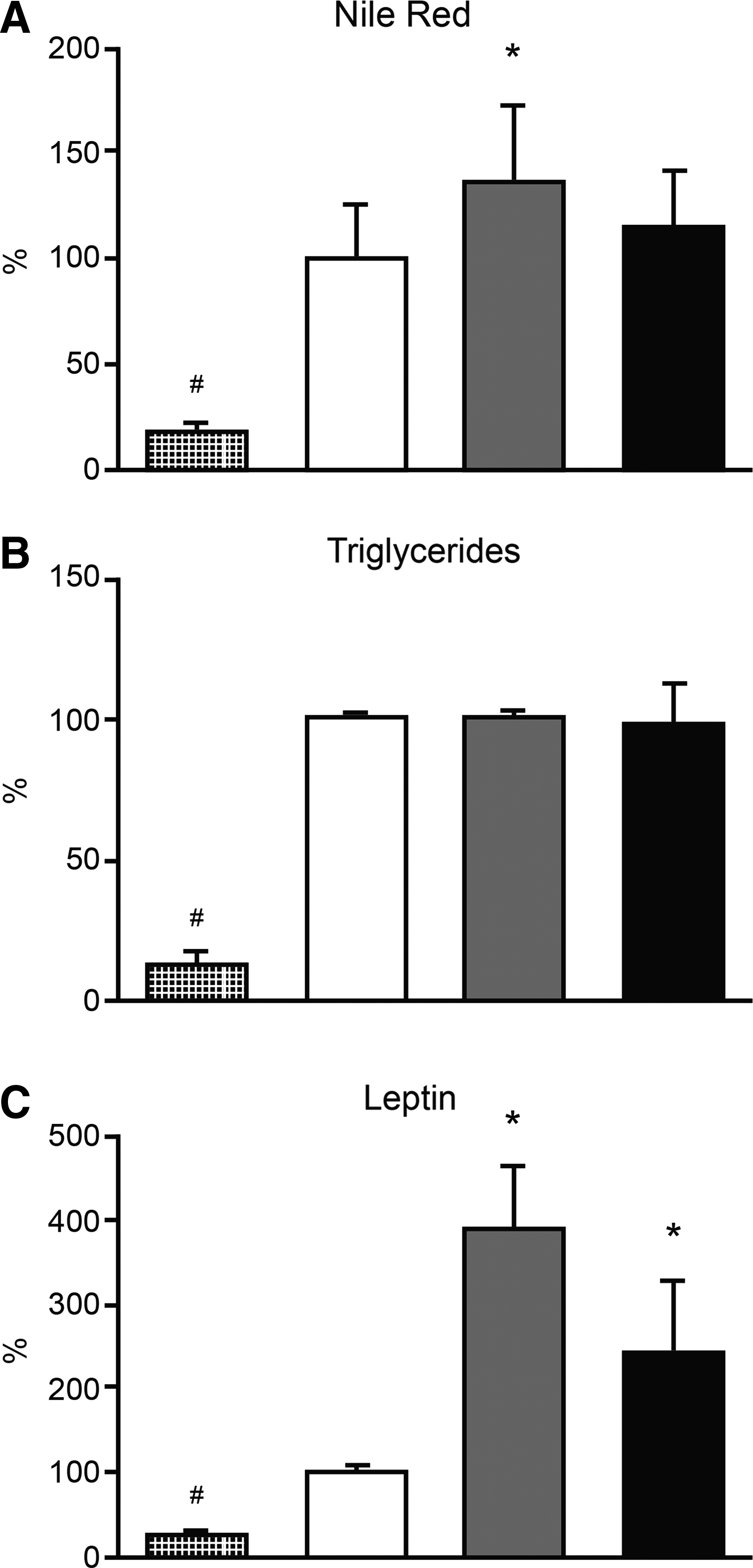
Adipocyte markers. Dot, white, gray, and black bars code for undifferentiated, differentiated, penicillin–streptomycin–amphotericin-treated differentiated, and gentamicin-treated differentiated human adipose tissue-derived stem cells (hASCs), respectively. The mean value for differentiated hASCs has been represented as 100%. #* denote significant differences versus differentiated hASCs.
At concentrations used in cell culture, the penicillin 100 U/mL–streptomycin 100 μg/mL–amphotericin 0.25 μg/mL mix significantly increased Nile Red staining (136%, p=0.0274) and leptin secretion (390%, p=0.0027), although it did not affect triglyceride levels (Fig. 1). During the adipocyte differentiation, gentamicin 5 μg/mL significantly increased leptin production (244%, p=0.0441), but it had no effect on triglyceride levels (Fig. 1). This gentamicin concentration is within the range of concentrations (0.9–11.4 μg/mL) found in the blood of some patients, for example, in neonates with hypoxic ischemic encephalopathy treated for presumptive infection, and therefore, it could have physiologic consequences on medicated individuals. 13
Although considered selective for the A-site of the prokaryotic ribosome, most aminoglycosides also bind to the A-site of the eukaryotic cytosolic ribosome, but with lower affinities. Nevertheless, their binding to the A-site of the eukaryotic mitochondrial ribosome, whose sequence is very close to the bacterial one, might inhibit mitochondrial protein synthesis and negatively affect mammal cells. 14 However, the CIV-specific activity and quantity, an enzyme with mitochondrial DNA (mtDNA)-encoded subunits and, therefore, potentially more susceptible to aminoglycosides, were not affected in hASCs differentiated in a culture with the aminoglycoside streptomycin or gentamicin (Fig. 2). Maybe, there was a relative decrease in the CIV activity and quantity but compensated, in some way, by a general mitochondrial biogenesis. However, these antibiotics did not affect the CS-specific activity, a nuclear DNA-encoded enzyme that is a marker of mitochondrial biogenesis (Fig. 2).
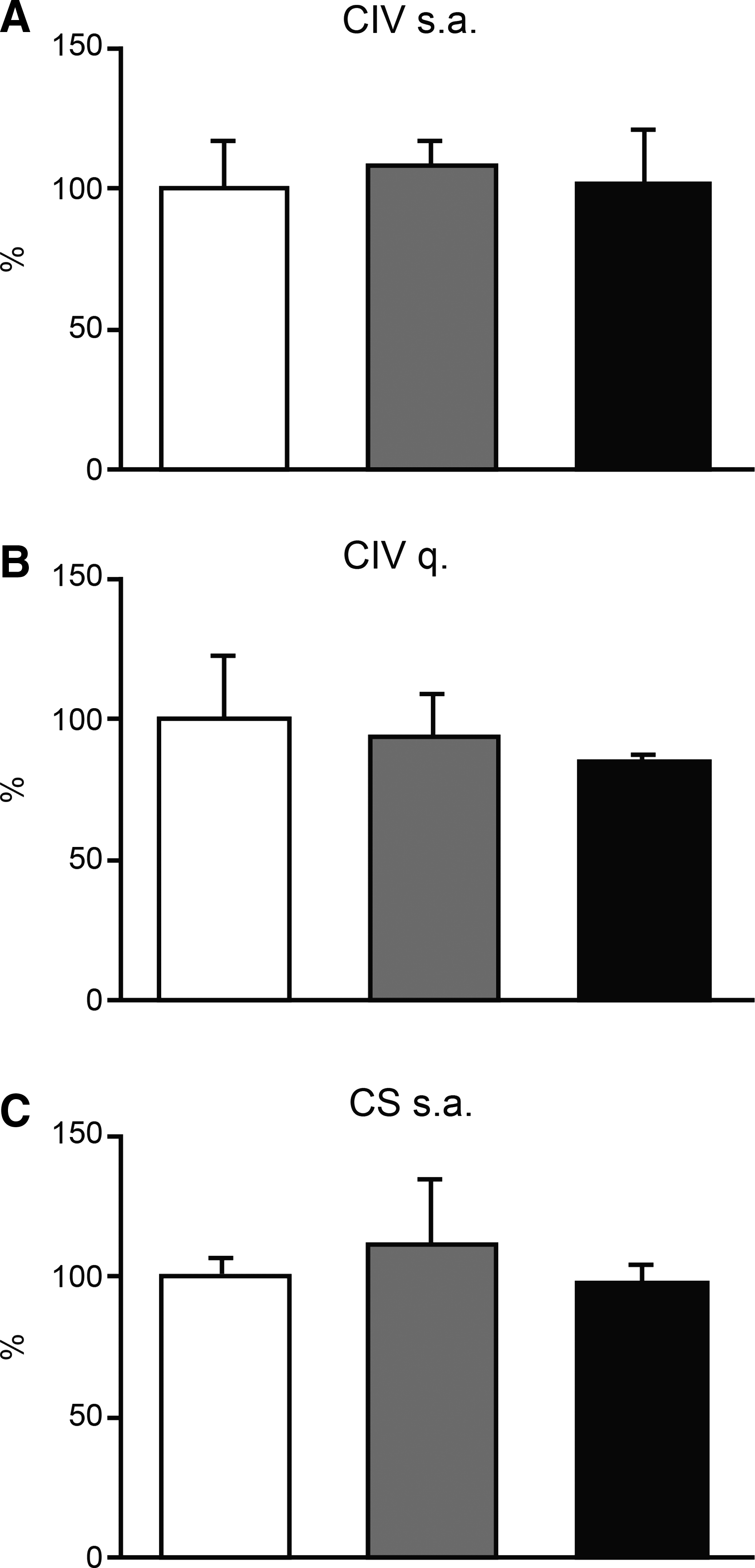
Mitochondrial enzymes. White, gray, and black bars code for differentiated, penicillin–streptomycin–amphotericin-treated differentiated, and gentamicin-treated differentiated hASCs, respectively. The mean value for differentiated hASCs has been represented as 100%.
Experimental evidence in animals has indicated that reactive oxygen species (ROS) may be one of the factors responsible for the development of aminoglycoside toxicity.15,16 Therefore, we checked the ROS amount in hASCs differentiated in the presence/absence of antibiotics. The antibiotics mix, or gentamicin, provokes a significant increase in ROS production (∼140%, p≤0.0238) (Fig. 3A). Many evidences from the literature claim that ROS are responsible for rise in different variables related to adipogenic differentiation.17–24 Therefore, we differentiated hASCs in the presence of these antibiotics with or without NAC. This antioxidant significantly decreased Nile Red staining (>70%, p<0.0001) and secreted leptin concentrations (>60%, p<0.0001) (Fig. 3B, C).

Reactive oxygen species effects on adipocyte differentiation. White, gray, stripped gray, black, and stripped black bars code for differentiated, penicillin–streptomycin–amphotericin-treated differentiated, penicillin–streptomycin–amphotericin-N-acetylcysteine (NAC)-treated differentiated, gentamicin-treated differentiated, and gentamicin-NAC-treated differentiated hASCs, respectively. The mean value for differentiated hASCs has been represented as 100%. *denotes significant differences versus differentiated hASCs. #denotes significant differences between untreated and NAC-treated hASCs differentiated in the presence of antibiotics.
Discussion
At very high concentrations, penicillin G [≥730 μg/mL or ≥1300 U/mL 25 ] had an inhibitory effect on mouse embryonic stem cell (mESC) differentiation into cardiomyocytes, as determined by contracting cardiomyocytes and quantitative expression of the myosin heavy chain (MHC) gene. This penicillin concentration also inhibited neuronal, osteoblast, and chondrocyte differentiation as determined by quantitative expression of the neurofilament 160, osteocalcin, and aggrecan genes, respectively. 26 However, these concentrations are not relevant for normal cell cultures.
At concentrations usually found in cell culture, the combination of penicillin 200 U/mL–streptomycin 50 μg/mL, streptomycin 50 μg/mL, or gentamicin 50 μg/mL interfered with the synthesis of sulfatide, a component of the myelin membrane and indicator of cell differentiation, in brain cell cultures of newborn mice. 27 The accumulation of MHC, often used as a marker of myogenic differentiation of chick embryo myogenic cells, was significantly reduced in cultures with streptomycin 100 μg/mL. 28 Penicillin 100 U/mL–streptomycin 100 μg/mL or gentamicin 100 μg/mL decreased the growth rate of mESC and, after their differentiation to type II pneumocytes, the mRNA levels for surfactant protein C. 29
Our results confirm that antibiotics commonly used in cell culture, at concentrations usually employed, can affect cell differentiation. Moreover, they extend previous results obtained in other animal cells to encompass human cells.
Compounds from the aminoglycoside family are the antibiotics shared in all these reports. It has been previously shown that particular mtDNA mutations can increase aminoglycoside toxicity, but the absence of a CIV defect rules out an effect on mitochondrial protein synthesis as the cause for the altered cell differentiation. However, in the absence of these pathologic mtDNA mutations, high concentrations or after long periods of exposure, aminoglycosides are also usually toxic. It has been proposed that ROS can act to promote both the initiation of adipocyte lineage commitment of stem cells and the terminal differentiation of preadipocytes to mature adipose cells.17–24 Our results on the adipocyte differentiation in NAC presence support that the increase in ROS levels provoked by aminoglycosides is responsible for the rise in other variables related with adipogenic differentiation, such as Nile Red staining and leptin secretion. The origin of aminoglycoside side effects on adipocyte differentiation and general cell differentiation probably results from a combination of different factors. Aminoglycosides are positively charged molecules that can interact with other negatively charged molecules, such as nucleic acids or phospholipids, or inhibit phospholipases.30–32 Moreover, it has been shown that streptomycin ≥80 μg/mL increases read through of stop codons and gentamicin 5 μg/mL induces read through of a premature termination codon found in an expressed human pseudogene.33,34
Therefore, aminoglycosides can modify cell physiology in unknown ways.30–32 To avoid undesired phenotypic effects, in general, cultured cells should be grown without antibiotics. The use of aseptic techniques makes antibiotics unnecessary.
Footnotes
Acknowledgments
We would like to thank Santiago Morales for his help with the figures. This work was supported by grants from the Instituto de Salud Carlos III (FIS-PI14/00005 and PI14/00070); Departamento de Ciencia, Tecnología y Universidad del Gobierno de Aragón y Fondo Social Europeo (Grupos Consolidados B33); and FEDER Funding Program from the European Union. CIBERER is an initiative of the ISCIII.
Disclosure Statement
No competing financial interests exist.