
Abstract
Introduction:
New cells/hydrogel-based treatments for intervertebral disc (IVD) regeneration need to be tested on animal models before clinical translation. Ovine IVD represents a good model but does not allow the injection of a significant volume into intact IVD. The aim of this study was to compare different methods to create a cavity into ovine nucleus pulposus (NP) by enzymatic digestion (E), mechanical nucleotomy (N), or a combining technique (E+N), as a model to study IVD regeneration strategies with intact annulus fibrosus (AF) in functional spinal units (FSUs) in vitro.
Methods:
The transpedicular approach via the endplate route (2 mm tunnel) was performed on ovine FSU (IVD and superior and inferior endplate) to access the NP. FSUs were treated by N (Arthroscopic shaver), E (Trypsin/Collagenase), or E+N. Treatments were evaluated macro- and microscopically. The degradation of proteoglycan (PG) around the cavity was assessed by gel electrophoresis. Cell viability was evaluated using the lactate dehydrogenase (LDH) assay. Cavity volume was quantified through computerized tomography after injection of agarose gel/contrast agent.
Results:
A cavity with intact AF was successfully created with all three methods. The N group showed high reproducibility, low PG degradation, and no endplate thinning. Histological analysis demonstrated NP matrix degradation in enzyme-treated groups, while the PG content was homogenous using mechanical discectomy. Cell viability was affected only in the E group. The cavity volume normalized to the total IVD volume was 5.2%±1.6% in E, 5.0%±1.4% in E+N, and 4.2%±0.1% in N.
Conclusions:
Mechanical nucleotomy leads to a more reproducible and less destructive cavity in the NP. Enzymatic methods perform better in terms of cavity volume; however, the cells and PG of the surrounding tissue may be affected. The mechanical nucleotomy enables the creation of a cavity into the IVD while keeping the AF intact, allowing the injection of reproducible volumes of hydrogel and tissue engineering construct for preclinical tests.
Introduction
I
Several animal species have been used to address questions regarding both etiopathogenesis and the development of therapeutic strategies. Most of them are intended to reproduce human IDD in small animals by creating a degenerative cascade through an acute injury, such as scalpel cut or a needle puncture of the annulus fibrosus (AF).4–6 However, none of these approaches will finally result in a condition identical to that found in human disorders. 3
Several important variables have to be considered when using animals for studying the IVD, such as development and anatomy of the spine; loading and size differences; and mechanical, biochemical, and nutritional conditions. Therefore, to answer specific scientific questions, such as cell/biomaterial engraftment, cell survival and differentiation, extracellular matrix (ECM) neo-synthesis, biomechanical restoration, disc function, and safety, a valid large animal model is necessary. 3
To study nucleus pulposus (NP) regeneration strategies, it is paramount to maintain an intact AF. It has recently been demonstrated that even small needle punctures of AF may lead to further degeneration and disc herniation at the site of injection. 7 Indeed, AF puncture may alter the disc biochemically and biomechanically, making the tissue unable to support physiological intradiscal pressure and avoid the leakage of NP and/or engineering constructs with biomaterials and cells. 8 As an alternative route to the disc, the transpedicular approach allows access to the NP, while maintaining the structural integrity of the AF (Fig. 1).9,10
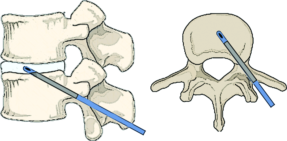
Schematic image of the transpedicular approach to the nucleus pulposus (NP) in the human spine. Color images available online at
The sheep represents a suitable model for both in vitro and in vivo studies, since ovine discs show physicochemical features similar to the human IVD.3,11,12 In fact, sheep discs resemble human ones in the lack of notochordal cells in adulthood 13 and their biomechanical behavior in vitro appears to be qualitatively similar. 14 However, the high swelling pressure of nondegenerative (healthy) IVDs does not allow the injection of significant volumes into intact discs unless they undergo treatments.15,16
The aim of this study is to develop a disc model to test injectable NP regenerative constructs by means of creating a cavity within the NP, while maintaining an intact AF. We characterized and compared IVD properties of ovine spinal segments that underwent enzymatic digestion, mechanical nucleotomy, and a combination of both treatments using the transpedicular approach.
Material and Methods
Ovine spinal segment harvest
Ovine lumbar functional spinal units (FSUs—IVD and superior and inferior endplate) were harvested from 12 skeletally mature sheep spines. All soft tissues were dissected. FSUs (levels L1–L2 to L5–L6, n=60) were obtained by parallel cuts, normal to the axis of the spine through the lumbar vertebrae, preserving the transverse processes of the lower vertebra. Under fluoroscopy, the transpedicular approach was performed to access the NP as previously described. 10 Briefly, a 2 mm Kirschner wire (K-wire) was inserted through the pedicle and the vertebral body to the inferior endplate to reach the center of the NP.
IVD cavity model
FSUs were divided into three groups (n=18/group): disc cavity obtained by enzymatic digestion (E), mechanical nucleotomy (N), and enzymatic digestion followed by mechanical nucleotomy (E+N).
In the E group, a 14G cannula was introduced in the 2 mm tunnel of the FSU and 100 μL of a solution containing 50 mg/mL trypsin (activity 10,000 units/mg; Sigma-Aldrich) and 50 mg/mL collagenase II (activity=330 units/mg; Worthington Biochemical Corporation) in phosphate-buffered saline (PBS) was injected into the disc space. Maintaining the cannula in place, FSUs were immersed in PBS and maintained at 37°C for 24 h.
The N group was obtained by introducing a 2 mm diameter shaver blade (Wolf 8564.011), powered by an arthroscopy shaver unit (Wolf 2302 Riwo Drive) and connected to a vacuum pump, through the transpedicular tunnel to the NP. NP tissue was removed, while maintaining the shaver blade in the disc space oscillating at 4000 rpm for 5 min, rotating on its own axis of 360° continuously.
The E+N group underwent a combination of both treatments. A lower concentration of enzymes was used for the combined approach compared to the enzymes concentration used in E. Briefly, the enzymatic digestion was performed through the application of 100 μL solution of 20 mg/mL trypsin and 20 mg/mL collagenase II in PBS. The FSUs were maintained at 37°C for 24 h and then the mechanical nucleotomy was performed. Both procedures were performed as described above.
Macro- and micro-evaluation
Gross anatomy
Cavities created were aspirated, washed thrice with PBS through the cannula, and filled with 100 μL 3% agarose/dimethyl methylene blue (DMMB) gel at 37°C. Following agarose gelation, IVDs were dissected with a scalpel and samples were imaged.
Histological Analysis
Following creation of the cavities, FSUs were dissected to remove excess of bone tissue. IVDs were fixed in 10% neutral buffered formalin, decalcified in ethylenediaminetetraacetic acid (EDTA) for 4 weeks, and processed for paraffin sectioning (5 μm thickness). Sagittal sections were stained with Safranin-O/Fast Green using standard techniques, and analyzed under light microscope (Nikon Eclipse E800).
Quality assessment of proteoglycans
The degradation of proteoglycans (PGs) within the tissue around the cavity (remaining NP and inner AF tissue), following treatment (E, N or N+E), was assessed (n=3/group). Agarose gel electrophoresis was used to determine the molecular size of PGs compared to fetal aggrecan and papain-digested aggrecan, which indicate the migration position of intact aggrecan and free chondroitin sulfate chains, respectively. Briefly, after cavity preparation, samples were dissected by an axial cut through the IVD and the remaining NP and inner AF tissue was harvested and weighted. PGs were extracted from each sample using 4 M guanidinium chloride (GuCl) in 100 mM sodium acetate as previously described, 17 then dialyzed into water. The glycosaminoglycan (GAG) content of the samples was then determined by the DMMB dye-binding assay. 18 To determine the size of PGs in the remaining NP and inner AF tissue samples, aliquots containing 5 μg GAG were analyzed by gel electrophoresis on a 1.2% agarose gel. 19 Gels were stained with toluidine blue for the detection of GAG chains on the PGs.
Cell viability around the defect
Cell viability was assessed using the lactate dehydrogenase (LDH) method, as previously described. 20 Briefly, following treatment (E, N, or N+E), lumbar ovine discs with endplates (n=3/group) were cultured for 1 week in Dulbecco's modified Eagle's medium (Invitrogen) containing 4.5 g/L glucose and supplemented with 1% insulin/transferrin/selenium (Sigma-Aldrich), 2% fetal calf serum (PanBiotech), and 0.1% primocin (Invivogen). Intact discs were used as controls and were cultured under the same conditions. Following culture, the intact endplate was removed and each disc frozen in cryocompound. Cryosections (10 μm thick) were cut and stained with ethidium homodimer and LDH. 20
Volume measurements of the NP cavity
Cavities previously created in FSUs (n=3/group) were aspired, washed thrice with PBS through the 14G cannula and then filled with 3% agarose/barium sulfate gel at 37°C. Following gel polymerization at room temperature, FSUs were evaluated through computer tomography. High resolution peripheral quantitative computed tomography (HR-pQCT) measurements (XtremeCT; SCANCO Medical AG) were performed at 82 μm isotropic resolution. The X-ray tube was operated at 60 kVp, 900 μA. A region of interest (ROI) was defined for the IVD to investigate the cavity volume in the NP as the signal intensity of the contrast agent. A threshold range of [1056–3000] mgHA/cm3 was used to calculate the contrast agent (Barium sulfate+Agarose) percentage in the investigated ROI. The contrast agent volume fraction (Contrast Agent Volume/IVD Total Volume, in %) was also determined.
Statistical analysis
The experiment was repeated thrice for each outcome measurement. Volumes of the cavities are expressed as mean and standard deviation (SD). Differences were evaluated by one-way analysis of variance followed by the post hoc multiple comparisons according to Dunnett's test (significance level: p<0.05).
Results
Macro- and micro-morphology evaluation
Cavities into the NP were successfully created through all the methods described as shown by the gross anatomy of IVDs filled with agarose/DMMB gel. No macroscopic differences were noted among different groups (Fig. 2).
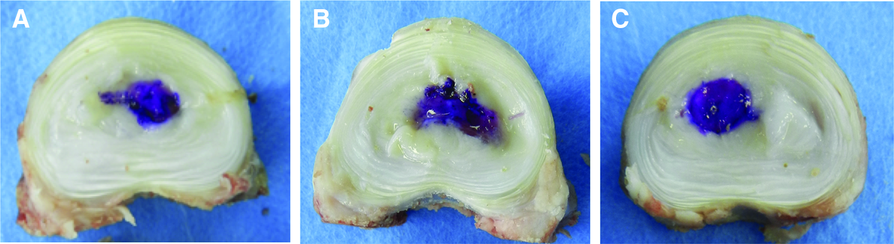
Representative images of ovine lumbar intervertebral discs (IVDs) with cavities. The cavity in the NP was successfully created using three different methods with no differences among the groups.
Figure 3 shows representative histological sections of ovine IVDs treated by E, N, E+N, and a control disc. Safranin-O/Fast Green staining showed that the intensity of the red staining of NP was significantly reduced in E and E+N groups compared with the N group and an intact disc, indicating loss of PGs. E and E+N groups also showed a less intense staining of the AF while it was conserved in N. The NP matrix was fibrillated and depleted in the E and E+N groups, while the N group had a compact and homogeneous structure (Fig. 4A–C). The GAG content of the endplate was strongly reduced in the E group appearing just as a thin layer at the bony junction (Fig. 4D). The endplate in the E+N group was also thinner compared with the N group, but maintained a higher GAG amount, as attested by the stronger Safranin O staining at the bony junction (Fig. 4E, F).
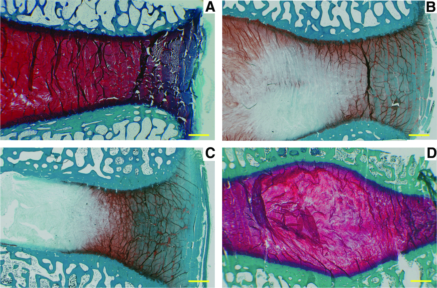
Representative histological sections of ovine lumbar IVDs treated with enzymatic

Representative histological sections of ovine lumbar IVDs treated with enzymatic digestion (E)
PG quality assessment
Agarose gel electrophoresis, used to determine the molecular size of PGs, revealed differences in PG size among groups. As shown in Figure 5, the size of the PGs in the remaining NP and inner AF tissue samples surrounding the cavity demonstrated a decrease in size going from the N to E+N group, passing through the E group. In all groups, the PGs were smaller than intact aggrecan but larger than chondroitin sulfate chains.

Gel electrophoresis. Proteoglycan (PG, +control): intact fetal aggrecan. CS (−control): free chondroitin sulfate chains. The degradation of the matrix is higher in E+N and E than N. Color images available online at
Cell viability around the defect
Ethidium homodimer and LDH staining of ovine cryosections showed the presence of a cavity, where no cells and ECM was observed in all treatment groups. As reported in Figure 6 A, E, I, the pure enzymatic treatment resulted in a damaged zone ∼500 μm thick where only dead cells were found in a loose ECM. Further away from the defect (>500 μm), the viability of the cells was unaffected. Interestingly, when a lower concentration of enzyme was used in combination with mechanical removal of NP tissue (Fig. 6B, F, J), cell viability was maintained. A region characterized by living cells in a loose ECM was observed (∼900 μm thick). Finally, when a mechanical approach was used for NP tissue removal (Fig. 6C, G, K), cell viability and ECM were not affected in the region surrounding the defect. Indeed, the tissue surrounding the defect was similar to the intact control group (Fig. 6D, H, L).

Representative histological sections of ovine lumbar IVDs treated with E
Cavities volume
Under HR-pQCT, the agarose/contrast agent gel was used as a tool to measure the volumes of cavities and its distribution within the IVD. Representative CT scans of the lumbar FSUs from different treatment groups and three-dimensional reconstruction of the cavities are shown in Figure 7. The mean and SD of cavity volumes were 37.1±9.4 mm3 in the E group, 27.6±6.8 mm3 in the N group and 29.7±10.0 mm3 in the E+N group. The percentage of contrast agent gel compared to the whole IVD volume were 5.3%±1.6% in the E group, 4.3%±0.2% in the N group, and 5.0%±1.5% in the E+N group (Fig. 8). No statistically significant differences were detected in these data.
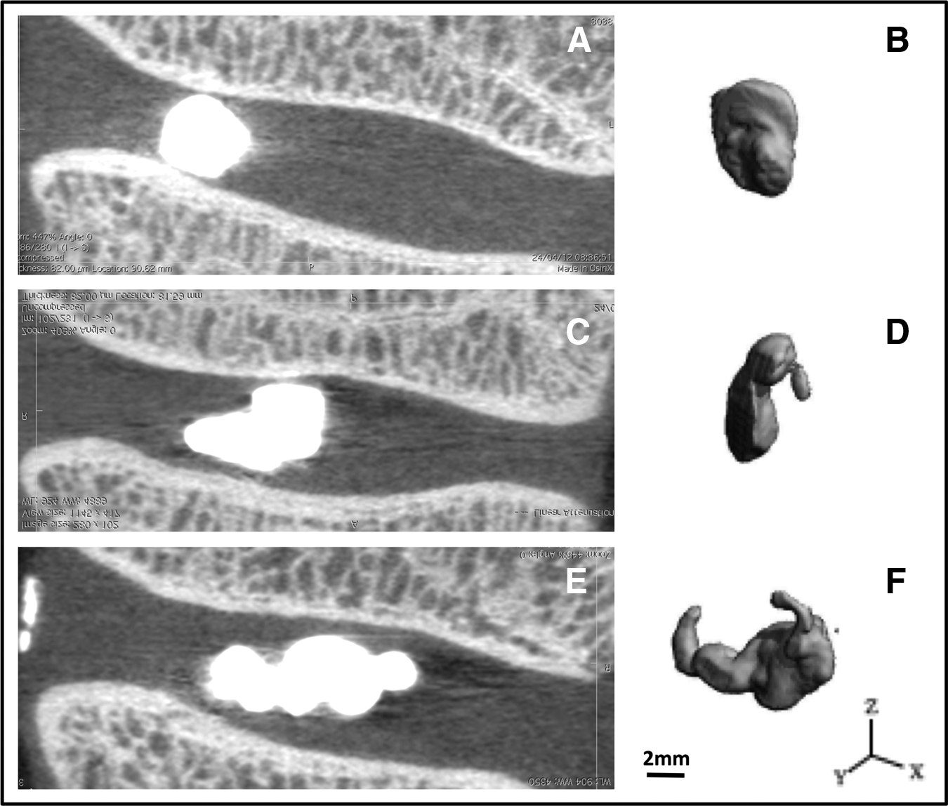
Representative computer tomography (CT) scans of cavities created in ovine lumbar IVDs filled with agarose-barium sulfate and their relative three-dimensional (3D) reconstructions
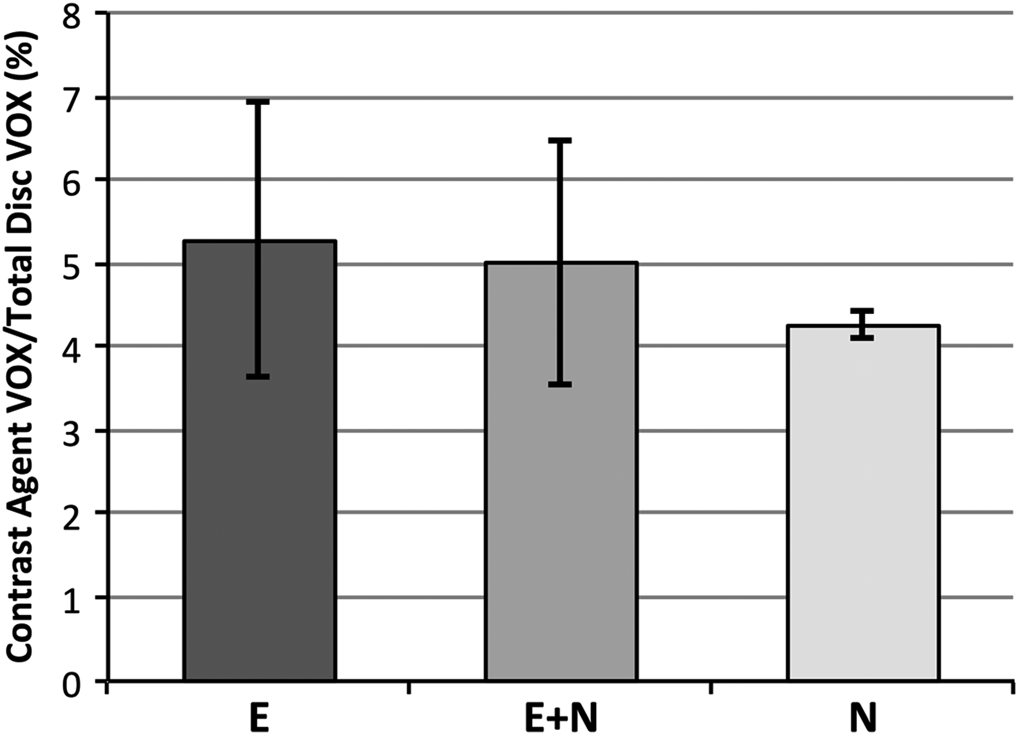
Cavity volumes (VOX) for different groups of treatment, plotted as mean and standard deviation (n=3) expressed as the percentage of the contrast Agent Volume of the IVD Total Volume.
Discussion
Nowadays, the international scientific community is strongly interested in developing new and effective tissue engineering and regenerative strategies to solve degenerative and traumatic diseases including IDD. 2 Tissue regeneration has made rapid progress in recent years, making possible the synthesis of constructs that closely mimic the structure and properties of native NP. Many new promising biomaterials and tissue engineering strategies have recently been described. 21 However, they need to be tested in viable animal models. Regulatory agencies for the evaluation of medicinal products need evidence of successful application within an animal model to proceed to human work. 3
Current animal models for the evaluation of new therapeutic approaches are questionable due to the fact that the development, nutrition, and mechanical function differ between species. Moreover, IDD animal models may not successfully replicate events typical of human disc disease. In fact, IDD is a slow and progressive degenerative process almost impossible to be reproduced by an acute injury, drug, or mechanical stress as performed in animal models. This study aimed to develop a new IVD cavity model, maintaining the AF intact, rather than an IDD model. This novel preclinical tool would enable the biological and biomechanical study of novel therapies based on cells/hydrogels for NP regeneration.
As shown by the injection of an agarose gel, all the different methods were able to create a cavity in the NP. No significant differences among groups were observed in the volumes of the obtained cavity. Even though a slightly lower volume of the cavity was observed in the nucleotomy group (N) compared to the others, the lower SD demonstrated its high reproducibility. Moreover, the enzymatic groups showed a higher breakdown of NP matrix, endplate thinning, and PG degradation in both NP and AF compared with the nucleotomy group.
The effect of enzymatic digestion and mechanical removal of tissue has been widely studied in cartilage. 22 Mechanical trauma and enzymatic treatment can both lead to a necrotic region surrounding the defect. Nevertheless, enzymatic treatment of cartilage has been reported to promote integration and chondrocyte mobility. However, little is known on the effect of mechanical or enzymatic removal of NP tissue on the viability of cells in IVDs. Our findings indicate that least damage was observed with the mechanical removal of tissue and that a combined approach of mechanical removal and small amounts of enzymes could increase the mobility of living cells by creating a looser ECM; however, stronger concentration of enzymes may be lead to decreased viability around the defect.
All these biological, biochemical, and morphological alterations make the enzyme-based strategies weaker and less suitable to study matrix regeneration. Moreover, enzyme-based methods require a two-step procedure in vivo, which increases both animal discomfort/morbidity and research costs.
Proteolytic enzyme digestion of the IVD has been widely studied both in vitro and in vivo mostly to develop a disc degeneration model.16,23–26 Lately, proteolytic enzymes including chymopapain, trypsin and papain, and chondroitinase ABC have been used for various applications.16,25,27 Chan et al. optimized enzymatic concentrations of papain to obtain an IDD model on bovine tail discs to be used in a mechanical loading bioreactor in vitro. By varying the concentration of papain, an increasing amount of GAG and water loss could be induced to simulate the different stages of IDD. 24 However, consistent with our data, the proteinase also digested matrix components in the AF rather than only PGs in the NP. In our short-term investigation with ovine skeletally mature sheep discs, culture under free-swelling conditions successfully maintained cell viability of the discs. Previous studies have indicated that dynamic culture conditions favor the exchange of metabolites to and from the IVD, which in turn results in higher cell viability compared with unloaded or static loading conditions. 28 Therefore, it can be expected that dynamic culture conditions will also favor exchange of metabolites in nucleotomized discs in long-term cultures. The nucleotomy will affect the mechanical response of the disc under loading; specifically, the range of motion will be larger in nucleotomized discs compared with intact discs. Therefore, this should be taken into account when choosing the loading regime to be applied to nucleotomized discs using bioreactors.
The importance of the endplate for IVD nutrition is well known and a lesion created by the proposed approach might induce Schmorl's Nodes, altering the nutrition supply to the center of the disc with further degeneration.29–31 However, the tunnel diameter is small (2 mm) compared with the size of the disc and the endplate hole itself can be sealed using the harvested bone cylinder by bone biopsy tools, polymethyl methacrylate cement, scaffolds inductive for bone and cartilage regeneration, 9 or screws. Nevertheless, in vivo studies are needed to better characterize the effects of the transpedicular approach on the endplate, NP matrix metabolism, cell behavior, and biomechanical function.
The model, allowing injection of a significant amount of biomaterials (viscous hydrogels) or tissue engineered constructs into the disc space without affecting the AF integrity, will be a useful tool for in vitro (organ culture in loading bioreactor) or in vivo experiments. It allows novel cell/hydrogel constructs to be transplanted in the harsh environment of the NP with low oxygen and glucose concentration under dynamic loading condition without leakage from the AF.
Conclusions
A novel NP cavity model for the preclinical tests of tissue engineering-based therapies for NP regeneration can be achieved approaching the NP via the endplate route through the transpedicular approach. The mechanical nucleotomy enables the creation of a cavity into the IVD while keeping the AF intact, allowing the injection of reproducible volumes of hydrogel. This represents a significant contribution toward the clinical translation of new regenerative therapies to biologically restore the IVD in early/moderate degenerative stages.
Footnotes
Acknowledgments
The authors are grateful to Boyko Gueorguiev-Rüegg for providing help in the biomedical services facilities of the ARI; to Markus Wilke for providing the shaver unit; and to Nora Goudsouzian for her valuable help in the histology analysis. The support by the Research Grant of the Italian Minister of Instruction, University and Research (MIUR-PRIN-200938NT8Z), the Research Grant for Young Investigator of the Italian Minister of Health (GR-2010-2318448), the BIOSPINA award of the Italian Society of Spine Surgery (GIS), and AOSpine are gratefully acknowledged.
Disclosure Statement
No competing financial interests exist.