
Abstract
The extracellular matrix provides both mechanical support and biochemical cues that influence cellular behavior. Matrix stiffness, in particular, has been found to regulate cellular morphology, motility, proliferation, differentiation, and drug responses among other behaviors. Thus, biomaterial platforms that exhibit wide range of stiffness and are available in a semi high-throughput format such as a multiwell plate would be useful for elucidating cell–substrate relationships. Polyacrylamide (PA) gels have been widely used as cell platforms since they span a range of stiffness between 0.3 and 300 kPa in Young's modulus, which encompasses all soft tissues. However, PA gels are time consuming and labor intensive to prepare, and are not amenable to a multiwell plate format. In this study, we present a novel custom multiwell plate design that allows for a one-step stiffness assay assembly that reduces preparation time and labor intensity by several fold. Gel stiffness is controlled by ultraviolet light intensity and exposure time to achieve a wide stiffness range from a single gel precursor solution. The geometry of the gels is defined by a custom photomask and gel thickness is controlled by spacers. A multiwell plate upper structure is designed similar to a regular multiwell plate such that a gel fits in each well and cells and media are added on top. The upper structure design allows for adequate gas exchange and minimum evaporation. Comparison between cell behaviors seeded in the custom and a standard multiwell plate demonstrated the suitability of the design as a cell culture platform. In summary, we describe and validate a novel custom design for an easy and rapid assembly of photopolymerizable PA-based stiffness assay.
Introduction
M
Currently, PA gels are mostly prepared in small batches because the preparation protocols are time and labor intensive. 16 Several groups, including ours, have attempted to produce PA gels for cell culture in a semi high-throughput format. Developed methods include cutting gels from a thick gel sheet, which is technically challenging for soft gels, 18 cutting gels from a gel sheet preadhered to a flexible plastic support, which can accommodate gels of any stiffness,19,20 or sandwiching gel precursor solution with a custom coverglass array directly into a well plate. 9 While enabling PA gel assembly in a multiwell plate format not possible by traditional methods for PA gel preparation, the above techniques are still time consuming.
In this study, we present a novel custom multiwell plate design that allows for a single-step PA-gel stiffness assay assembly that reduces preparation time and labor intensity by several fold. Gel stiffness is controlled by ultraviolet (UV) intensity and exposure time to achieve a wide stiffness range from a single gel precursor solution. The geometry of the gels is defined by a custom photomask and gel thickness is controlled by spacers. We further confirmed that the custom plate exhibited proper gas exchange, minimum evaporation, and allowed for cell growth, spreading, and proliferation comparable to a standard 96-well plate. While the current design is focused on assembling PA gels in a multiwell plate format, the method itself is versatile and can be extended to other photocrosslinkable hydrogels.
Device Description
The device described here consists of three main components: a glass base lined with a gel-adhesive flexible plastic support, a custom-designed upper structure containing the wells, and a custom-designed lid (Fig. 1). The base and upper structure were held together by clamps. Proper seal between the upper structure and the base was achieved by silicone o-rings. Biocompatible materials of desired mechanical and physical properties were chosen for each component. The design described here has overall dimensions as well as well volumes similar to a standard 96-well plate. However, to allow space for the clamps, a 48-well upper structure was designed. Device components and pertinent design specifications are discussed in detail in the Materials and Methods Section.
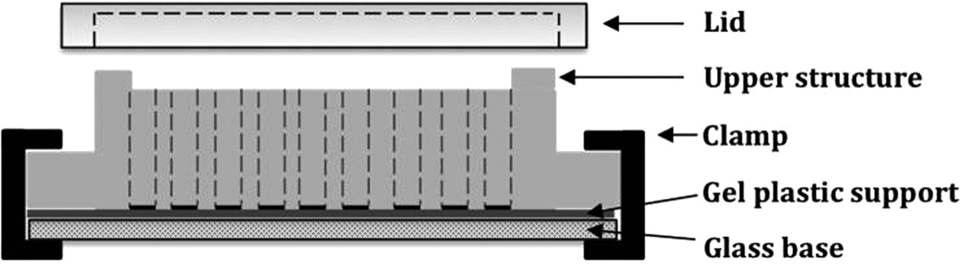
Schematic representation of the custom multiwell plate device.
The gel-containing multiwell plate was assembled as depicted in Figure 2. Briefly, the glass base was lined with a flexible plastic support that allows covalent attachment of PA gels. Silicone spacers were added to determine the gel thickness. PA gel precursor solution of desired composition was pipetted between the spacers and sandwiched with the photomask. The whole system was then subjected to UV polymerization: however, due to the presence of the photomask, only the exposed areas of the gel precursor solution (illustrated as gray circles on Fig. 2, step 4) solidified. The excess gel solution was then washed away leaving behind cylindrical hydrogels that fit precisely into the custom-designed upper structure.

Schematic representation of polyacrylamide-based stiffness assay assembly.
This design has several advantages: (1) any photopolymerizable hydrogel can be used, (2) any hydrogel geometry can be used as long as the photomask and upper structure are designed accordingly, (3) multiple hydrogels (defined by the photomask) of uniform thickness and flat surface are prepared in a single step in a matter of minutes, and (4) the flexible plastic support containing the permanently adhered gels can be handled separately upon device disassembly to allow for more involved hydrogel and/or cell manipulations at the completion of an experiment. The device can be reused multiple times since the gels are only attached to the disposable plastic support. Lastly, various degrees of gel stiffness can be achieved from a single precursor solution by tuning the duration and intensity of UV exposure during the polymerization step.
Materials and Methods
All materials were purchased from Sigma-Aldrich (St. Louis, MO), unless otherwise specified.
Device Components
Device base
The glass base consists of a rectangular glass piece with dimensions of 4" × 5" × 0.25" (Gate City Glass & Co, Inc., Kansas City, MO). The width and length were designed to be similar to the base of a standard multiwell plate and the height was chosen such that buckling of the base is negligible. Buckling is undesirable for two main reasons: (1) it can cause fracture of the base due to increased stress, and (2) it can compromise the seal with the upper structure resulting in leakage. The glass base was lined with a flexible plastic support (GelBond PAG Film; GE Healthcare Life Sciences, Pittsburg, PA). The use of the flexible plastic support GelBond offers the following advantages: (1) it is disposable, (2) it enables PA hydrogels to adhere to it permanently upon polymerization, thus keeping them anchored to the bottom of the well upon device assembly, and (3) it enables easy gel handling upon device disassembly.
Note that the GelBond plastic support does not interfere with fluorescent cell imaging, thus allowing for various cell staining protocols to be carried out directly on the GelBond-attached hydrogel. 20 GelBond has two surfaces—a hydrophobic one, which repels the gels and a hydrophilic one to which the gels adhere. To assemble the device, GelBond is positioned onto the glass base hydrophobic side down.
Upper multiwell plate structure
The upper structure is the most complex component of the device. High-density polyethylene (HDPE) (
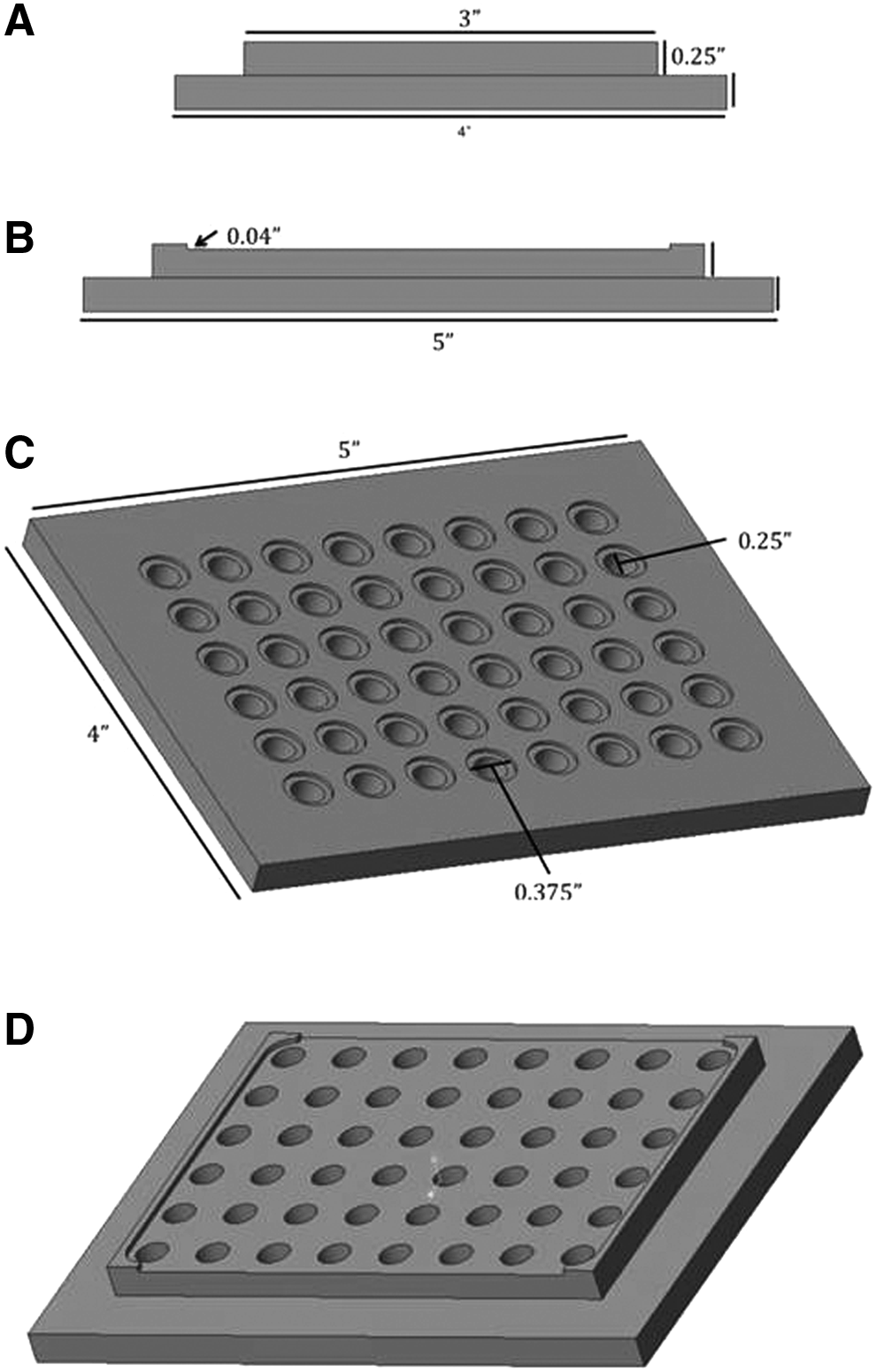
Schematic of upper structure drawn on Creo (PTC, Needham, MA) from:
There is also a lip on two sides of the middle portion of the upper structure that rises 0.04" (Fig. 3D). The lip is meant to separate the upper structure from the plate lid to allow for proper gas exchange.
Photomask
To photopattern the gels to match the wells geometry of the upper structure, a photomask is placed directly over the gel solution before exposure to UV light. The polyester Mylar photomask (Fig. 4) was custom ordered from CAD Arts Services, Inc., Bandon, OR.

Schematic of the photomask with dimensions.
Lid
The lid was made from a clear acrylic plastic (

Schematic of the lid with dimensions. The schematic was drawn on Creo.
Device Assembly
Preparation of the gel precursor solution
To prepare PA gel precursor solution, acrylamide (40% w/v; Bio-Rad, Hercules, CA), bis-acrylamide (2% w/v; Bio-Rad), and deionized water were added at a desired ratio, which determined the overall stiffness of the gel. For example, for some of the data presented here, 12% acrylamide and 0.25% bis-acrylamide in deionized water were used. The precursor solution was degassed for 30 min before gelation. The photoinitiator, Irgacure 2959 (BASF Corporation, Florham Park, NJ), was added to the solution at 0.1% w/v, mixed gently by pipetting up and down five times and pipetted onto the hydrophilic side of GelBond between spacers. Silicone spacers of 0.5 mm (Grace Bio-Labs, Bend, OR), were placed around the edges of the GelBond to assure uniform gel thickness. A 10 mL gel precursor solution was used to make the custom stiffness assay described in this study.
Photopatterning of the PA hydrogel
To photopattern the PA hydrogel, the photomask (Fig. 4) was positioned on the spacers directly over the gel precursor solution. The solution was then exposed to UV light (IntelliRay 600; UviTron, West Springfield, MA, 15 mW/cm2, 365 nm) to initiate crosslinking. To achieve varying stiffness from the same precursor solution, different parts of the gel were exposed to UV light for different amounts of time. This was accomplished by positioning an opaque shield, such as aluminum foil, over the wells intended for shorter exposure times and then moving the shield in step increments at predetermined times (Fig. 6), where longer exposure times resulted in stiffer gels. After crosslinking, the mask was removed and the excess uncrosslinked solution rinsed away with deionized water leaving behind cylindrical gels permanently adhered to the GelBond, which were then fit to the wells of the upper structure.
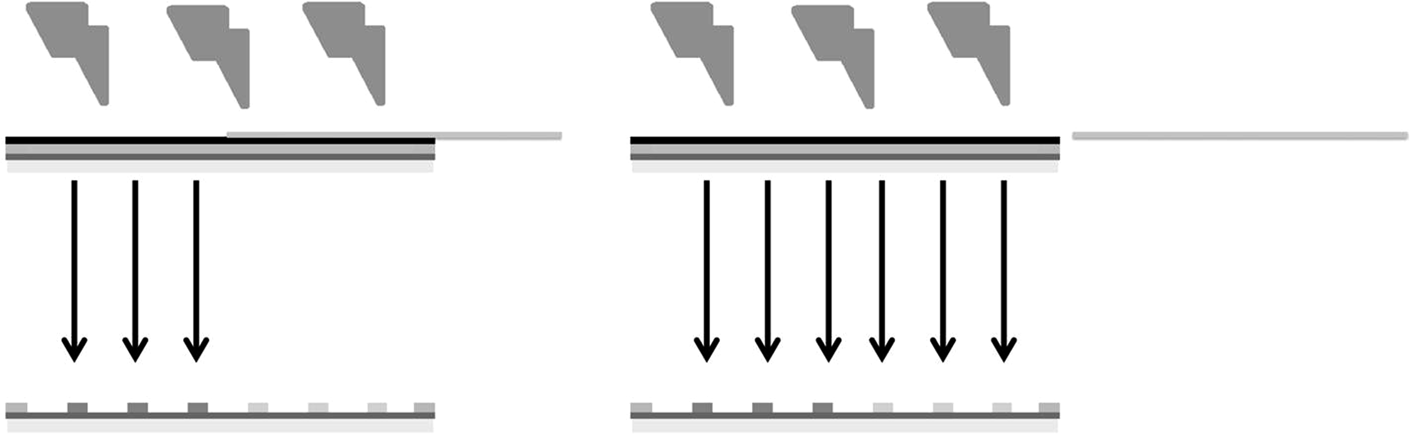
Schematic representing the process of UV crosslinking of the hydrogels to achieve hydrogels of varying stiffness from a single hydrogel precursor solution. The schematic represents UV light being shone over the hydrogel solution. An opaque piece (e.g., aluminum foil) positioned over the photomask is used to selectively block UV light. As the piece is moved, it allows parts of the gel to be exposed to UV light for longer periods of time, resulting in higher stiffness (as depicted by the darker gray circles) than the gels that were exposed to UV for shorter time. This creates the varying stiffness across the plate.
Assembly of the device components
A general schematic of the device assembly is shown in Figure 2 above and a cross-section of the assembled device is depicted in Figure 1. Briefly, the glass base was lined with a GelBond piece of matching dimensions. The GelBond was positioned hydrophobic side-down and then 0.5 mm silicone spacers were lined along the edges. The gel precursor solution was pipetted onto the hydrophilic side of GelBond between the spacers and sandwiched with the photomask. After UV exposure, the photomask was removed and the excess uncrosslinked solution rinsed off. The upper structure was positioned carefully over the gel such that a gel fitted into each well. The glass base and upper structure were then clamped together with eight binder clips (2" wide, 1" capacity; Officemate OIC, Edison, NJ)—two clips on each side. Image of the final assembled device is depicted in Figure 7.
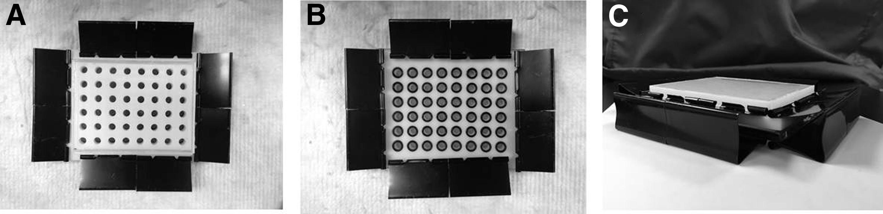
Image of the assembled device:
Collagen coating of the PA gels
After device assembly, the PA gels were coated with Rat Tail Collagen Type I (BD BioSciences, Bedford, MA) at a concentration of 0.2 mg/mL in PBS to allow for cell adhesion. To apply the collagen coating, the gels were first derivatized with Sulfo-SANPAH (Thermo Scientific, Redford, IL) dissolved in dimethyl sulfoxide (DMSO): deionized water at a ratio of 4:96. To prepare the Sulfo-SANPAH solution, the reagent was first dissolved in DMSO at 10% w/v and stored at −80°C in 20 μL aliquots until further use. Each aliquot was thawed and diluted in deionized water immediately before use. Approximately 50 μL of the Sulfo-SANPAH solution was placed on top of each gel and activated by exposure to high-intensity UV light (365 nm) for 5 min. The unreacted crosslinker was removed by rinsing with PBS twice.
Collagen solution was then added on top of each gel at ∼50 μL per well and allowed to react for 2 h at room temperature. The excess unreacted collagen was then rinsed with 10 mM PBS twice and sterilized under UV (∼302 nm) in a tissue culture hood for 2 h before cell seeding. Coated gels were used immediately or stored hydrated at 4°C for up to 2 days.
Sterilization of the device components
Different sterilization procedures were observed for each device component as dictated by the component properties, geometry, and opacity. The glass base, which is transparent, was sterilized by wiping with 70% ethanol followed by a 30 min UV exposure (302 nm). Because of their opacity, the o-rings and the upper structure were sterilized by soaking in 70% ethanol for 30 min and then rinsing with phosphate-buffered saline (PBS) once to remove residual alcohol. The PA gels and the GelBond were sterilized by a 30 min UV exposure (302 nm) only.
Device Testing
Rheology
For rheology testing, the gels were prepared in the form of slabs of 20 mm diameter and 0.5 mm thickness and swollen in PBS for 24 h before measurements. The gel stiffness was measured by rheology (AR 2000ex Rheometer; TA Instruments, New Castle, DE) with a 20 mm upper parallel plate geometry, oscillatory frequency sweep test 1–10 Hz, and 2% constant strain. Young's modulus was related to the storage modulus by the following equation:
where E is Young's modulus and v is the Poisson's ratio, which was approximated to 0.5 for PA gels. 22
Leakage, evaporation, and gas exchange tests
The device was tested in the absence of hydrogels for leakage, evaporation, and gas exchange and compared with a standard 96-well plate. Detailed methods are provided in the Supplementary Data (Supplementary Data are available online at
Cell maintenance
Nonsmall lung cancer cell line A549 (obtained from NCI-DCTD Repository; NCI, Frederick, MD) and mouse fibroblast cell line NIH 3T3 (a generous gift from Grant Kolar, Saint Louis University) were cultured in RPMI 1640 medium supplemented with 10% fetal bovine serum and 1% penicillin/streptomycin and incubated in a humidified incubator at 37°C and 5% CO2. Media were changed every 48 h. Cells were harvested by a 5 min exposure to trypsin/EDTA and passaged every 4–5 days (at ∼80% confluency).
Cell proliferation
To assess the ability of the device to support cell growth and proliferation, the device was assembled with PA gels of 2.2 ± 0.1 kPa and 93.8 ± 1.8 kPa in Young's modulus. A standard 96-well plate with PA gels of the same stiffness was used as a control. The gels in the standard 96-well plate were assembled on top of GelBond as described by us previously.19,20 Also, as described previously,19,20 all gels were UV sterilized for 2 h, collagen coated with 2 mg/mL Collagen Type I (BD Biosciences, San Hose, CA) in PBS using the crosslinker Sulfo-SANPAH (Thermo Scientific), washed two times with PBS and equilibrated in complete cell medium for a minimum of 4 h to remove residual acrylamide monomer and bis-acrylamide crosslinker, which are known toxins. 23
To compare cell viability in our device to the standard 96-well plate, 104 cells/well were seeded in three random wells in the center of both plates and cultured for 72 h. Cell metabolic activity was tested at 24, 48, and 72 h with a Resazurin assay as described by us previously. 24 Briefly, a working solution of 50 μM Resazurin sodium salt (ACROS Organics, Morris Plains, NJ) was prepared in 10 mM PBS (pH 7.4). This was then dispensed at volumetric ratio of 1:10 Resazurin:media into each well of the custom device or the standard 96-well plate, where cells were seeded. The plates were incubated for 2 h in a humidified incubator at 37°C and 5% CO2. Upon incubation, 80 μL of the Resazurin:media solution was transferred from each well to a well of a new 96-well plate and fluorescence was measured at excitation and emission wavelengths of 560 and 610 nm, respectively (SpectraMax i3; Molecular Devices, Sunnyvale, CA).
Immunocytochemistry
A549 cells were cultured on standard and custom 95 kPa PA hydrogels, and for NIH 3T3, cells were cultured on standard and custom 2 kPa PA hydrogels for 4 h. Samples were fixed for a minimum of 1 h at 22°C in PBS containing 4% paraformaldehyde and 0.1% Triton-X 100. After fixation, samples were washed in deionized water and blocked with 2% bovine serum albumin for 15 min and then incubated with Alexa Fluor 546 phalloidin (Invitrogen, Carlsbad, CA) and Hoechst 33258 (Sigma-Aldrich) for 20 min at 37°C. Samples were washed in deionized water and mounted with hydrogel side up onto glass slides using Aqua-Poly/Mount (Polysciences, Warrington, PA). Images were taken using a Leica DMIRE2 HC inverted epifluorescence microscope fitted with a 12-bit grayscale CCD camera using an oil-immersion 63× objective.
Statistical analysis
The results of all experiments are the mean values (±SD) of three to eight samples per condition, performed in three to six independent experiments. Comparisons between multiple samples were performed with single-factor analysis of variance and comparisons between two samples were performed with two-tailed Student's t-test, followed by post hoc analysis. In all cases, differences between data sets were considered significant when p < 0.05.
Results and Discussion
In this study, we describe the design and assembly of a custom multiwell plate device for the simple and quick assembly of PA gels. Traditionally used in electrophoresis, 25 PA gels are becoming increasingly important as substrates for cell culture due to the similarity between their viscoelastic properties and modulus to soft tissues in the body. 19 However, preparing PA gels is time and labor intensive and thus, they are typically made in small batches. 16 Due to their prevalence as a substrate in various studies of stiffness-dependent cell behaviors,26,27 new methods are required for the reproducible and quick fabrication of PA as well as other hydrogel-based stiffness assays. In lieu of this need, our design aimed at developing a custom device that allows for the assembly of a robust PA-based stiffness assay in one easy step. While this study is focused on PA, note that the developed device should be compatible with other photo-crosslinkable hydrogels.
The device design and assembly were discussed in detail in the previous sections (Figs. 1 and 2). Briefly, the device consists of a glass base, a gel adhesive flexible plastic support (GelBond), a custom-designed upper structure (Fig. 3), and a custom-designed lid (Fig. 5). Uniform PA solution was poured between spacers onto the GelBond-lined glass base and sandwiched with a photomask (Fig. 4). Gels were formed upon UV illumination and then the upper structure was fitted such that a gel fitted into each well. The base and upper structure were then clamped to avoid leakage. O-rings were also used on the upper structure to secure the seal. The hydrogels were then washed to remove residual toxic polymer monomers, and equilibrium-swollen in media before cell seeding.
Note that because of the strong adhesion of the gels to the GelBond flexible plastic support, the gels fitted the wells of the upper structure not only before, but also after swelling in media. While hydrogels typically show a large volume increase upon swelling, unconstrained and constrained hydrogels exhibit different swelling behaviors. 28 Specifically, unconstrained gels swell proportionally in all directions preserving their original shape, whereas constrained gels swell along the unconstrained dimension only 28 ; the PA hydrogels constrained to the bottom of the well, increased preferentially in height and not in diameter upon swelling. We have previously demonstrated similar behavior for GelBond-adhered PA gels, where the GelBond/PA constructs were glued to the well bottom of multiwell plates.19,20
To achieve gels of varying stiffness from a single gel solution, different parts of the plate could be illuminated for different amounts of time (Fig. 6). This strategy is currently used for the production of PA gels of stiffness gradients 10 ; here it was used in a similar fashion, but for the production of a stiffness assay, where each well contained a gel of a single stiffness. Figure 8 shows the Young's modulus of a PA gel made from 12% acrylamide and 0.25% bis-acrylamide as a function of UV exposure time. Data indicate that ∼10-fold difference in stiffness could be achieved from a single gel precursor solution. For example, 25 s illumination resulted in ∼1 kPa hydrogel, whereas 2500 s illumination resulted in ∼95 kPa hydrogel, effectively covering the whole range of soft tissues in the body. 1
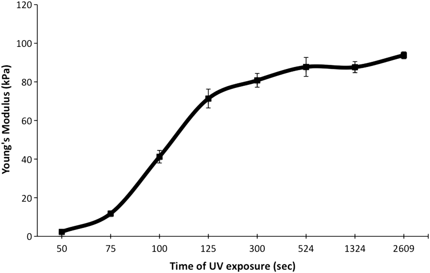
Increasing exposure time to UV light increases the stiffness of polyacrylamide gels.
Note that for rheology testing, PA hydrogels were prepared as 20 mm disks rather than in the specified device geometry and that continuous rather than intermittent UV light exposure was used. While we confirmed that intermittent UV light exposure with five cycles of 1 min on/5 s off did not significantly affect the resultant hydrogel modulus compared with continuous UV exposure (data not shown), several important considerations need to be taken into account. Photopolymerization and, hence, modulus of PA, depends on processes such as the diffusion of ambient oxygen into the polymer precursor solution, as well as the diffusion of free radicals from the illuminated into the dark region (refer to photomask image) and the diffusion of additional acrylamide monomers from the dark into the illuminated region, which consequently participate in the polymer chain. 29 Since these are diffusion-driven and, thus, time-dependent processes, total illumination time (including “off” periods), spacing between the wells and the volume of the gel precursor solution, would all have an impact on the final modulus of the hydrogels.
The developed device geometry, in terms of well spacing and hydrogel precursor solution volume, was optimized to enable the desired gel pattern. However, further optimizations of the system are possible. For example, an automated stage for sliding an opaque mask could be employed, as opposed to manually moving the opaque mask (aka foil) as discussed here, which necessitated intermittent light exposure. Sliding an opaque mask between a photopolymerizable hydrogel precursor solution and a light source has previously been used to produce hydrogels with stiffness gradients.10,30 Such optimization should alleviate concerns related to photopolymerization kinetics due to intermittent UV exposure as well as shorten the total exposure time by negating the need for “off” cycles.
Next, we compared the device performance to that of a standard 96-well plate. We confirmed that no leakage was observed long term from the wells of the device and that the custom device exhibited similar evaporation (Supplementary Table S1) and gas exchange (Supplementary Table S2) as a standard 96-well plate.
Finally, we investigated whether the custom device can support cell viability similarly to a standard multiwell plate. Figure 9 depicts the fold change in A549 cell number (as compared with the cell number at 24 h of culture) as a function of culture time as measured by a Resazurin assay—an indicator of cell metabolic activity. Our results demonstrated that the custom device was able to support cell viability and proliferation as well as a standard multiwell plate. We would like to note that cells were cultured on top of collagen-coated PA gels in both plates to simulate the device's intended use.
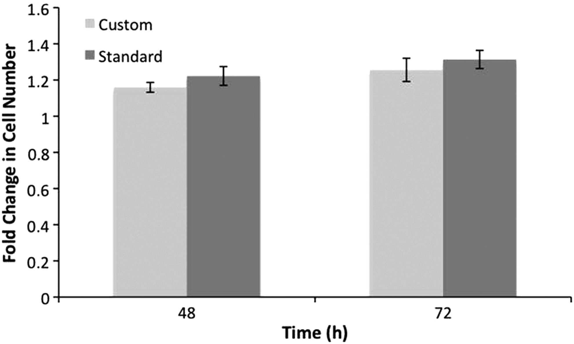
Fold change in cell number for A549 cells in custom and standard well plates for 48, and 72 h indicates that there is no significant difference in cell viability between the custom and a standard well plate for the course of the experiment (n = 3, p < 0.05).
We also investigated whether the custom device can support cell culture in comparison to a standard multiwell plate by utilizing immunocytochemistry. Figure 10 shows the actin cytoskeleton in A549 cells on a standard and custom 95 kPa PA hydrogel. We did not observe a difference in the actin of the A549 cells between the standard and custom gel, which validates that the custom device supports cell culture similarly to a standard multiwell plate. Further analysis was taken from the actin images to provide information on the cell area, actin intensity, shape factor, and cell length (Table 1). The length was measured as the longest span between two points in the cell perimeter. The shape factor is depicted as a value ranging from 0 to 1, representing how closely the object represents a circle, where 1 illustrates a perfect circle and values approaching 0 represent a flattened cell. Our results demonstrated that there was not a significant difference between each of the aforementioned variables for the standard and custom plate.

A549 cells cultured for 24 h onto a 95 kPa standard
Same analyses were performed for NIH 3T3 cells seeded on a softer 2 kPa PA hydrogel (Fig. 11; Table 2). As expected, 5 cells exhibited a rounder morphology and, hence, lower cell length, as well as less spreading on the softer hydrogel as compared with the stiffer one. Importantly, no significant difference was observed between the cells seeded in the custom as compared with the standard multiwell plate. These results further validate that the custom device is suitable for the support of cell culture.
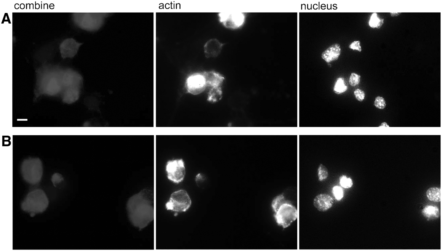
NIH 3T3 cells cultured for 24 h onto a 2 kPa standard
In summary, we demonstrated that upon assembly, the novel device described in this study, performed as well as a standard multiwell plate in terms of liquid evaporation, gas exchange, and the ability to support cell culture. The developed device enables assembly of PA-based stiffness assays in one single step and could be invaluable in the study of stiffness-dependent cell behaviors. Importantly, the design is very versatile: while the work described here is focused on a device for the assembly of PA gels, the same device could be used for other UV crosslinkable hydrogels such as acrylate- and methacrylate-terminated polymers.
Footnotes
Acknowledgments
This work was funded by start-up funds provided to Dr. Zustiak by Saint Louis University as well as by a President's Research Fund (PRF) grant and Stroble Award in Health Sciences awarded to Dr. Zustiak by Saint Louis University.
Disclosure Statement
No competing financial interests exist.
References
Supplementary Material
Please find the following supplemental material available below.
For Open Access articles published under a Creative Commons License, all supplemental material carries the same license as the article it is associated with.
For non-Open Access articles published, all supplemental material carries a non-exclusive license, and permission requests for re-use of supplemental material or any part of supplemental material shall be sent directly to the copyright owner as specified in the copyright notice associated with the article.