
Abstract
Centrifugation based on density gradients is a general methodology for isolating human bone marrow (hBM)-derived mesenchymal stem cells (hBMSCs). The mononuclear cell (MNC) layer can be obtained using a density gradient solution in the conventional protocol, but it is not suitable for direct transplantation due to the possible toxicity of this solution. The results obtained are also influenced by the skill level when applying the technique, which involves time-consuming processes. We have developed a novel protocol for isolating hBMSCs using hyaluronic acid (HA), which is the most widely used injectable biomaterial in clinical settings and a major component of the extracellular matrix. Laying hBM over the HA and then applying centrifugation yielded three separate layers, with the HA layer, including MNCs being the most superficial one. Increasing the volume of HA and/or its crosslinking rate enhanced the yield of MNCs from hBM, and the cell yield was also significantly higher for a lower centrifugal acceleration (530 g) than for a higher one (1500 g). Isolated hBMSCs by HA exhibited similar biological characteristics such as in terms of their proliferation rate, fibroblast-like morphology, cell-cycle status, immunophenotype, and multipotency. The use of either type of hBMSC confirmed the regenerative potential of bone and bone marrow-like tissue in ectopic transplantation models. This is the first report of a novel protocol for isolating hBMSCs that utilize HA. We suggest that this novel isolation technique can be used for the direct application of autogenous MSCs with advantages of being less time-consuming and involving steps that are easier to perform.
Introduction
S
Hyaluronic acid (HA) is the main component of the extracellular matrix (ECM) and is one of the natural polysaccharides composed of alternating (1 → 4)-β-linked N-glucuronic and (1 → 3)-β-linked N-acetyl-

Schematic diagram showing the isolation of MNCs by centrifugation with Ficoll and HA hydrogel.
Herein, we report a novel protocol for isolating MSCs using HA hydrogel. The technique utilizes physical features of HA such as its viscosity and its adherence to cells. We also investigated the isolation conditions for HA that would obtain the highest yield of MSCs, and characteristics of the MSCs isolated using HA hydrogel were compared with using the conventional protocol.
Materials and Methods
Preparation of human BM aspirates
Human BM (hBM) aspirates (80–100 mL) were collected in a heparin-containing syringe from the vertebrae of eight volunteers (two males and six females, median age 52 years, age range 37–59 years). The donors submitted to orthopedic surgery (Naeun Hospital, Anyang, Korea) after providing informed consent using guidelines approved by the Institutional Review Board, College of Dentistry, Yonsei University.
Isolation of hBMSCs by density gradient centrifugation using Ficoll
hBM aspirates (10 mL) were diluted 1:1 in Dulbecco's phosphate-buffered saline (PBS) and layered onto Ficoll-containing density gradient medium (Histopaque; Sigma-Aldrich). After centrifugation (1500 g for 20 min) at room temperature, the buffy coat was collected in a new tube and washed in PBS (Fig. 1B). It was then centrifuged at 530 g for another 5 min, the supernatant was washed, and the remaining pellet was resuspended in 2 mL of culture medium. The culture medium consisted of α-minimum essential medium (α-MEM) (GIBCO) supplemented with 10% fetal bovine serum (GIBCO), 20 μg/mL gentamicin (GIBCO), 2 mM GlutaMAX-1 100× (Invitrogen), and 100 μM
Isolation of hBMSCs by density gradient centrifugation using HA hydrogel
The carrier used is a stabilized gel type of HA that was pharmaceutically avalilable (Hyal-Forte; Shin Poong Pharm). HA raw material is produced in cultures of Streptococcus equi subsp. zooepidemicus (ATCC 39920) by fermentation. The fermented products are purified through precipitation, filtration, and freeze-drying to produce a sponge form. Crosslinking type of HA was also provided from the same manufacturer (Shin Poong Pharm), which the purified HA is stabilized by butanediol diglycidyl ether crosslinking. The degree of chemical modification can significantly affect the properties of HA due to differences in the crosslinking ratio (0%, 30%, and 50%), which was controlled by mixing the purified noncross-linked and cross-linked HA with the proportion of 0%, 30%, and 50%, respectively. It was then formulated into the gel type in PBS in sterile conditions authorized by guideline for Good Manufacturing Practice (Shin Poong Pharm).
hBM aspirates (10 mL) were layered onto HA without PBS dilution and then centrifuged for 5 min at room temperature. This step was performed repeatedly to assess the effects of various conditions such as the type and volume of HA and the centrifugal acceleration for optimization of the condition for BMSC isolation through HA. After separating the different layers, those cells with very low densities combined with HA were collected (Fig. 1C). This collected layer was then lysed with hyaluronidase (Malinda Inj.; BCworld Pharm) and washed with 10 mL of culture medium. The remaining pellet was resuspended in 2 mL of culture medium. The resulting cells in 1.5 mL of solution were counted and seeded at 5–10 × 106 cells/20 mL with culture medium in T75 culture flasks. The cells in the remaining 0.5 mL of solution were assessed based on the CBC.
Comparison of characteristics of BMSCs isolated using Ficoll and HA hydrogel
The characteristics of MSCs in two experimental groups were compared by previously described protocols in our other studies.36–38
hBMSCs were isolated from aspirated hBM by density gradient centrifugation using two types of media, Ficoll and HA hydrogel. Cell morphology and proliferation rates were compared, and the number and size of colony-forming units were calculated for determining/comparing the presence of putative BMSCs. Osteogenic, adipogenic, and chondrogenic differentiations were induced, and in vitro differentiations and their related gene expressions were analyzed for evaluating multipotency of isolated hBMSCs in both control and experimental groups. Cell surface markers were evaluated by fluorescence-activated cell sorting, and in vivo differentiation was assessed by the ectopic transplantation model using immunodeficient mice. All procedures of aforementioned experiments were described in Supplementary Data (Supplementary Data are available online at
Statistical analyses
All of the in vitro experiments were performed in technical triplicate, and mean and standard deviation values of the three results were calculated. Unpaired t-tests were used to analyze the differences between the two groups. Repeated-measures analysis of variance followed by Scheffé's comparison was used for multiple analyses. Differences were considered statistically significant at p < 0.05.
Results
Assessment of the MNC layer in the HA hydrogel
When the hBM was separated into three layers using the novel HA-mediated method, the superficial layer comprised HA hydrogel, including some of the BM content. Fluorescence-activated cell sorting analysis was applied to the cultivated cells from the HA layer to confirm whether or not it included putative stem cells. More than 90% of the cells expressed the typical MSC marker CD44, whereas the cells displayed limited expression of the hematopoietic marker CD34. The expression levels of these markers were the same as for hBMSCs isolated using the conventional Ficoll-mediated method (Fig. 2). The results showed that putative stem cells were present in the HA layer.
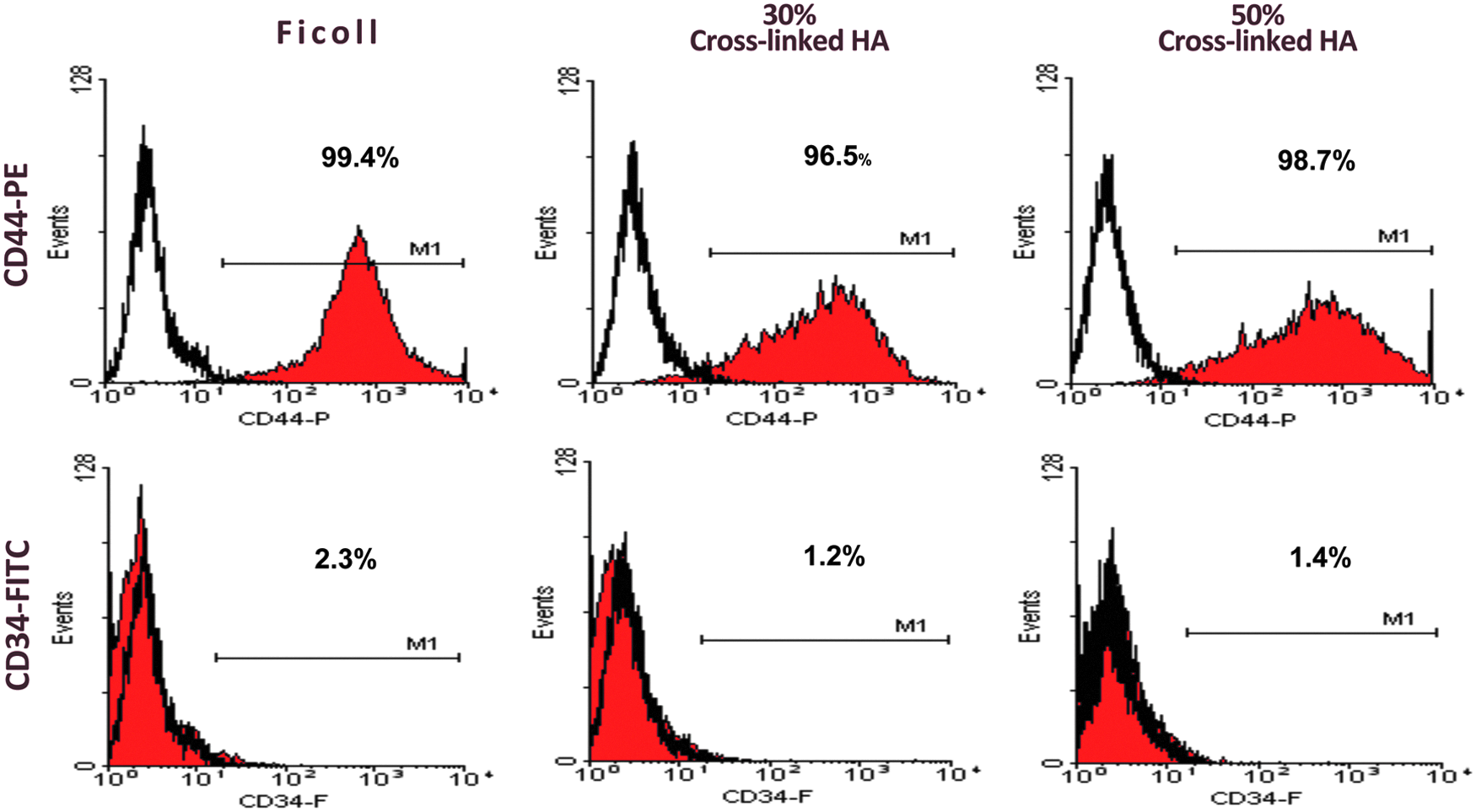
Immunophenotype analysis of MNCs isolated using Ficoll and two types of HA hydrogel. The expression rate of CD44 was more than 90%, whereas that of CD34 was lower than 3%. The tendency was the same in all of the groups (Ficoll, 30% cross-linked HA, and 50% cross-linked HA). CD44, MSC marker; CD34, hematopoietic stem cell marker.
Optimization of the condition for BMSC isolation through HA
Assessment of cell yield and proliferative activities depending on the crosslinking rate of HA
HA takes several different physical forms depending on its concentration, aquation rate, and chemical structure. These subtypes of HA have different physical properties, and thus, also different capacities for isolating BMSCs. One of the subtypes was categorized according to a component ratio of cross-links. Three types of HA hydrogel composed of 0%, 30%, and 50% cross-linked HA were used to determine the optimal cross-link rate for obtaining the highest cell yield from BM. The cell count and CBC differed significantly immediately after isolation among the three groups, with the number of white blood cells (WBCs) increasing with the cross-link ratio. Relative to the 0% and 30% cross-linked HA groups, the numbers of WBCs were 6.3-fold and 1.6-fold higher in the 50% cross-linked HA group (p < 0.05; Fig. 3A), while the values in the CBC test were 11.5- and 1.7-fold higher (p < 0.05; Fig. 3B). hBMSCs from each HA layer were expanded to allow comparison of their biological characteristics, which revealed that the proliferation rate of the isolated BMSCs did not differ significantly between the three groups (p = 0.836; Fig. 3C).
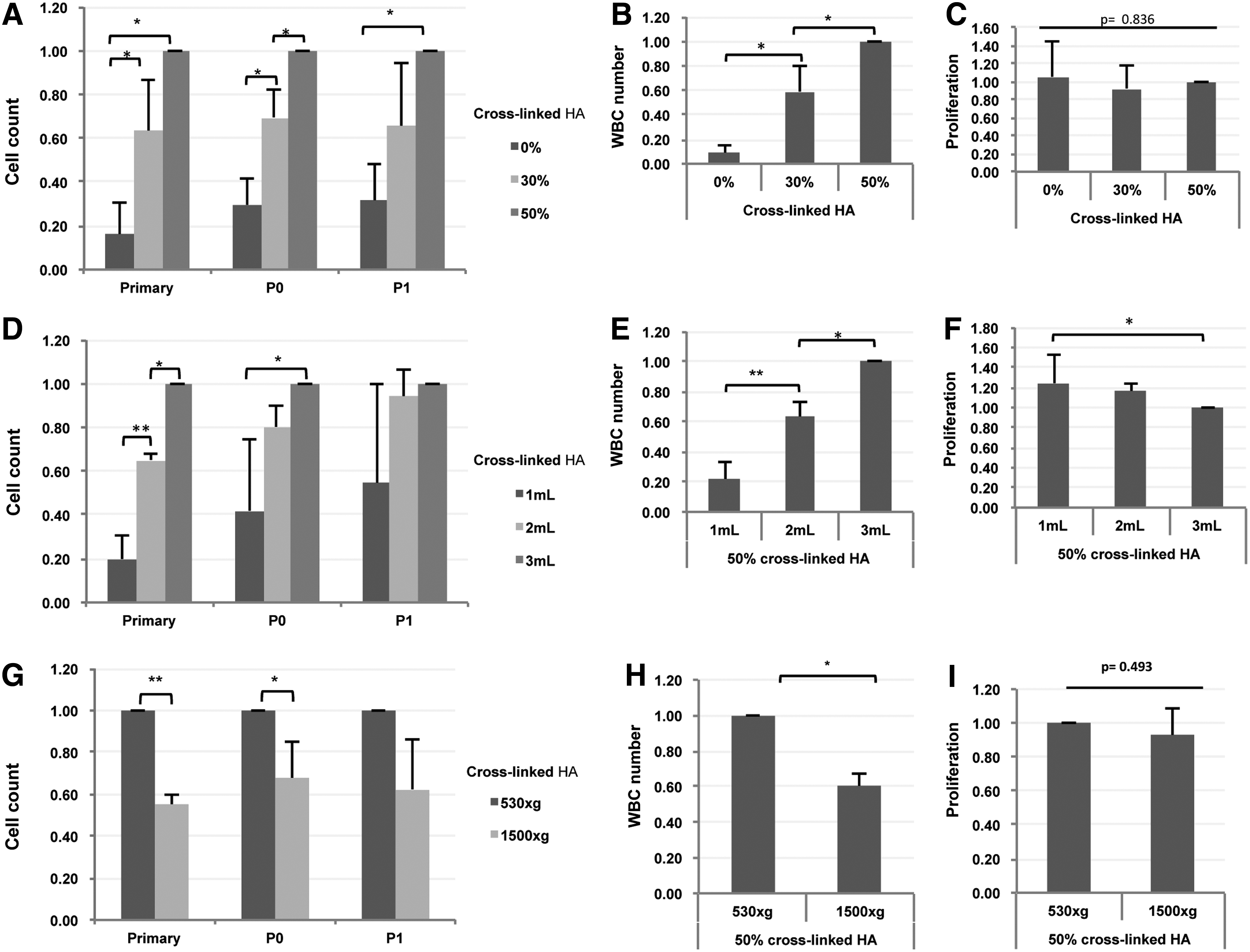
Results from optimization of the HA hydrogel condition for BMSC isolation. The total yield of MNCs was derived from data obtained for the crosslinking rate [
Assessment of cell yield and proliferative activities depending on the isolation conditions
When performing isolation experiments for an equal amount of hBM, it is important to consider the effects of conditions such as different volumes of HA and different centrifugation accelerations on the MNC yield. Three different volumes (1, 2, and 3 mL) and centrifugal accelerations (170, 530, and 1500 g) were used to determine the optimal conditions for a fixed hBM volume of 10 mL. The cell yield was assessed by the CBC and cell count immediately after performing the isolation. The numbers of WBCs for each MNC tend to increase with the HA volume: in the 3-mL-HA group, it was 5- and 1.5-fold higher than in the 1-mL-HA and 2-mL-HA groups (p < 0.001; Fig. 3D). In addition, the pattern of the MNC density did not differ from that of the WBCs (p < 0.001; Fig. 3E). The proliferative potential of cells from each HA layer was assessed by counting the cells, which revealed a significant difference at P1 between using 1 and 3 mL of HA (p < 0.05; Fig. 3F): using 3 mL of HA showed a low proliferation activity compared to using 1 or 2 mL of HA. The assessment of the cell proliferative activities revealed that the cell population was more heterogeneous when using the larger volume (i.e., 3 mL) of HA. Comparing the three centrifugation conditions revealed that the cell yield was 1.8-fold higher (p < 0.001; Fig. 3G) and the number of WBCs in the CBC test was 1.7-fold higher (p < 0.05; Fig. 3H) in the 530 g group compared to the 1500 g group. Meanwhile, measurements could not be made for the 170 g group because the 3 mL of HA remained in suspension—comprising a mixture of HA and BM—after centrifugation (data not shown). The proliferation of adherent cells obtained from each MNC layer at P1 did not differ significantly between the 530 and 1500 g groups (p = 0.493; Fig. 3I).
Comparison of biological characteristics of BMSCs isolated using Ficoll and HA hydrogel in vitro
Isolation efficiency
The cell yield for the optimal condition of the novel HA-mediated method, which was centrifuging 1 mL of BM at 530 g with 2 mL of 50% cross-linked HA, was compared with that when using the conventional Ficoll-mediated method. The numbers of MSCs obtained using the HA-mediated method were 60% and 70% of that obtained by Ficoll-mediated isolation according to direct cell counting and the CBC test, respectively (p < 0.05; Fig. 4A). Although this indicates that smaller numbers of MSCs were yielded by the HA-mediated protocol than the conventional method, the former can be considered a candidate method for direct clinical application since it does not require a washout procedure that is necessary to avoid Ficoll contamination (this step can result in a substantial loss of isolated cells).

Comparison of in vitro characterization of hBMSCs isolated using Ficoll and 50% cross-linked HA.
Basic characteristics
For the HA-mediated isolation technique to be considered a suitable alternative method for isolating hBMSCs, it is necessary to evaluate the effect of HA on the biological characteristics of hBMSCs. hBMSCs isolated using HA were compared with BMSCs isolated using the conventional Ficoll-mediated method in terms of the known characteristics of stem cells. First, the cell morphology indicated a fibroblast-like appearance that did not differ between the two methods (Fig. 4B). The proliferation activities, as determined from the doubling time and the CCK-8 assay, also showed similar patterns. The doubling time of both cell types shortened until P3 and then lengthened from P3 to P6 (Fig. 4C). In one passage, P5, the CCK-8 assay was conducted after 3 and 5 days of cultivation, which revealed no significant difference between the two groups (Fig. 4D). To additionally verify the proliferation, we evaluated the cell cycles present at P5 by using flow cytometry analysis to determine the percentages of cells in particular cell cycle stages. MSCs in the HA group were in the G0/G1 phase, with 15.01% in the S phase and 9.36% in the G2/M phase; the corresponding percentages did not differ significantly for MSCs in the Ficoll group, at 17.7% and 8.45%, respectively (Fig. 4E).
The colony-forming abilities were also measured at P3 and P5. Spindle-like cells were observed in both groups after 14 days (Fig. 4F). The colony-forming units (P3, p = 903; P5, p = 0.777; Fig. 4G) and colony diameter (P3, p = 0.757; P5, p = 0.856; Fig. 4H) did not differ between the two groups.
These results indicate that there were no significant differences between BMSCs in the two groups in terms of the basic characteristic of stem cells.
Immunophenotype characterization
Immunophenotype testing was conducted to show that the isolated cells included MSCs. The expression of putative human MSC markers (CD44, CD73, CD105, and CD90) was observed in the HA group, whereas the expressions of a common leukocyte marker (CD45), monocyte marker (CD14), and hematopoietic cell marker (CD34) were not observed. In addition, the overall expression patterns of cell surface markers were similar in the two groups (Fig. 5A).

Comparison of the immunophenotype and differentiation potential of hBMSCs isolated using Ficoll and 50% cross-linked HA.
Differentiation potential
The capacity for differentiation into different mesenchymal tissue was assessed for BMSCs isolated using the Ficoll-mediated and HA-mediated methods, in terms of the extents to which the BMSCs were directed toward the osteogenic, adipogenic, and chondrogenic lineages. Osteogenic differentiation was confirmed by the detection of an osteogenic phenotype consisting of an Alizarin red stained mineralized nodule. The total area of mineralized nodules did not differ significantly between the two groups (Fig. 5B, C), as did the osteogenic differentiation-related mRNA expression such as of ALPL and Runx2 (Fig. 5D). Furthermore, BMSCs were capable of undergoing adipogenic differentiation, developing into Oil-Red-O+ cells. The total area of lipid vacuoles did not differ between the two groups (Fig. 5E, F). This result was also confirmed by the expression of adipogenic differentiation-related mRNA such as of LPL and PPAR-γ (Fig. 5G). Chondrogenic differentiation was evident using Alcian blue staining, which detects cartilage-specific proteoglycans (Fig. 5H). The stained area and the expressions of Aggrecan and SOX9 as a chondrogenic differentiation-related gene did not differ significantly between the two groups (Fig. 5I, J).
In vivo mineralized tissue formation in transplanted hBMSCs isolated using Ficoll and HA hydrogel
To verify whether the two isolation methods affect the in vivo multipotency, in vivo transplantation of BMSCs with hydroxyapatite/tricalcium phosphate carrier was used to observe the regeneration of calcified tissue such as mineralized bone and BM-like tissue. At 8 weeks after transplantation, abnormal findings such as local inflammation at the surgical site were not observed in the in vivo ectopic transplantation model.
A histological study of these structures using hematoxylin and eosin staining revealed the presence of mineralized bone and BM-like tissue formation in BMSCs isolated using both methods along the surface of the carrier (Fig. 6A), while the negative control group showed fibrous tissue without any bone or BM-like structure formation. BM-like tissue possesses hematopoietic cell-like components, and the amounts of bone and BM formation did not differ significantly between the two groups (Fig. 6A, B).
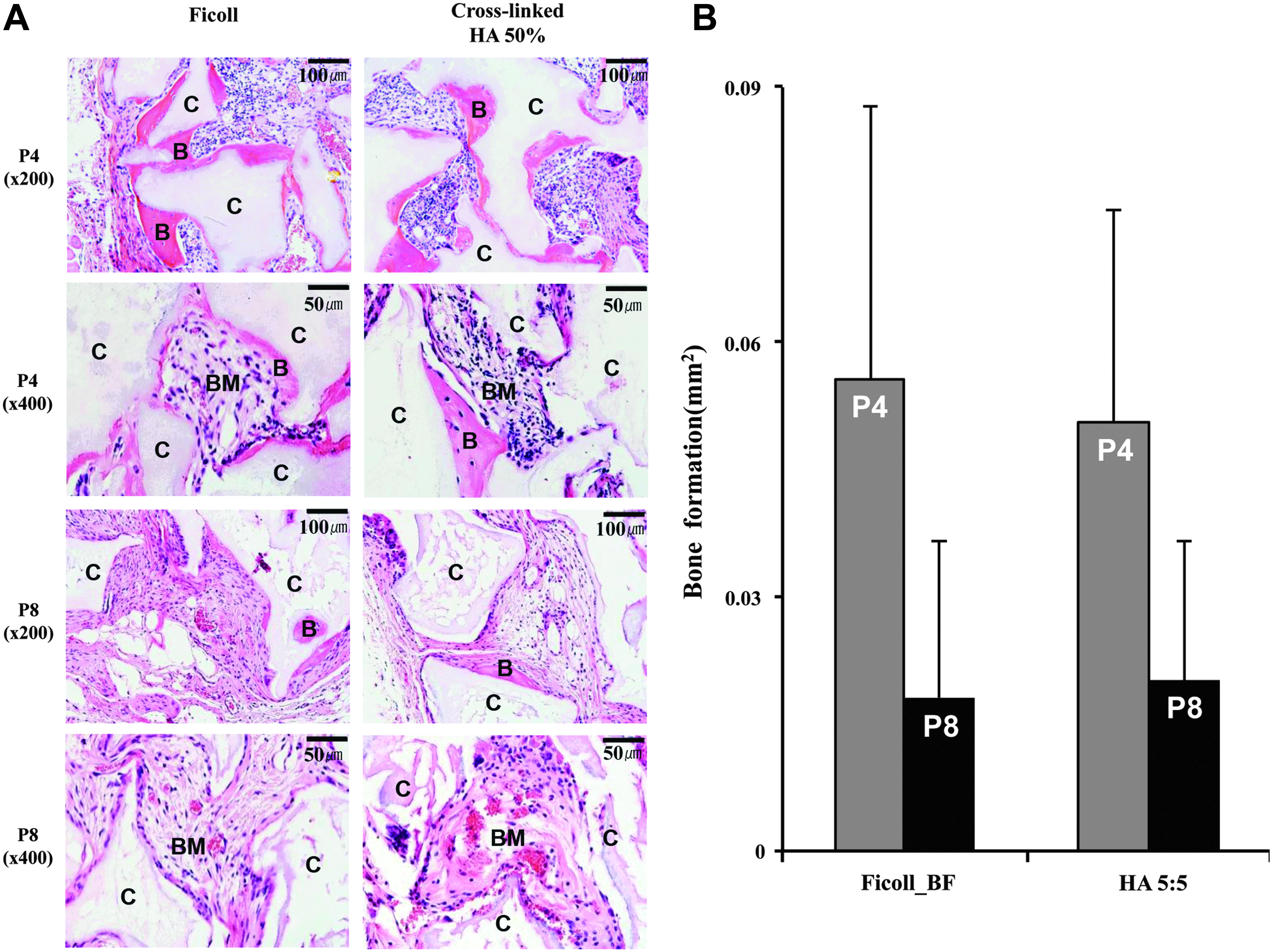
Histological analysis of ectopic transplantation of BMSCs isolated using Ficoll and 50% cross-linked HA.
Discussion
The novel HA-mediated isolation technique described herein is easy to implement directly in clinical applications, and it provides advantages of being less time-consuming and involving steps that are easier to perform compared with the conventional method using a density gradient solution. In the conventional technique, the layer for MNC acquisition is too thin to allow it to be removed separately and it is also sandwiched between the other layers. In addition, the thorough washing-out step required to avoid contamination with the density gradient solution includes a possibility resulting in substantial loss of the MNC layer. In contrast, in our novel technique the MNCs can be easily isolated from the most superficial layer of HA, and this layer could be used directly in clinical applications without requiring additional purification steps.
The present study has shown that the biological characteristics of BMSCs isolated using HA are similar to those of BMSCs isolated using gradient solutions, including in terms of their morphology, proliferation, cell-cycle status, adhesion, immunophenotype, differentiation potential, and in vivo biological capacity in ectopic transplantation models. This reflects the regenerative potential of the BMSCs isolated using HA as stem cells, because the efficacy of BMSCs isolated with conventional protocols has been widely researched in regenerative medicine using cell transplantation.39–41
The degree of crosslinking is one of several key factors that influence changes in the physical properties of HA. HA consists of repeating disaccharides of glucuronic acid and N-acetyl-
The density of HA (0.03–0.2 g/mL) is lower than those of BM and their component cells (WBCs, 1.06–1.08 g/mL; platelets, 1.05–1.07 g/mL; and red blood cells, 1.1 g/mL).45,46 When the BM was applied over the HA layer and centrifugated in the present study, the HA layer of the superficial layer included WBCs, which have an intermediate density. These results further indicate the presence of adhesion between HA and WBCs. Subjecting the HA layer to a large centrifugal acceleration resulted in a poor cell yield due to the weak interactions between HA and the cells. However, when the HA layer was subject to a centrifugal acceleration that was too small (less than 530 g), the HA and BM remained as a suspended solution that did not divide into multiple layers (data not shown).
The physiological function of HA is well established, especially in terms of the CD44 receptors. 47 Zhu et al. showed that CD44+/+ BMSCs adhered strongly to HA, whereas CD44−/− BMSCs did not. 48 We therefore propose that CD44+/+ cells are selectively picked by HA-mediated methods and also confirm that the WBCs present in the HA layer after the immediate isolation step will stain positive against CD44. Binding of CD44 on the surface of MSCs with the CD44 receptor on HA enhances the expression of HA synthases, and this results in a thick HA coat surrounding MSCs. MSCs ultimately maintain their stemness due to the presence of this thick HA coat. 49 Furthermore, HA accelerates wound closure by the CD44 receptor and induces early-stage wound healing.33,48,50 Therefore, the clinical use of MSCs immediately after their isolation using our novel HA-mediated method can show promises in regenerative medicine applications.
Conclusions
The present study investigated a novel isolation technique for MSCs using HA and evaluated its efficacy compared to the conventional technique using a density gradient solution. The cell yield was dependent on the centrifugal acceleration, HA volume, and crosslinking structure. While applying the novel isolation method with optimized conditions produced a significantly smaller number of MNCs, their characteristics were similar to those obtained using the conventional technique with a density gradient solution. In addition, it is directly and clinically applicable with no additional loss of cells because of no need for an additional processing to prevent contamination by the density gradient solution. Therefore, this novel technique is a candidate MSC isolation method for the direct clinical application of autogenous MSCs with the advantages of being less time-consuming and involving steps that are easy to perform. Further studies are needed to determine optimized conditions for MNC isolation to increase the cell yield.
Footnotes
Acknowledgments
This work was supported by the National Research Foundation of Korea (NRF) grant funded by the Korea government (MSIP) (No. 2015R1A2A1A15053961) and was also supported by the Bio & Medical Technology Development Program of the NRF funded by the Korean government, MSIP (No. 2012M3A9B2052521 and 2012M3A9C6049862).
Role of Sponsor
The funding organization played no role in the design of the study, the choice of enrolled patients, the review and interpretation of data, or the preparation or approval of the manuscript.
Disclosure Statement
No competing financial interest exist.
References
Supplementary Material
Please find the following supplemental material available below.
For Open Access articles published under a Creative Commons License, all supplemental material carries the same license as the article it is associated with.
For non-Open Access articles published, all supplemental material carries a non-exclusive license, and permission requests for re-use of supplemental material or any part of supplemental material shall be sent directly to the copyright owner as specified in the copyright notice associated with the article.