
Abstract
Traditional methods of cartilage tissue engineering rely on the use of scaffolds. Although successful chondrogenesis has been reported in scaffold-based constructs, the use of exogenous materials has limited their application due to eliciting host immunogenic responses and potentially resulting in construct failure. As a result, tissue engineering approaches, which aim to generate scaffold-free cartilaginous constructs, have become of particular interest. Here, we generated stable three-dimensional scaffold-free cartilaginous constructs by cultivating expanded pediatric nasal chondrocyte multilayers in a slow turning lateral vessel bioreactor system under chemically defined media. Bioreactor cultivation resulted in increased construct cellularity, fourfold tissue thickness, and 200% sulfated glycosaminoglycan deposition with respect to static culture equivalent cultures. These improvements led to significantly enhanced mechanical and biochemical properties of bioreactor-cultivated constructs, allowing them to support their own weight, while static culture constructs remained fragile. Consequently, bioreactor-cultivated constructs closely resembled native nasal cartilage tissue histologically, mechanically, and biochemically. We propose that this method of cartilage construct formation could be used to obtain readily available human scaffold-free cartilaginous constructs.
Introduction
C
Traditionally, cartilage tissue engineering has often utilized scaffolds, in vivo grafting procedures, or animal cells.7–17 Although scaffolds can provide 3D structural information and aid in initial construct shape retention, their use commonly prevents construct integration and can elicit host-induced inflammatory and immunogenic responses and lead to postoperative complications.18,19 Alternatively, in vivo cartilage regeneration has yielded satisfactory results; however, it offers very limited experimental control and causes a severe burden on the patient due to prolonged subcutaneous grafting procedures required, and often does not offer three-dimensional (3D) shape specificity.1,8,12–14 Previous studies investigating scaffold-free cartilage tissue engineering have revealed that it is possible to generate scaffold-free cartilaginous tissue constructs in vitro; however, common methods demand complex and expensive preparation, and/or the use of animal model cells.15–17 Animal models have played a critical role in moving the field of tissue engineering forward, however, to ensure clinical application, human cells must be used. In this study, to address all three of these limitations together, we have used human chondrocytes in a simple layering approach with subsequent slow turning lateral vessel (STLV) bioreactor cultivation, a simple and conservative method for generating stable, 3D, and scaffold-free human cartilaginous constructs in vitro.
Materials and Methods
Pediatric nasoseptal chondrocyte harvest, isolation, and culture
This study was approved by the institutions' research ethics board. Ten nasoseptal cartilage samples that would have otherwise been discarded were obtained from male and female children 6 to 14 years of age. Four of these samples were sent for native tissue mechanical and biochemical analyses, two for native tissue histological and immunohistochemical analyses, and the other four were utilized for tissue engineering experiments. Cartilage samples were freed of surrounding soft tissue and washed with phosphate-buffered saline containing 1X antibiotic/antimycotic (Wisent Bio Products, Quebec, Canada), minced into 3 mm by 3 mm pieces, and digested with Dulbecco's Modified Eagle's medium (DMEM) containing 1X antibiotic/antimycotic and 0.25% collagenase type II (Worthington Biochemical Corporation, Lakewood, NJ, USA) overnight at 37°C and 5% carbon dioxide (CO2). Digests were passed through a 100 μm pore size cell strainer (Falcon, Franklin Lakes, NJ, USA), centrifuged, washed, and centrifuged again to obtain a small cell pellet free of digestive enzymes and undigested tissue. Viable cells were counted using a hemocytometer and the trypan blue dye exclusion method.
Chondrocyte multilayering
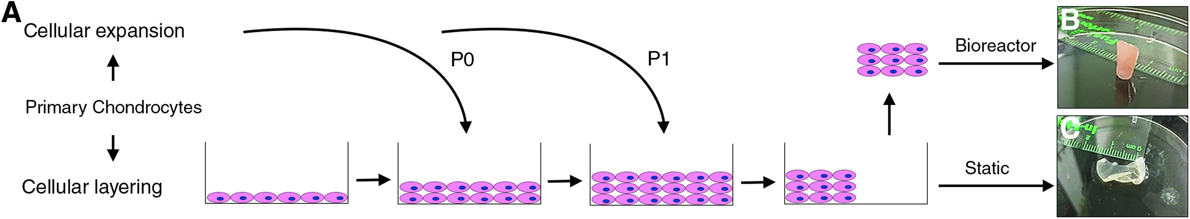
This process is outlined in Figure 1A. Freshly isolated nasal chondrocytes were split into two groups: (1) chondrogenic and (2) expansion. In the chondrogenic group, 2 × 10⁵ freshly isolated chondrocytes were seeded in a well of a six-well plate and were treated with DMEM/F12 supplemented with 1x ITS premix (Life Technologies), 0.1 μM dexamethasone, and 50 mg/mL ascorbic acid (Sigma-Aldrich, Oakville, ON, Canada). The expansion group cells were plated at a density of 5 × 10³ cells/cm2 in DMEM/F12 supplemented with 10% fetal bovine serum (Wisent Bio Products) and 1X antibiotic/antimycotic mix at 37°C and 5% CO2. On confluency, cells were trypsinized using 0.05% trypsin (Life Technologies), washed, counted, and 1 × 10⁶ cells were subcultured on top of the chondrogenic group, creating a layered culture system. The remaining cells were passaged at a 1:3 ratio. In total, two sequential layerings were performed, generating multilayers consisting of P0 and P1 expanded cells, respectively. 14 Following these layerings, the multilayer was lifted off the plate using a flat utensil and cut in half using a scalpel and one half was placed in the STLV bioreactor (Synthecon, Inc., Houston, TX, USA) in a chondrogenic medium consisting of DMEM/F12 supplemented with 1x ITS premix, 10 ng/mL transforming growth factor-β1, 0.1 μM dexamethasone, 50 mg/mL ascorbic acid, and 1X antibiotic/antimycotic mix at 37°C and 5% CO2, maintained under 15 × 10−3 g force. The other half was kept in the plate and cultured under static conditions using the same chondrogenic medium as that in the STLV at 37°C and 5% CO2, and was not attached to the plate to allow diffusion from both sides, similar to bioreactor conditions. Both treatments were carried out for 3 weeks, during which the medium was changed every other day for static cultures and once every 4 days for bioreactor constructs. In these 3 weeks, occasional monolayers, which would grow out of the static culture multilayers, were scraped off and suctioned out to disallow monolayer signaling within the well.
Dynamic rotational bioreactor
The rotary cell culture system utilizes an STLV, developed by National Aeronautics and Space Administration, to investigate zero-gravity conditions on tissues. This bioreactor provides a means of 3D culture in which the medium is in constant rotation, suspending the construct, mimicking a free floating (microgravity) culture condition, and thus providing fluid flow-enhanced flow diffusion. 20
Mechanical testing
Mechanical properties of bioreactor and static culture constructs as well as native pediatric nasal cartilage tissue were measured. Tissue thickness was determined using a digital caliper to measure three random locations of the constructs. Mechanical testing was performed using a Mach-1 Micromechanical Testing system (Biomomentum, Laval, QC, Canada) equipped with a 1 kg load cell. Tissue mechanical properties (bulk and elastic moduli at 37°C in DMEM/Ham's F12 media) were determined using a double compressive indentation method using two plane-ended indenters (2 and 4 mm diameter). Compressive indentations were conducted at a ramp rate of 10% strain/s to a maximum of 10% strain. The resulting force–deformation response (collected at a frequency of 10 Hz) was then used to determine the bulk and elastic moduli of native as well as engineered nasoseptal cartilage samples using a custom-designed code based on the theoretical model of cartilage indentation. 15
Histology
Cartilage tissue samples were processed for histology and immunohistochemistry after fixation in 10% formalin for 48 h. Histology and immunohistochemistry were performed on transverse sections of constructs to highlight thickness difference between bioreactor and static culture constructs. Tissues were then embedded in paraffin, sectioned into 5 μm sections, and placed onto treated microscope slides. Sections were deparaffinized by three 10-min xylene washes, hydrated through a graded series of ethanol (100%, 70%, and 50%) until brought into ddH2O. Slides were then stained with safranin O, toluidine blue, and alcian blue, which stain sulfated glycosaminoglycans (sGAG) red/orange, purple, and blue, respectively.
Immunohistochemistry
After deparaffinization and hydration, sections were subjected to 10 mM sodium citrate pH 6.0 antigen retrieval at 100°C for 15 min. Endogenous peroxidase activity was quenched with 3% hydrogen peroxidase in H2O treatment for 15 min. Tissue was then digested for 15 min with preheated (37°C) Pepsin digest mix (Life Technologies) and proteinase k (Sigma-Aldrich). Nonspecific binding was quenched via a blocking step with 5% bovine serum albumin blocking step for 1 h. Sections were then incubated with primary antibodies against collagen type I (Ab6308 at 1:400 dilution; Abcam, Cambridge, MA, USA), collagen type II (MAB8887 at 1:200 dilution, Millipore, Etobicoke, Canada), aggrecan (969D4D11 at 1:50 dilution; Thermo Fisher Scientific, Toronto, ON, Canada), or collagen type X (ab49945 at 1–1000 dilution; Abcam, Cambridge, MA, USA) at 4°C overnight, followed by the VECTASTAIN anti-mouse ABC kit conjugated with horseradish peroxidase (HRP) (Vector Laboratories, Inc., Burlingame, CA, USA). Finally, sections were incubated with the HRP substrate diaminobenzidine (DAB; Vector Laboratories) for 5 min. After DAB, sections were counterstained with Mayer's hematoxylin for 40 s, dehydrated via alcohol and xylene washes, and mounted.
Biochemical analysis
Biochemical properties of bioreactor and static culture constructs as well as native pediatric nasal cartilage tissue were measured. Weighed constructs were digested by papain (bioreactor samples: 40 μg/mL, native tissue samples: 80 μg/mL, in 20 mM ammonium acetate, 1 mM EDTA, and 2 mM dithiothreitol) for 72 h at 65°C. Aliquots of the digest were assayed for DNA and proteoglycan and collagen content. DNA content was quantified using Hoechst 33258 dye (Sigma-Aldrich) assay. The proteoglycan content was quantified by identifying the amount of sulfated glycosaminoglycans using 1,9-dimethylmethylene blue dye binding assay (Sigma-Aldrich). Total collagen content was quantified by identifying hydroxyproline content. Briefly, aliquots of the papain digest were hydrolyzed in 6 N HCl for 18 h at 110°C, and the hydroxyproline content was determined in the hydrolyzate using chloramine-T/Ehrlich's reagent assay. Total collagen content was estimated assuming that hydroxyproline accounts for 10% of the total collagen mass in cartilage. 15
Statistical analyses
All data are expressed as mean ± standard error of the mean. Samples from different patients were not mixed at any point in the study. Student's two-tailed t-test was used to compare means between bioreactor and static culture constructs, bioreactor constructs and native tissue, and static culture constructs and native cartilage tissue, for each mechanical and biochemical parameter, and p < 0.05 was considered significant.
Results
Physical properties of scaffold-free nasal cartilage constructs
After 3 weeks of bioreactor cultivation under chondrogenic conditions, a typical time frame for inducing chondrogenesis in vitro, 17 multilayers had developed into cylindrical 3D, opaque, cartilaginous constructs that did not deform under their own weight, whereas static culture counterparts remained clear and fragile (Fig. 1B, C). Bioreactor cultivation generated tissue constructs with 140–170% higher mass than static culture counterparts (Fig. 2A). Bioreactor constructs also displayed mechanical stability with a thickness of 0.90 ± 0.12 mm (50–72% of native tissue; p < 0.05), while static culture conditions resulted in fragile constructs with only 0.23 mm ±0.05 thickness (12–18% of native tissue; p < 0.05) (Fig. 2B). Comparing these measurements, STLV facilitated the formation of tissue-engineered construct, inducing a 290% increase in tissue thickness over traditional static culture (p < 0.05). As such, bioreactor-cultivated constructs exhibited similar bulk and elastic moduli to native tissue (65% and 66%, respectively), whereas static culture constructs did not display any mechanical stability (2% and 3% bulk and elastic moduli, respectively, of native pediatric cartilage) (Fig. 2C, D).
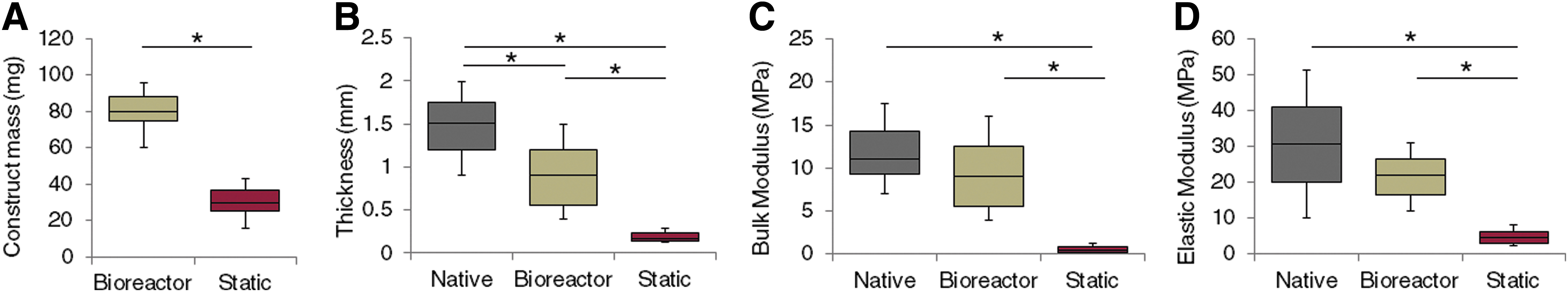
Physical properties of native pediatric nasal cartilage, as well as bioreactor- and static culture-generated cartilaginous constructs.
Histological and immunohistochemical evaluations of engineered constructs
Both bioreactor and static culture conditions demonstrated low levels of collagen type I (Figs. 3A–C and 4A–C) and high levels of collagen type II (Figs. 3D–F and 4D–F). In addition, bioreactor-cultivated constructs also exhibited regional staining resembling that of fibrotic perichondrium with the presence of Col I, and absence of Col II and sGAG, outlined in Figure 3A, B, and labeled as “P.” This was typically present on both sides surrounding the cartilaginous zone, however, more prominently the outer surface of the cylindrical construct that was most exposed to fluid flow. Statically cultured constructs exhibited a uniform matrix profile throughout (Fig. 3). Safranin O, toluidine blue, and alcian blue staining revealed that bioreactor-cultivated constructs showed the presence of high levels of sGAG, whereas static culture conditions showed very minimal staining (Figs. 3G–O and 4G–O). Additional staining of aggrecan and collagen type X reveals further similarities between bioreactor-generated cartilaginous constructs and native pediatric nasal cartilage (Figs. 3P–U and 4P–U). Analyzing cellular shape and organization via higher magnification images revealed that bioreactor-cultivated constructs consisted of more organized cell distributions in which the cells resided in large lacunae and abundant isogenous groups, indicative of cartilage tissue, whereas static culture constructs showed chaotic cellular organization, with far fewer cells in lacunae and isogenous groups (Fig. 4).
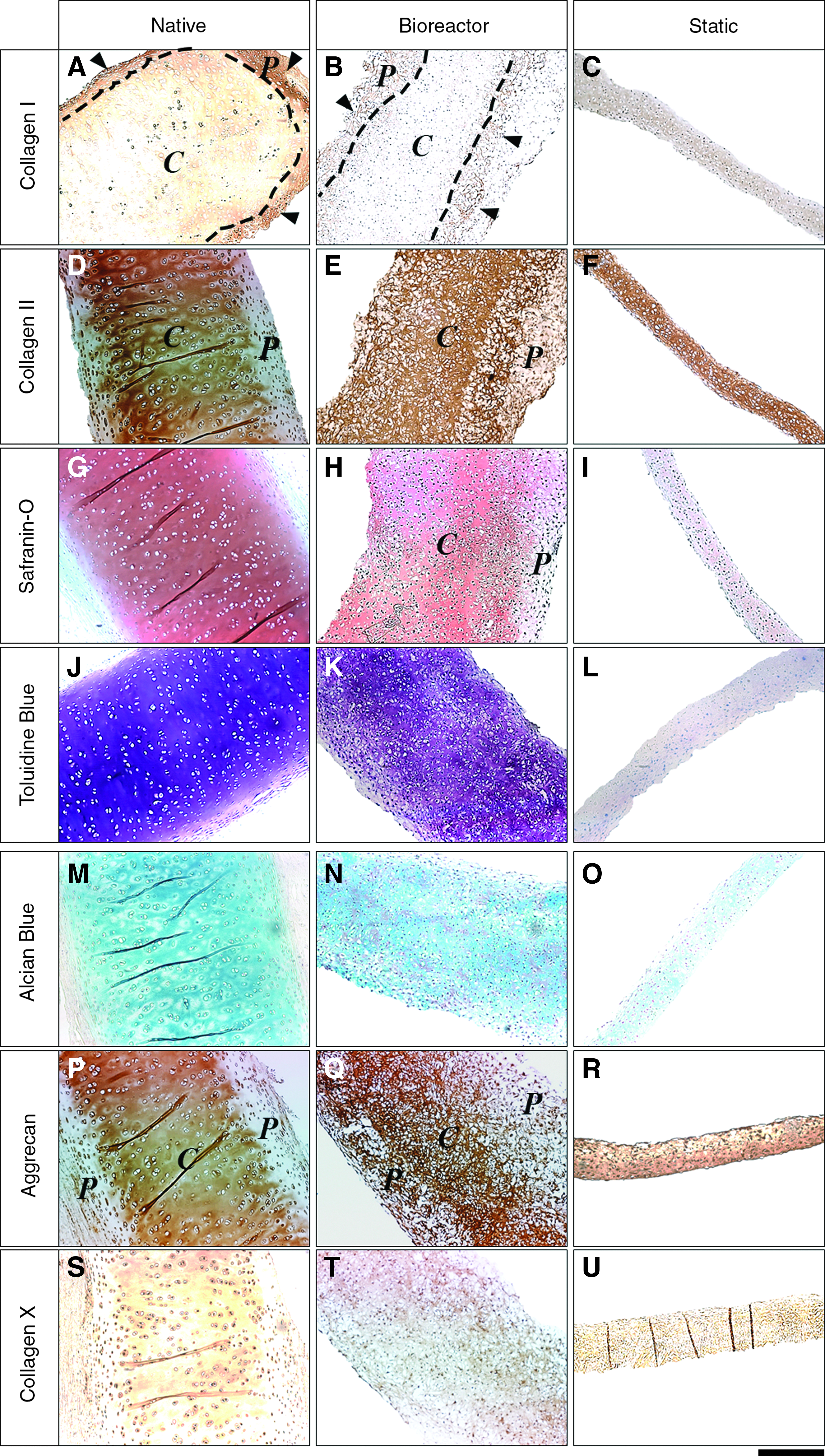
Histological and immunohistochemical comparison of native pediatric nasal cartilage with STLV bioreactor- and static culture-generated scaffold-free cartilaginous constructs.
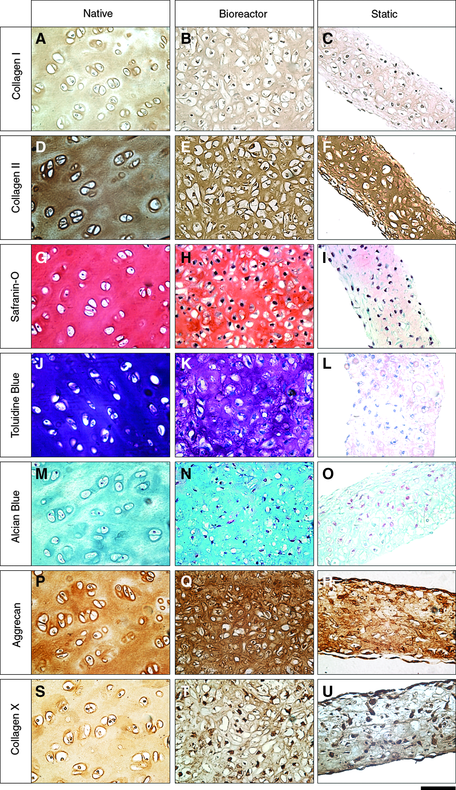
High-magnification histological and immunohistochemical comparison of native pediatric nasal cartilage with STLV bioreactor- and static culture-generated scaffold-free cartilaginous constructs.
Biochemical properties of native and engineered nasal cartilage constructs
Bioreactor cultivation resulted in a lower DNA/wet weight ratio (not significant), and 33–55% increase in total DNA content/construct (p < 0.05) (Fig. 5A, B). It is important to note that this difference was not due to cell death in static culture constructs, because using average construct masses (Fig. 2A), DNA/wet weight quantification (Fig. 5A), and a conversion factor of 6.6 pg of DNA per diploid human cell, 21 we identified that in static cultures total cell counts had increased by 15–25% from the original number of cells seeded in the multilayer. Bioreactor cultivation also resulted in a 200% increase in sGAG content compared to static culture normalized to construct weight (p < 0.05) (Fig. 5C). On normalization of sGAG content to DNA, to reveal the amount of sGAG produced per cell, it became clearer that chondrocytes within the bioreactor significantly secreted more sGAGs (Fig. 5D). In addition, no significant differences in collagen content were identified between the two conditions (Fig. 5E, F).
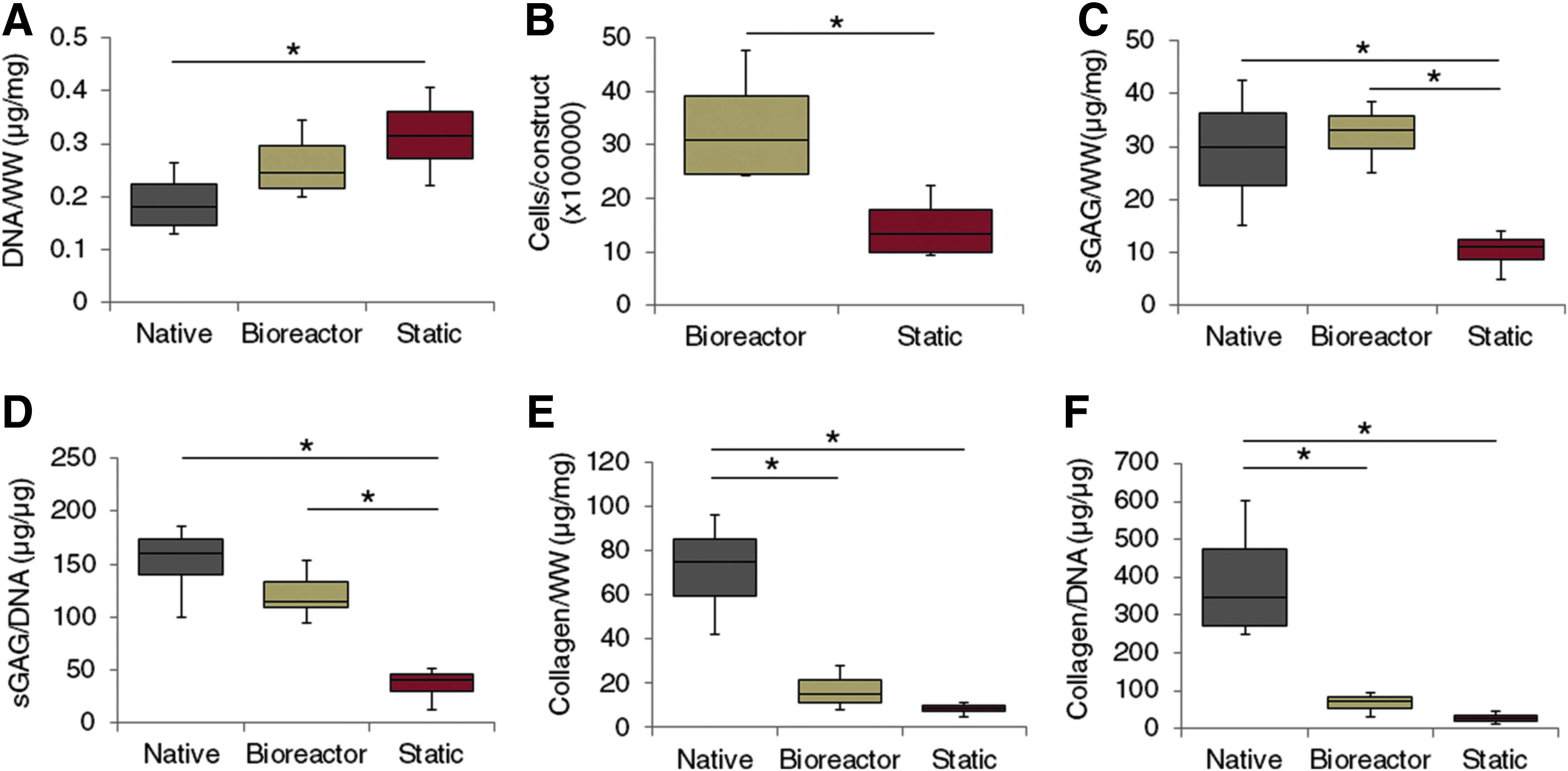
Biochemical quantification of native pediatric nasal cartilage with STLV bioreactor- and static culture-generated scaffold-free cartilaginous constructs.
Discussion
Finding robust methods of obtaining autologous tissue constructs has become a major focus in tissue engineering. However, generating functional tissue constructs ex vivo is challenging due to limited diffusion. To address this, bioreactors have been designed to facilitate tissue regeneration in vitro by providing fluid flow-enhanced diffusion.22,23 However, without the use of a scaffold, introducing cells to dynamic culture is often complicated as cells would have a limited support system. In this study, a multilayering approach was utilized to obtain a stable enough multilayer to be able to introduce into the bioreactor, as well as to help redifferentiate chondrocytes. A similar layering approach has been previously reported on auricular chondrocytes by Yanaga et al., where multilayers were aggregated and subcutaneously implanted into patients to develop the cartilaginous tissue. 14 In this study, we show that it is possible to develop mechanically stable cartilaginous constructs from scaffold-free chondrocyte multilayers, in vitro, via dynamic rotational culture, dispensing the need for subcutaneous grafting.
Typically, cartilage regeneration relies on the chondrogenic differentiation of a predetermined number of chondrocytes. Here, by introducing a very thin and fragile multilayer within the STLV bioreactor, we observe that chondrocytes further expand and differentiate within the bioreactor, and develop into thicker and more stable constructs not only via chondrogenesis but also via cellular expansion. We believe that this is primarily due to fluid flow-enhanced diffusion within the bioreactor. Giardini-Rosa et al. used an entirely different continuous flow bioreactor which demands large amounts of medium and maintenance to report cellular expansion and chondrogenesis simultaneously, although their report lacked static culture controls. 15 Previous reports investigating chondrogenesis using the STLV bioreactor system have reported enhanced chondrogenesis; however, they have typically utilized animal cells, exogenous materials, or pellet culture systems that offer little clinical relevance.11,24–26 In this study, our system solely comprised cells and intrinsic extracellular matrix (ECM), offering direct clinical application if used under good manufacturing practice.
Bioreactor cultivation resulted in significantly thicker tissue constructs with higher bulk and elastic moduli when compared to static culture counterparts. Since both conditions promoted cartilage-specific collagen expression profiles with high levels of collagen type II and aggrecan, along with low levels of collagens type I and X, similar to that observed in native cartilage, the differences in rigidity and mechanical stability are likely due to the significant increase of sGAG content and thickness in bioreactor constructs. This result is congruent with the literature, as it is known that proteoglycans provide mechanical resilience to cartilage. 27 It is important to note that although collagen/weight of bioreactor and static cultures were not statistically different, the amount of collage per surface area being tested would still be considerably more in bioreactor constructs due to the difference in thickness, which is likely the reason why bioreactor constructs displayed higher elastic moduli when compared to static cultures.
Although previous studies have demonstrated that sGAG production is enhanced by increasing cellular density, this could be problematic since increasing cell densities while maintaining limited diffusion could induce hypoxia within the center of the 3D construct and lead to necrosis and chemotaxis, ultimately debilitating matrix deposition and cartilaginous cartilage formation. 28 Finding a method that allows the development of higher cellular densities may be crucial for tissue regeneration. In this study, the seeding density was kept minimal during the in vitro layering process so that constructs could adjust cellularity to that permitted by the fluid flow-enhanced diffusion in the STLV bioreactor system. The fluid flow within the bioreactor enhances transport of nutrients and dissolved gasses by convection, allowing for the survival of higher cell numbers. Increase of cellularity within the bioreactor was confirmed by an overall 30–55% increase in DNA content compared to static culture constructs, whereas DNA/wet weight was not significantly different. As a consequence, bioreactor constructs also developed into much thicker tissue. Morphological evidence for this cellular expansion suggests interstitial growth, as numerous chondrocyte isogenous groups are present in bioreactor-cultivated constructs in contrast to those in static culture. In addition, the constant flow of medium against the construct also introduces fluid-induced shear stresses. Consequently, due to this shear stress and the fragile nature of the in vitro multilayers, the constructs adopted a cylindrical shape during the cultivation period. Although high amounts of shear stress have been associated with inducing osteoarthritis phenotype in engineered tissue (>1.6 Pa), 29 the shear stress induced by the STLV (∼0.15 Pa) is too little to elicit such responses. 30
Aside from characterizing physical conditions that promote chondrogenic differentiation, there is also an ongoing effort to optimize chemically defined chondrogenic medium for nasoseptal chondrogenesis. It remains to be explored whether additional chondrogenic factors such as bone morphogenetic protein-2 and protein-7, or insulin-like growth factor-1 and growth differentiating factor-5, which have been shown to enhance human nasal chondrogenesis, could further accelerate expansion and enhance chondrogenic differentiation in the STLV bioreactor system.31,32 Subsequently, it is crucial to assess the feasibility and long-term stability of scaffold-free cartilage tissue constructs in vivo via a subcutaneous xenografting approach in nude mice. This will aim to study construct–host integration and stability postimplantation, both crucial to the final stages of translating the results to the clinic. Studies investigating premature cartilage construct implantations have reported successful construct maturation and integration.1,9,14,33 We believe that such observations support the idea that our constructs could be readily translated to the clinic with predictable improved outcomes when including a step of prior maturation within the rotational bioreactor.
Finally, it is important to note that the pediatric source of the nasal chondrocytes used might have played a key role in their ability to generate sufficient ECM in vitro to allow construct mechanical stability within the bioreactor. Additional experimentation is required to identify whether or not adult-derived chondrocytes could behave similarly.
Conclusions
Human, scaffold-free cartilaginous tissue constructs exhibiting mechanical stability can be generated via cultivation of pediatric nasal chondrocyte multilayers in a dynamic STLV bioreactor system. This method of cartilaginous construct formation could potentially be applied clinically to generate tissue for use in major augmentative and reconstructive surgery.
Footnotes
Acknowledgments
Funding for this work was provided by the Department of Otolaryngology—Head and Neck Surgery, Transplant Center, and Division of Urology, all from the Hospital for Sick Children, Toronto, Canada.
Disclosure Statement
No competing financial interests exist.