
Abstract
Engineered heart tissues made from human pluripotent stem cell-derived cardiomyocytes have been used for modeling cardiac pathologies, screening new therapeutics, and providing replacement cardiac tissue. Current methods measure the functional performance of engineered heart tissue by their twitch force and beating frequency, typically obtained by optical measurements. In this article, we describe a novel method for assessing twitch force and beating frequency of engineered heart tissue using magnetic field sensing, which enables multiple tissues to be measured simultaneously. The tissues are formed as thin structures suspended between two silicone posts, where one post is rigid and another is flexible and contains an embedded magnet. When the tissue contracts it causes the flexible post to bend in proportion to its twitch force. We measured the bending of the post using giant magnetoresistive (GMR) sensors located underneath a 24-well plate containing the tissues. We validated the accuracy of the readings from the GMR sensors against optical measurements. We demonstrated the utility and sensitivity of our approach by testing the effects of three concentrations of isoproterenol and verapamil on twitch force and beating frequency in real-time, parallel experiments. This system should be scalable beyond the 24-well format, enabling greater automation in assessing the contractile function of cardiomyocytes in a tissue-engineered environment.
Introduction
H
Several devices have been developed to grow engineered heart tissue and measure either beating frequency, or both beating frequency and twitch force.20,21 Frequency-only systems include devices that measure beating rates of tissues in microfluidic channels 22 or devices that use microelectrode arrays. 23 There are also several devices that can measure twitch forces of tissue-like structures in vitro, including sheets of cells on flexible flaps,24,25 sheets of cells on force transducers, 26 tissues on polyacrylamide gels,27,28 and tissues suspended between load transducers29,30 or polymeric posts.31,32 Engineered heart tissues suspended between flexible, silicone posts enable one to detect the twitch forces generated by the tissues.33,34 With silicone posts, some aspects of measuring twitch force and beating frequency can be automated. However, in our experience, they require dedicated microscopy equipment and custom image analysis routines to ensure accurate and consistent results.
In this article, we describe a novel tool for measuring the beating frequency and twitch force of myocardial tissues in situ through the use of magnetic field sensing. The system consists of engineered heart tissues suspended between silicone posts, where one post has an embedded magnet in its tip and the other is rigid from an embedded glass capillary tube (Fig. 1a). Tissue constructs on posts are placed into a 24-well tissue culture plate with giant magnetoresistive (GMR) sensors positioned below the plates. When a tissue construct beats, the force of its contraction cause the flexible post to deflect, thereby moving the position of the embedded magnet. The movement of the embedded magnet causes a voltage change at the GMR sensor, which in turn, is used to measure the force and frequency of the contractions.
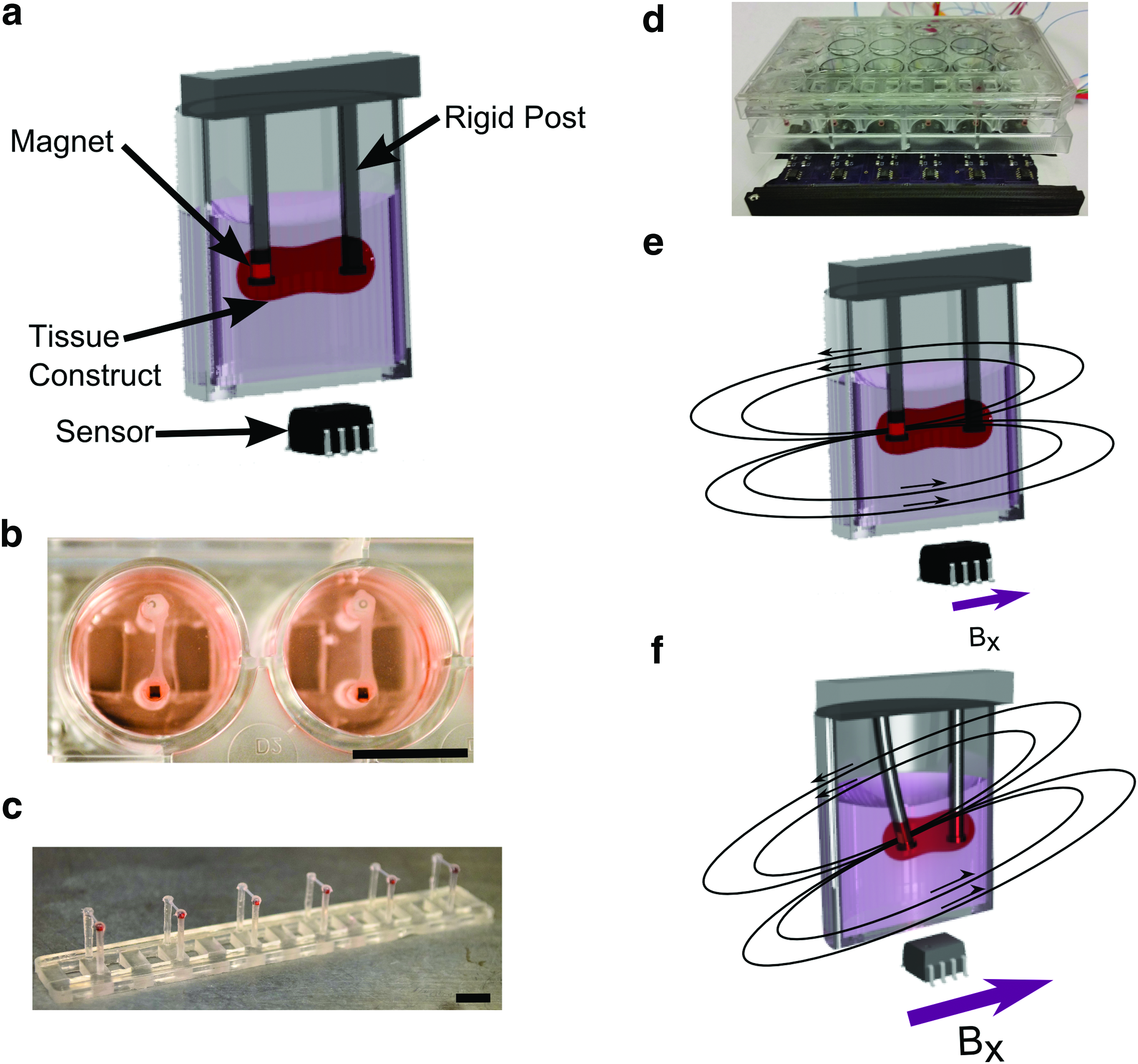
Magnetic sensing used to determine twitch force and beating frequency in engineered heart tissue.
Materials and Methods
Post fabrication
The posts were constructed using polydimethylsiloxane (PDMS, Sylgard 184; Dow Corning, Midland, MI) in a custom mold (Limited Productions, Inc., Bellevue, WA). First, a small amount of PDMS was poured into the mold and baked at 65°C to form the caps at the tips of the posts. After forming the caps, 1 mm3 cubic magnets (MyMagnetMan, Largo, FL) were placed into sections of the mold that formed the flexible posts. Glass capillary tubes (1.1 mm diameter; Drummond Scientific Co., Broomall, PA) were placed into sections of the mold that formed the rigid posts. PDMS was then poured into the entire mold and cured overnight at 65°C. External magnets were used to hold the embedded magnets in place and maintain their orientation at the bottom of the posts while the PDMS cured. After the posts were removed from the mold, excess PDMS was trimmed away, yielding silicone posts that were 12.5 mm tall (including the 0.5 mm tall caps), 1.5 mm diameter, and spaced 8 mm apart (Fig. 1b, c).
Magnetic sensor array fabrication
GMR sensors (AAH002-02; NVE, Eden Prairie, MN) were arranged on printed circuit boards (OSH Park, Lake Oswego, OR) with other circuitry for filtering and amplifying the signal (Supplementary Fig. S1; Supplementary Data are available online at
Magnetic model
A model of the system was developed in Matlab (Mathworks, Natick, MA) by treating the embedded magnet as a point dipole. A dipole approximation was used because the distance from the magnet to the sensor was much larger than the size of the magnet. The dipole of a magnet in a silicone post was determined experimentally to have a strength of
where
Device characterization
Devices were characterized by deflecting individual posts with a linear micro-positioner (SM-50; Newport Corp., Irvine, CA). The deflection of each post was viewed on an optical microscope (LV-100; Nikon, Tokyo, Japan) with a camera (OrcaFlash 2.8; Hamamatsu, Hamamatsu City, Japan). Images were recorded simultaneously with voltage readings from each GMR sensor using LabView software. Five different magnetic posts were used to characterize each sensor. The data were used to obtain a calibration constant for voltage-to-position measurements using linear-regression analysis with a least squares approach. The same configuration for characterization was used for all subsequent experiments with engineered heart tissue.
Engineered heart tissue generation
Undifferentiated human induced pluripotent stem cells (hiPSCs) from the IMR90 line, were maintained in mTeSR (Stemcell Technologies, Vancouver, Canada) on tissue culture dishes coated with Matrigel (Corning, Corning, NY). An established protocol for directed differentiation was used to derive cardiomyocytes (CMs), as previously described. 36 Differentiation yielded greater than 80% cardiomyocytes that were positive for cardiac troponin T by flow cytometry. hiPSC-CMs were cast into engineered heart tissue on day 24 after induction of differentiation. Tissue constructs were generated following a similar protocol to Schaaf et al., 37 where each tissue was cast from a 100 μL fibrin gel solution containing 5 × 10⁵ hiPSC-CMs and 5 × 10⁴ supporting stromal cells (line HS-27a 38 ). Agarose wells were formed by pouring a 2% agarose solution into a 24-well plate fitted with custom built spacers. A pair of posts was positioned into each agarose well, and the cell-laden gel solution was poured into the well to create a tissue construct bound onto the tips of each post. After 80 min of time for gelation, the newly formed tissues were transferred to a fresh 24-well plate. Engineered heart tissues were maintained in a 24-well plate and were supplied with fresh RPMI media (11875-119; Invitrogen, Carlsbad, CA), supplemented with B-27 plus insulin (17504044; Life Technologies, Carlsbad, CA), and 5 mg/mL aminocaproic acid (A2504; Sigma-Aldrich, St. Louis, MO), three times per week. The tissue constructs were maintained in culture for 2 weeks before testing, for sufficient compaction and development of a stable beating pattern. From our observations, tissues achieve a maximal force after 2–3 weeks in culture. We did not observe any muscle fatigue in the tissues during the course of our experiments.
Frequency and force plotting
Voltage outputs from the magnetic sensors were recorded using LabView and displayed on a screen for real-time monitoring during experiments. Further analysis was performed on all experiments to assess the frequency and magnitude of contractions over time. The data were filtered with a low-pass filter to remove measurement noise using an eighth order Butterworth filter with a cutoff frequency of 7 Hz. Only the peak-to-peak amplitudes were analyzed for force measurements, so low-frequency fluctuations in baseline readings were eliminated from the signal. To eliminate low-frequency baseline drifts, an exponential moving average filter with an exponent constant of 0.0001 was used.
After filtering the data, custom-written code in Matlab for peak finding was used to identify the maxima and minima of the data to calculate twitch force and beating frequency. Twitch force was determined by subtracting the voltage at a maximum from an adjacent minimum, as long as the maximum and minimum were within 2 s of each other, and then converting from voltage to force based upon the calibration of the GMR sensor and the bending stiffness of the silicone posts. Beating frequency was determined based on the time between maxima.
A cutoff reading of 7 mV was chosen based on analysis of the signal produced by nonbeating tissues. Below this threshold, there was an increased chance of registering a false beat due to noise in the system. Furthermore, during drug testing, a tissue was determined to have stopped beating when the frequency of beats fell below 0.2 Hz. To reduce errors due to a disturbance in the posts during the addition of reagents, a 4-s window of time was removed from the averages for both twitch force and beating frequency. A combined 10-s moving average of the twitch force and beating frequency was obtained for each of the four experiments.
Pacing experiments
To compare the waveforms of tissues measured magnetically to tissues measured optically, tissues were paced at 1.5 and 2 Hz in both platforms. Both measurements were taken with the tissues and sensors at 37°C. Carbon rods were used as electrodes and affixed with PDMS to the bottom of 24-well plate dishes. Platinum wire was wrapped around the electrodes, and connected to a stimulator (S88X; Astro-Med, Inc., West Warwick, RI). Biphasic pacing was used with a pulse duration of 5 ms and pulse amplitude of 5 V for both conditions. The tissues were observed to follow the electrical stimulation and produced twitch forces with consistent amplitude during pacing. Optical measurements were taken with an inverted microscope (Eclipse Ti; Nikon, Inc., Melville, NY) using a CCD camera (Clara, Andor, Belfast, Northern Ireland). The position of the flexible post as a function of time was determined using custom-written code for image analysis developed in Matlab. Determination of the location of the post using magnetic sensing was performed using the methods described above.
Pharmacological testing
Experiments were performed using verapamil hydrochloride (CAS 152-11-4; Tocris Bioscience, Bristol, United Kingdom) and isoproterenol hydrochloride (CAS 5984-95-2; Sigma-Aldrich, St. Louis, MO). Verapamil is a calcium channel blocker, 39 while isoproterenol is a β-adrenergic agonist, 40 and both alter twitch force and beating frequency in the adult heart. All tissues were treated with 50 μL of a mixture of the relevant drug in DI water. A batch mixture of 2.5 mM of each drug in DI water was passed through a sterile filter and portioned into the appropriate dilution based on the final concentration. The media volume in each well during the experiment was 2.5 mL, and 50 mL of the drug solution was added to yield the desired final concentration of the drug in the complete media. The control case had no treatment added to the media during the experiment. The sham control had 50 μL of DI water added to the media. All experiments with drugs were performed at 37°C. Drugs were washed out with new media and tissues were allowed to reset for 2 days before undergoing further treatments. The twitch force and frequency was measured for four tissues per condition.
Results
Engineered heart tissue fabricated onto magnetic post arrays
The twitch forces and beating frequency of engineered heart tissue have been measured previously using pairs of silicone posts, where both posts deflect when a tissue undergoes a twitch contraction.33,34 Here, we built a similar array of posts, but where one post was flexible and magnetic, while the other was rigid and nonmagnetic. When a tissue contracts, it causes a deflection in the magnetic post with negligible deflection in the rigid post (Fig. 1e, f). The deflection of the magnetic post changes the magnetic field at the GMR sensor, which reports the change as voltage signal. Posts were made out of polydimethylsiloxane (PDMS) and were evenly spaced as an array for insertion into a 24-well plate (Fig. 1b, c). Caps on each post were used to prevent the detachment of the tissues during culture. Based upon the dimensions of the posts and its behavior as a cantilever, there is a linear relationship between the deflection of the post and the force of the tissue. The dimensions of the post, along with a Young's modulus of 2.5 MPa for PDMS, resulted in a bending stiffness of 0.98 μN/μm, calculated by assuming the net tissue force acts at the tip of the post, and using the method described by Schoen et al. 41 Thus, we developed an approach where twitch force of an engineered heart tissue causes its magnetic post to deflect linearly, which can be detected by the reading of the GMR sensor underneath the culture dish.
Magnetic sensing of post deflections
GMR sensors were soldered to printed circuit boards containing circuitry for high-pass filters and instrumentation amplifiers. After signal conditioning, the voltage reading from the GMR sensors were passed to a data acquisition system running custom software to record the deflections of the magnetic posts. In addition to recording the data, it was possible to monitor the deflection in real-time.
The location with the highest measurement sensitivity for the GMR sensors was determined by modeling of the magnetic field produced by the neodymium magnet. Specifically, we analyzed the change in the magnetic field due to a 300 μm deflection in a magnetic post (Fig. 2a), which is on the larger end of observed post deflections. Our results indicated that in a layout for a 24-well culture plate, there is minimal crosstalk between the magnetic post and its neighboring sensors in adjacent wells, for this 300 μm deflection. In addition, the optimal location for the GMR sensor in the plane 14 mm below the TC dish could be determined with this model, which was approximately a 3.5 mm offset from the point directly below the magnetic post (Fig. 2b). This offset location was due to the translation and rotation of the magnetic dipole during the post motion. The model further predicts a linear response for displacements up to 2 mm, which indicates that the upper limit in a reading is 1.96 mN (Supplementary Fig. S2). Based upon a cutoff reading of 7 mV, the lower limit is 26 μN. Thus, these results demonstrate that there is an ideal location for the magnetic sensor and that there is little interference in detecting the beating frequencies and twitch forces in tissues that neighbor each other in a 24-well plate.
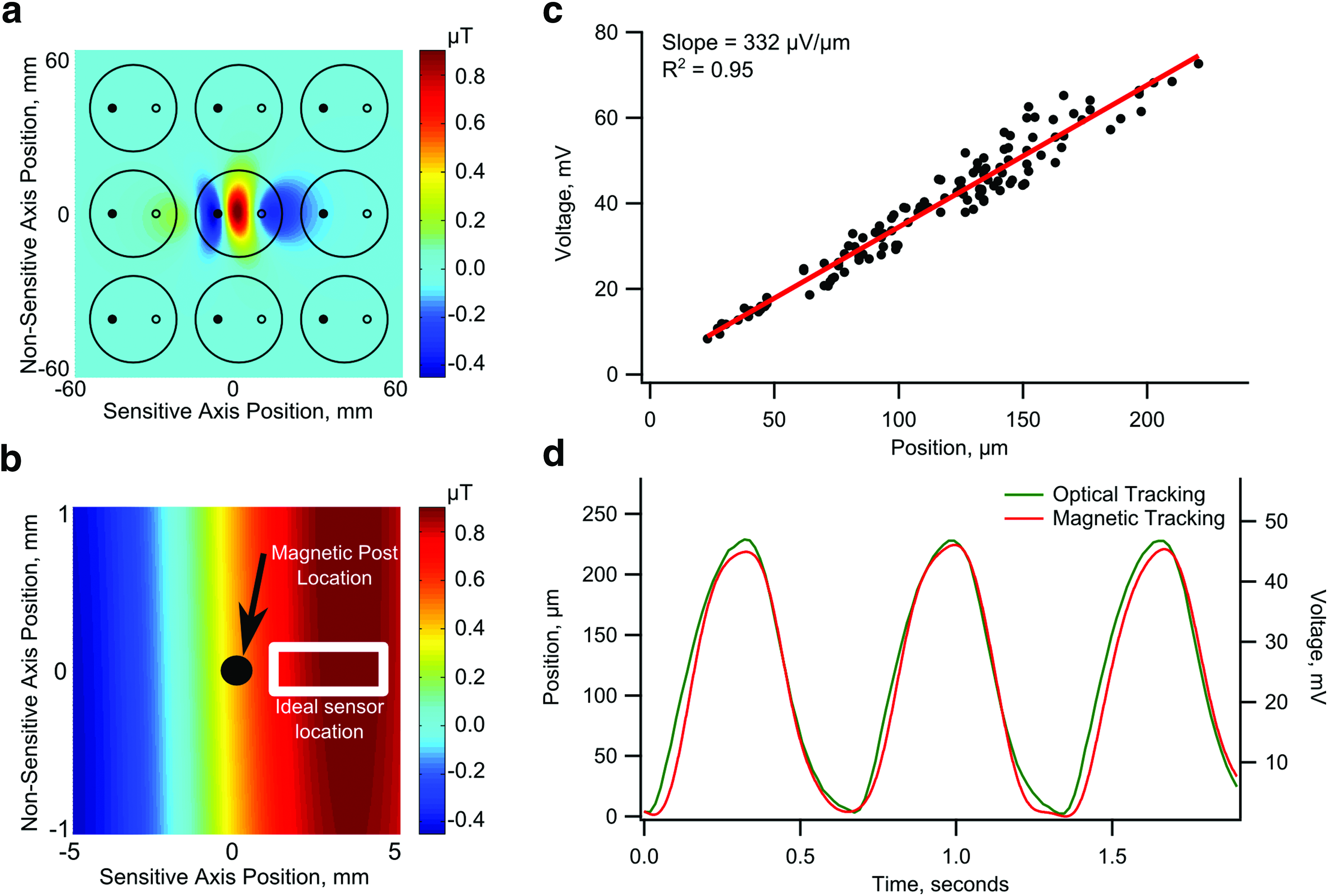
Calibration of magnetic sensing.
Characterization of magnetic sensing
A custom manipulator system was built to move the posts manually while tracking the position and measuring the voltage changes. Due to a 0.16 Hz cutoff frequency on the high-pass filters integrated into the circuit design, dynamic motion of the posts was required to properly calibrate the system. We calibrated the relationship between the motion of the posts and the voltage output by placing the arrays under a reflective microscope. Posts were dynamically moved using a linear micro-positioner while simultaneously monitoring the voltage outputs.
Five different arrays of magnetic posts were used to calibrate the system (Supplementary Fig. S3). The trend for each sensor was quite linear (R2 >0.90) and produced a signal within the expected range based on the models and the expected motions of the posts with engineered heart tissue (Fig. 2c). Each sensor had a different calibration coefficient, likely due to variability in the sensor fabrication, circuitry, and layout of the sensors on the device. Although the sensors did have minor variations, the trend for each individual sensor was linear. Hence, this characterization showed that we can model the force of each tissue by directly relating the voltage change to post deflection.
Comparing optical and magnetic readings of engineered heart tissue
Engineered heart tissues containing hiPSC-CMs and stromal cells spanned between the magnetic and rigid PDMS post pairs within 24-well tissue culture dishes. After 2 weeks of culture, the tissue constructs were beating with uniform and stable contractions, which deflected the tip of the flexible posts between 100 and 300 μm during each twitch. Each tissue construct showed consistent force production when electrically paced. Within a 10-min window, the contractions of a tissue construct were recorded under optical microscopy and then transferred to the GMR sensors for magnetic tracking. A strong correlation of the waveform for the deflection of the post was observed between optical tracking and magnetic tracking (Fig. 2d). This result demonstrated that the magnetic system is suitable for measuring the beating frequency and twitch force as optical tracking.
Pharmacological testing
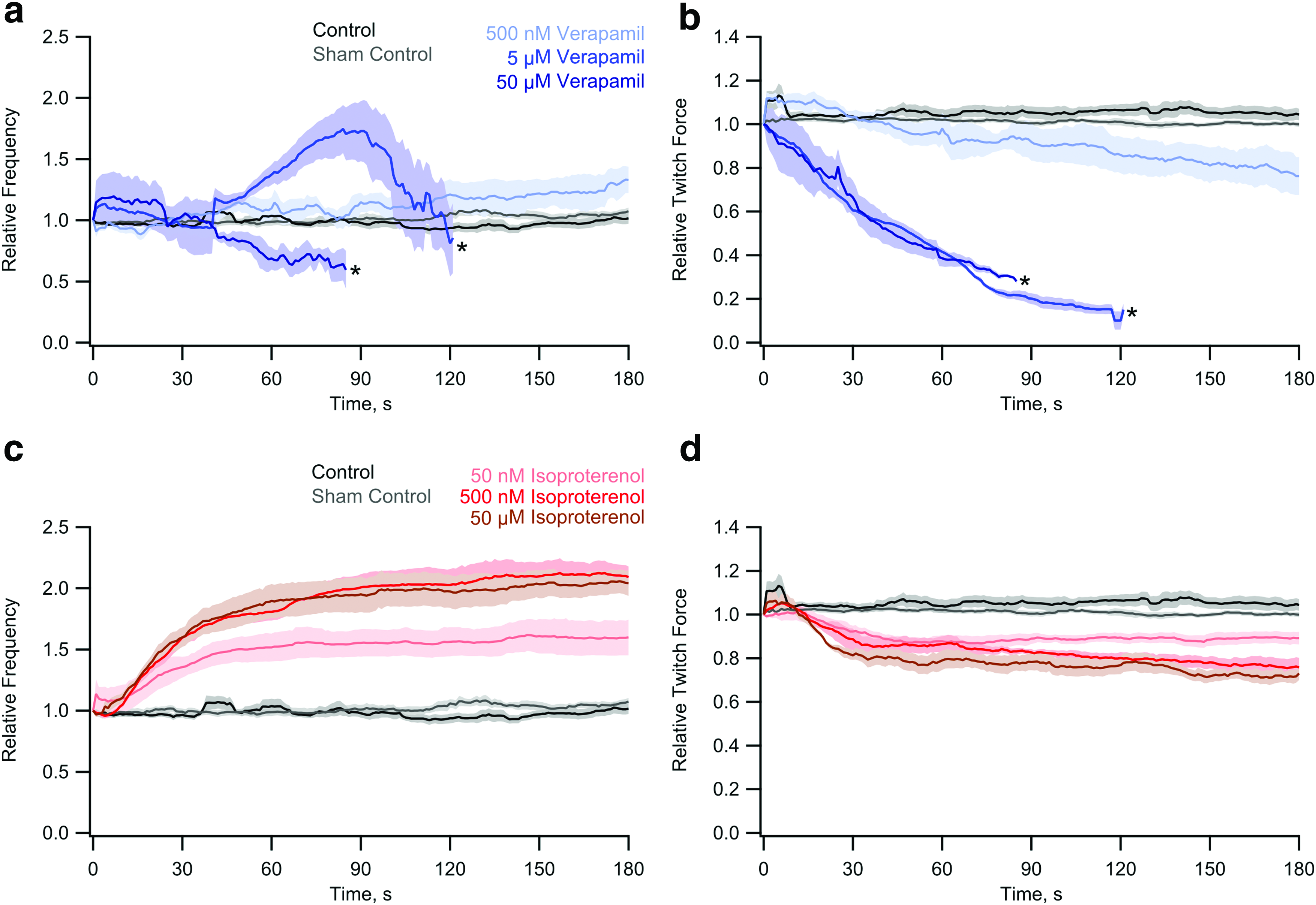
One of the benefits of this device is the ability to simultaneously study the effects of drugs on the twitch force and beating frequency of tissues in real time. The ability for parallel studies was demonstrated using three concentrations of both isoproterenol and verapamil. Isoproterenol, a β-adrenergic agonist, and verapamil, a calcium channel blocker, were chosen for their well-studied effects on cardiomyocytes. In general, verapamil lowers twitch forces, 42 while isoproterenol increases beating frequency and, for adult cardiomyocytes, beating force. 40 All of the tissues were measured on the magnetic sensors for 1 min before adding drugs to the system. Twitch force and beating frequency of the tissues was monitored for at least 3 min after the addition of drug or vehicle. We noted that there was variation in the baseline twitch force and beating frequency between tissues before the addition of drugs.
In order to compare the effects of the drugs, relative twitch force and beating frequency was used to determine the percent change of each tissue over time. Each study generated complete waveforms of the twitch contractions over the course of at least 3 min (Fig. 3). A set of tissues were not given any drug as a control or given water as a sham control. In both cases, the spontaneous beating of the tissues had little variation throughout the experiments.
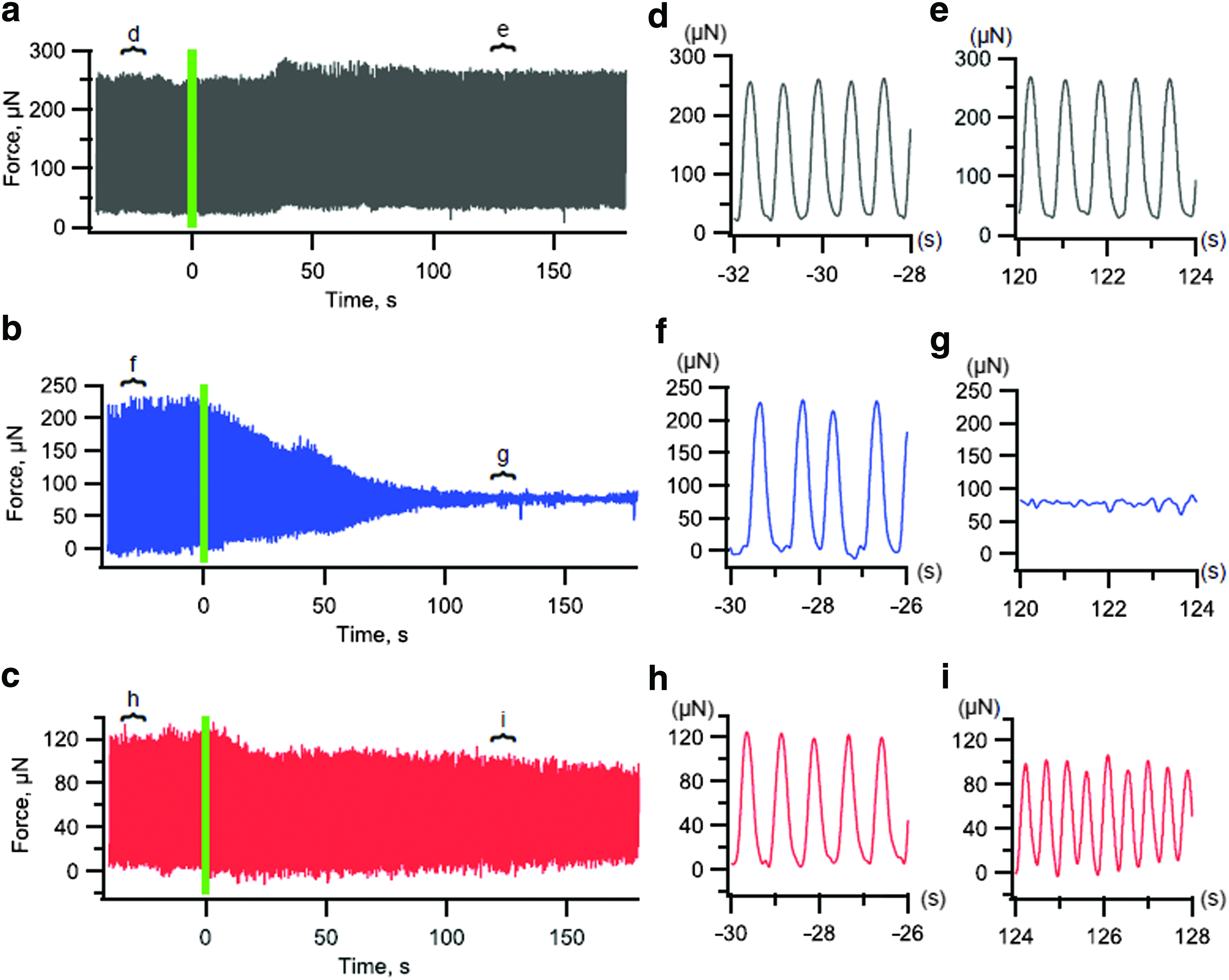
Real-time analysis of tissue forces (representative force-time profiles) in response to pharmacological compounds.
Verapamil had a large effect on both the beating frequency and the force generation of the tissue constructs (Fig. 4). Increasing concentrations of verapamil caused lower forces and lower frequencies. The effect of verapamil on force was nearly immediate, although there typically was an initial increase in beating frequency before it was reduced. A low dosage of verapamil caused a slight increase of the beating frequency from the baseline frequency. There were some fairly large variations in the verapamil beating frequency and force magnitude, some of which can be attributed to the choice of a 7 mV cutoff for force generation. This was due to the amount of noise in the system.
Combined data from multiple experiments with verapamil and isoproterenol.
As expected, isoproterenol increased the beating frequency as the concentration was increased. The increase in beating frequency started quickly, and appeared to level off within 2 min after adding the drug, (Fig. 4). A decrease in contractile force was observed with increasing concentrations of isoproterenol. The decrease in force is typical for immature myocardium, which has not yet expressed phospholamban, the Ca2+-regulatory protein that is targeted by beta-adrenergic stimulation.33,43,44 These results indicated that our system is sensitive to changes in twitch force and frequency to measure the effects of drugs and could be used in a drug screening assay.
Discussion
In this article we have developed a novel platform for real-time magnetic measurements of twitch forces and beating frequencies of engineered heart tissues derived from hiPSC-CMs. We designed our system so that the tissue constructs spanned between a pair of silicone posts, where one was rigid and another was flexible and contains an embedded magnet. The motion of the magnet was tracked using a GMR sensor located underneath a 24-well plate to determine the force of the tissue. We characterized the system against optical tracking of the post deflection and showed a linear response of voltage to tissue force. Finally, we used different concentrations of isoproterenol and verapamil to demonstrate the utility of the device to measure twitch force and frequency in real-time parallel experiments.
Several tools have been developed for monitoring the beating frequency and twitch force production of EHTs, but thus far, none have produced simple measurements to enable real-time contractile information. Previous devices have looked into electrical measurements of force using PDMS devices, 45 and other devices have embedded magnetic material into PDMS cantilever devices.46,47 Few devices have used magnetic field changes to measure forces, 48 and we believe that this is the first device to measure tissue forces using embedded magnets in a PDMS device. The device provides some key advantages as it is able to continuously monitor the forces of several tissue constructs throughout an experiment. Real-time monitoring can ensure that tissues are behaving properly and it can aid in performing massively parallel experiments. In this article, the system was limited to six simultaneous measurements, but, as shown in Figure 2a, the system could be expanded to simultaneously monitor an entire 24-well plate. Even with only six simultaneous measurements, four arrays of posts were tested with two controls and four measurements in rapid succession without the need to position the posts or find the posts in a microscope objective. This rapid screening can be useful for high-throughput, low-cost monitoring of engineered heart tissues for drug screening or tissue replacement applications.
To demonstrate the utility of this device, two different drugs were used to alter the frequency and force of tissue contractions. Verapamil was chosen as a calcium channel blocker to reduce contractile force and frequency while isoproterenol was chosen as a β-adrenergic agonist with the expected effect of increasing contraction frequency. We found verapamil to lower both contractile force and frequency, and temporarily stop the tissue construct from beating. Furthermore, we found that the frequency of contractions increased before the contractions ceased to beat with detectable forces for higher concentrations of verapamil. For a low concentration of verapamil, we found that the frequency increased, while the magnitude of forces had a slight decrease. The increased frequency and lowered force response of EHTs to verapamil has been seen before,49–51 and is now able to be measured in situ and in real time.
Isoproterenol increased the frequency along the same order as what had been seen previously.27,52 The response was a steady over the course of ∼90 s. After 90 s, the response leveled off to the new frequency and twitch force. Somewhat surprisingly, the force of contractions decreased, however, it has been shown that EHTs derived from induced pluripotent stem cells have different responses than that of native heart tissues. 53 The structure of induced pluripotent stem cells used in these experiments is likely much more immature than adult cardiomyocytes, 44 and it has been shown that immature cardiomyocyte tissues do not produce as strong a force response to isoproterenol. 43
This system was used to simultaneously measure the response of six different tissues at a time, and four replicates were easily measured in the time it took the tissues to respond to the forces. Little adjustment or calibration was necessary once the device was built, and postprocessing analysis could easily be integrated into the data acquisition program to produce immediate results from experiments using this system. Furthermore, the tissue constructs and multi-well dishes are all disposable, while the sensors and electronics are free from contacting the biological media, and thus do not require sterilization between each use. This enables testing of devices over time for studies that have slower-acting drugs, or to monitor the twitch forces of tissues in an incubator over time. This system can be used to either rapidly screen for a number of different drugs or to monitor the contractile forces of EHTs being developed for potential therapeutic replacements.
To expand the utility of the device for rapid and simultaneous measurements of twitch force and frequency, some aspects of the device presented here would need improvement. Currently, this system is able to measure twitch forces, but is not able to measure passive tension, that is, when the tissue is in diastole, due to the high-pass filter. The system could alter the filtering design to measure passive tension of the tissues. In addition, placement of sensors could be further improved to reduce variance between each sensor. These improvements can be engineered to provide more information about cardiomyocytes and provide an inexpensive and autonomous platform for functional assessment of hiPSC-CMs or other cardiomyocytes.
Footnotes
Acknowledgments
The authors wish to acknowledge support from the National Science Foundation (CBET-1509105, CMMI-1402673 to N.J.S.), the National Institutes of Health (F32 HL126332 to A.L.; R01 HL084642, P01 HL094374, P01 GM81619, U01 HL100405 to C.E.M.), and the Fondation Leducq Transatlantic Network of Excellence (to C.E.M.).
Disclosure Statement
K.S.B. has equity in Stasys Medical Corporation. C.E.M. is a cofounder and has equity in BEAT Biotherapeutics. N.J.S. is a cofounder and has equity in Stasys Medical Corporation.
References
Supplementary Material
Please find the following supplemental material available below.
For Open Access articles published under a Creative Commons License, all supplemental material carries the same license as the article it is associated with.
For non-Open Access articles published, all supplemental material carries a non-exclusive license, and permission requests for re-use of supplemental material or any part of supplemental material shall be sent directly to the copyright owner as specified in the copyright notice associated with the article.