
Abstract
Three-dimensional (3D)-printed constructs made of polycaprolactone and chondrocyte-impregnated alginate hydrogel (hybrid cartilage constructs) can mimic the biphasic nature of articular cartilage, thus offering promise for cartilage tissue engineering applications. Notably, the regulatory pathway for medical device development requires validation of such constructs through in vitro bench tests and in vivo preclinical examinations for premarket approval. For this, noninvasive imaging techniques are required for effective evaluation of the progress of these cartilage constructs, especially when implanted in animal models or human subjects. However, characterization of the individual components of the hybrid cartilage constructs and their associated time-dependent structural changes by currently available noninvasive techniques is challenging as these constructs contain a combination of hydrophobic and hydrophilic biomaterials with different refractive indices. In this study, we report the use of a novel synchrotron radiation inline phase contrast imaging computed tomography (SR-inline-PCI-CT) approach for noninvasive (in situ) characterization of 3D-printed hybrid cartilage constructs that has been implanted subcutaneously in mice over a 21-day period. In parallel, traditional invasive assays were used to evaluate the in vivo performance of the implanted hybrid cartilage constructs with respect to their cell viability and secretion of cartilage-specific extracellular matrix over the 21-day period postimplantation in mice. SR-inline-PCI-CT allowed striking visualization of the individual components within the 3D-printed hybrid cartilage constructs, as well as characterization of the time-dependent structural changes after implantation. In addition, the relationship between the implanted constructs and the surrounding tissues was delineated. Furthermore, traditional assays showed that cell viability within the cartilage constructs was at least 70% at all three time points, and secretion of alcian blue- and collagen type 2-positive matrices increased progressively over the 21-day period postimplantation. Overall, these results demonstrate that the 3D-printed hybrid cartilage constructs have good in vivo performance and validate their potential for regeneration of articular cartilage in vivo. In addition, SR-inline-PCI-CT has demonstrated potential for longitudinal and noninvasive monitoring of the functionality of 3D-printed hybrid cartilage constructs in a way that is translatable to other soft tissue engineering applications.
Introduction
C
Confocal microscopy, optical coherence tomography, and Raman spectroscopy have been studied as alternatives to 2D imaging techniques for TE applications.11–14 Unfortunately, these techniques have inadequate penetration depth and often require the use of contrast agents to enhance their sensitivity.15–18 Positron emission tomography and single-photon emission computed tomography have high penetration depth and have been praised for enabling successful tracking of cells in vivo.19–21 However, these techniques require the use of contrast agents that might have a negative effect on cell performance. In addition, these techniques often experience poor temporal and spatial resolution that consequently limit visualization of microstructural details, which is critical to tracking time-dependent biomaterial degradation and tissue growth.19–21 Radiography (such as micro-CT and CT) can assess structural details within tissue constructs,22,23 but poor contrast from highly hydrophilic materials with low attenuation coefficients, such as cartilage and hydrogels, is their major drawback for CTE applications. 24 Magnetic resonance imaging (MRI) is an established preclinical and clinical technique, well known for its ability to delineate soft tissue contrast; therefore, it is often used for visualizing cartilage damage, tissue remodeling, soft tissue constructs, and, recently, with contrast agents to track cells in constructs.25–30 However, MRI has poor spatial resolution, provides poor imaging contrast for hydrophobic materials and tissues, and may require contrast agents to boost sensitivity. These drawbacks are the bottlenecks that make it challenging to use MRI for noninvasive characterization of thin growing neotissues and our multimaterial cartilage constructs.26,31 Overall, the trade-offs of currently available imaging techniques made their utilization challenging with respect to noninvasive characterization of the biphasic cartilage constructs investigated in this study and their time-dependent structural changes.
An effective noninvasive 3D imaging technique intending to monitor the functionality of the hybrid cartilage constructs should have the capability to (1) visualize the hydrophobic and the hydrophilic components of the hybrid cartilage constructs; (2) enable visualization of the progression of the newly forming neotissues within the constructs and the biodegradation profiles of all the components of the cartilage constructs; and (3) delineate the relationship between the cartilage constructs and their surrounding host tissues without destroying or posing any risk to the host animal model or human subject.16,18,30,32 Owing to the presence of multiple attenuation coefficients, densities, and other material properties (i.e., the combination of hydrophobic PCL and hydrophilic cell-impregnated alginate hydrogel) within the hybrid cartilage constructs, their noninvasive characterization is challenging.10,16,18,33 The novel method of synchrotron radiation inline phase contrast imaging computed tomography (SR-inline-PCI-CT) circumvents most of the limitations of the other imaging techniques discussed above and, therefore, have potentials for noninvasive monitoring of the 3D-printed hybrid cartilage constructs and their performance over time either in situ, ex vivo, or in vivo. Although at least six specific PCI techniques are being explored, SR-inline-PCI-CT has the simplest experimental setup, uses no optical element (e.g., gratings or diffracting crystals), and was the first phase-contrast technique to be pioneered.18,34–37 SR-inline-PCI-CT uses variations in phase shifts of X-rays passing through the samples to visualize materials with different refractive indices, electron densities, and atomic numbers present within the samples without requiring the use of exogenous contrast agents.18,34–36 At diagnostic X-ray energies, SR-inline-PCI-CT produces its image signal from refraction generated from the real part of the material refractive index, which is up to 1000 times greater than the absorption signal used by conventional absorption-based imaging techniques.34–36,38 Due to the high lateral (spatial) coherence of synchrotron X-rays, SR-inline-PCI-CT translates variations in densities and refractive indices of different materials into edge enhancement at their interfaces in the images. Thus, SR-inline-PCI-CT offers a robust capability to simultaneously characterize newly growing or native hard and soft tissues. SR-inline-PCI-CT can also visualize time-dependent structural changes in multiple biomaterials over time postimplantation.7,10,30,39–41 In relation to this present study, we have recently reported that SR-inline-PCI-CT enabled unparalleled and noninvasive visualization of the 3D-printed hybrid cartilage constructs submerged in fluid (to mimic physiological conditions) and their associated time-dependent subtle structural changes over a 42-day period of in vitro culture. 7
In the present study, we used in vivo evaluation of the 3D-printed hybrid cartilage constructs as therapeutics for damaged articular cartilage and assessment of their performance using both traditional invasive and synchrotron-based noninvasive techniques. 3D-printed hybrid cartilage constructs (PCL/alginate/cell constructs) were implanted subcutaneously in the backs of nude mice and tracked over a 21-day period. Invasive assessments of the functionality of these constructs demonstrated high cell viability and secretion of cartilage-specific ECM over the 21-day postimplantation in mice. Importantly, SR-inline-PCI-CT enabled in situ noninvasive characterization of the individual components of the 3D-printed hybrid cartilage constructs, their surrounding tissues, and associated structural variations over 21-day postimplantation in mice. Summarily, these data further validate the potentials of the 3D-printed hybrid cartilage constructs for CTE applications (this time in vivo) and proved that noninvasive longitudinal monitoring of the functionality and degradation of the multicomponent tissue constructs might be possible using SR-inline-PCI-CT.
Materials and Methods
Materials
PCL (average Mw ∼45,000), alginic acid sodium salt, medium viscosity alginate (MVA), mouse chondrogenic cell line ATDC5, calcium chloride dehydrate (CaCl2), Stemline® Keratinocyte Medium II-Calcium free (SKM), 4-(2-hydroxyethyl)-1-piperazineethanesulfonic acid (HEPES) buffer, phosphate buffer saline Tween-20 (PBST), and ethylenediaminetetraacetic acid (EDTA) were purchased from Sigma-Aldrich, St. Louis, MO. Dulbecco's modified Eagle's medium (DMEM)/Ham's F-12 (1:1), fetal bovine serum (FBS), penicillin, streptomycin, buprenorphine, glutamine and ascorbate-2-phosphate, and insulin-transferrin-selenium plus (ITS+) liquid media supplement were purchased from Life Technologies, Carlsbad, CA. Purified anti-Col2 antibody (CIIC1) was purchased from Developmental Studies Hybridoma Bank, Iowa City, IA, and goat anti-mouse IgG (H+L) secondary antibody and Alexa Fluor® 488 conjugate were purchased from EMD Millipore, Temecula, CA. Twenty adult male athymic immunodeficient nude (Crl: NU(NCr)-Foxn1nu) mice were purchased from Charles River Laboratories, NY.
Cell culture/expansion
Frozen ATDC5 cells were thawed and 2D cultured in tissue culture dishes for 1 week in a humidified incubator with 5% CO2 at 37°C. Culture media were changed every 2–3 days to enable cell expansion. The culture medium consisted of DMEM/Ham's F-12 (1:1) supplemented with 5% FBS, 100 U/mL penicillin, 100 μg/mL streptomycin, 10 mg/mL glutamine, and 0.05 mg/mL ascorbate-2-phosphate. After 1 week, expanded cells were collected from tissue culture plates, counted, and resuspended in culture medium for fabrication of the hybrid constructs.
Design and fabrication of 3D-printed hybrid cartilage constructs
Design and fabrication of the 3D-printed hybrid cartilage constructs were conducted as previously described.6,7 The geometry of the designed model of the constructs was 10 × 10 × 0.96 mm and consisted of four layers of cylindrical strands with 1 mm interstrand spacing and an alternating 0–90° perpendicular pattern from one layer to the other. For the fabrication, PCL pellets were loaded into the high-temperature dispensing head of a 3D Bioplotter system (EnvisionTEC, Germany), heated up to 150°C for 20 min to destroy potential microbial contamination and cooling to the dispensing temperature of 85°C. MVA was dissolved in SKM medium to obtain 3.3% w/v alginate hydrogel. Then, alginate-cell solution was prepared by suspending 8 × 10⁶ cells per millimeter of alginate hydrogel solution for the preparation of a final concentration of 2.5% w/v. The prepared alginate-cell solution was loaded into the low-temperature dispensing head and maintained at 10°C. During the printing, melted PCL strands were dispensed from a 300 μm inner diameter cylindrical metal needle at deposition speed (the same as the needle's horizontal speed) of 1 mm/s and a pneumatic pressure of 0.08 MPa, and cell-laden alginate strands were printed within two adjacent PCL strands from a 200 μm inner diameter conical needle at deposition speed of 25 mm/s and pneumatic pressure of 0.03 MPa. The side-by-side and layer-by-layer alternating deposition of PCL and alginate strands continued for each construct until four hybrid layers were printed. Strand thicknesses were 0.3 mm for the PCL strands and 0.2 mm for the alginate strands, respectively. After printing each layer, alginate strands were partially cross-linked using fumes released from a nebulizer, which contained 170 mM CaCl2 in 4.2 mM HEPES (in 0.35 M sucrose). Once a construct was completely printed, it was immediately transferred into 100 mM CaCl2 (in 4.2 mM HEPES and 0.35 M sucrose solution [pH 7.4]) for 20 min to further cross-link the alginate-cell strands within the constructs. After cross-linking, the constructs were washed in DMEM solution for 5 min twice and placed in 12-well culture plates with culture medium and, subsequently, transferred into an incubator operating at 37°C and 5% CO2. The culture medium was changed every 2 days to enhance cell differentiation and consisted of DMEM/Ham's F-12 (1:1) supplemented with 5% FBS, 100 U/mL penicillin, 100 μg/mL streptomycin, 10 mg/mL glutamine, 10 mg/mL ITS+ liquid media, and 0.05 mg/mL ascorbate-2-phosphate. The constructs were cultured in vitro for 2 weeks in the incubator to initiate cell differentiation before implantation into the mice. Hybrid scaffolds with no impregnated cells were also fabricated, as described above, to serve as controls.
Surgical implantation of 3D-printed hybrid cartilage constructs
The Animal Research Ethics Board of the University of Saskatchewan, SK, Canada, approved the protocol for implantation of the 3D-printed hybrid constructs in mice in compliance with guidelines for humane animal care set by the Canadian Council on Animal Care. Upon their arrival, the 20 adult male nude mice (Crl: NU(NCr)-Foxn1nu) were acclimatized for 2 weeks before scheduled surgical implantation. The mice were housed in groups of three in individual Plexiglass cages, lined with sterile sawdust in a room with a controlled photoperiod (lights on from 06:00 through 18:00), at a constant 21°C, and 60% humidity. The mice were handled aseptically, provided ad libitum with sterile water and standard mouse chow, and were randomly divided into control and treatment groups. Hybrid construct-implanted mice were prepared for (1) histological assessments and (2) in situ SR-inline-PCI-CT. For the surgery, the mice were anesthetized in a biosafety cabinet and kept warm using a heated pad. Small incisions were created along the backs of the mice for four constructs to be implanted in their dorsal subcutaneous pockets: two on the left and two on the right sides (Fig. 1). All four constructs implanted into the mice for histological analysis were cell laden, and the mice prepared for SR-inline-PCI-CT imaging were each implanted with two cell-laden constructs on the right and two cell-free hybrid constructs on the left sides. The animals were injected with 0.1 mg/kg subcutaneous (sc) of buprenorphine as an analgesic once immediately after the surgery, again 12 h postoperatively, and followed by its addition into their drinking water for a period of 1 week.
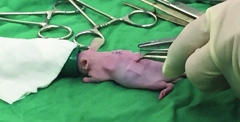
Image of a nude mouse undergoing surgical implantation of 3D-printed hybrid cartilage constructs. Subcutaneous pockets were created lateral to a midline incision, through which the constructs for the left and right sides were inserted. 3D, three-dimensional. Color images available online at
Invasive assessments of hybrid cartilage constructs
At each of the three time points (7, 14, and 21 days) postimplantation, three mice were sacrificed by decapitation for histological analysis of the 3D-printed cartilage constructs. All four cell-laden constructs were excised from the mice, and each construct was cut into four square sections, and each of them was used for the different histological and biological analyses, as described below (each assay had n = 12 per time point).
Cell viability of cells within the 3D-printed hybrid cartilage constructs
For the cell viability assay, two-color florescence LIVE/DEAD® Kit (Molecular Probes, OR) was used. At each time point, the samples were stained as previously described.6,7 Briefly, samples were washed with DMEM and submerged in staining solution containing 2 μM calcein-AM and 0.5 μM ethidium homodimer (EthD-1) in DMEM and kept in the dark for about 1 h in an incubator running at 37°C and 5% CO2. After staining, photomicrographs of the horizontal and cross-section views of the stained constructs were acquired to visualize live cells (fluorescing green) and dead cells (fluorescing red) using a DP70 camera attached to a Nikon fluorescent inverted microscope (Nikon, Eclipse E600, SPOT Insight™ Camera). For quantification of cell viability, constructs were submerged in 50 mM EDTA solution to dissolve the alginate strands and release the cells. Then, 10 μL of the cell suspension was gently pipetted onto a glass slide and covered with a coverslip to acquire photomicrographs of live and dead cells at different locations on the glass slide. Live and dead cell numbers were counted in randomly selected regions of interest cropped from photomicrographs in ImageJ software. 42
In vivo secretion of sulfated GAGs within 3D-printed hybrid cartilage constructs
To estimate secretion of sulfated GAGs in the excised hybrid constructs, samples were stained with alcian blue and analyzed as previously described.6,7 Briefly, the samples were washed in DMEM, fixed in acetone: methanol (1:1), submerged in 0.5 mg/mL alcian blue in 3% acetic acid (pH = 1), and kept on rocking tray overnight at room temperature. The stained constructs were then destained in 25% ethanol in 3% acetic acid for 1 h and stored in 50% ethanol in 3% acetic acid before acquisition of photomicrographs of the constructs. For quantitative analysis, randomly selected regions of interest within the alginate hydrogel strands were cropped for each of the three time points using ImageJ software. 42 An appropriate threshold was applied to segment the alcian blue-stained region from the background for each image. The measured values of the stained region in relation to the entire region of interest were used to calculate the area covered by alcian blue staining in the constructs at 7, 14, and 21 days postimplantation and used to quantify the secretion of sulfated GAGs at each time point.
In vivo secretion of Col2 within the 3D-printed hybrid cartilage constructs
Immunofluorescent staining was used for analysis of the progression of secretion of Col2 within the hybrid constructs at 7, 14, and 21 days postimplantation as previously described.6,7 The specimens were fixed, washed thrice in PBST for 15 min each time, washed in 0.05% EDTA in PBS, and digested in trypsin solution (0.1% trypsin, 1 mM EDTA, and 1 × PBS). After the trypsin step, constructs were incubated for 2 h in 1:100 purified anti-Col2 (CIIC1) antibody in blocking buffer (4% normal goat serum and 2% normal sheep serum in PBST). The constructs were then washed six to eight times in blocking buffer over a 2 h period. For the second incubation, 1:1000 goat anti-mouse IgG-488 conjugate in blocking buffer was used. The constructs were washed again in PBST over another 2 h period to minimize background stains. Subsequently, photomicrographs of the horizontal and cross-sectional views of stained constructs at different time points were taken using a DP70 camera, as described above. For quantitative analysis, randomly selected regions of interest within the alginate hydrogel strands were cropped from photomicrographs per time point in ImageJ. 42 An appropriate threshold was applied to segment the Col2-stained region within the alginate strands from the background for each image in ImageJ as previously described. 7 The measured values of stained region in relation to the region of interest were used to quantify the area covered by Col2 stain in the constructs at 7, 14, and 21 days postimplantation.
Noninvasive visualization of 3D-printed hybrid cartilage constructs in mice using SR-inline-PCI-CT
Noninvasive SR-inline-PCI-CT imaging of the 3D-printed hybrid constructs at 7 and 21 days postimplantation in mice was performed at the Biomedical Imaging and Therapy facility 05ID-2 beamline at the Canadian Light Source (CLS), Saskatoon, Canada. Sacrificed animals in intact condition were transported to the CLS on ice for noninvasive in situ imaging. The imaging setup consisted of a double crystal bent Laue monochromator tuned to 30 keV imaging energy and a superconducting wiggler X-ray source with a beam of 220 mm horizontal size and 11 mm vertical size at a distance of 55 m from the source. 43 The mice were placed in sample holders and positioned on a rotating scanning stage for CT scanning. Based on our previous data, 7 tomographic data sets were collected at a 3 m sample-to-detector-distance using a beam monitor AA-60 (Hamamatsu) coupled to a Hamamatsu camera C9300-124 with effective pixel size of 8.47 μm and exposure time between 0.03 and 0.06 s. For each data set, 3000 projections were collected over a 360° rotation, and a set of 10 flat-field and 10 dark-field images were acquired before and after each scan to correct the acquired projections. Two to three scans were required to capture the entire mouse in each case.
Before the image reconstruction, the flat- and the dark-field images were used for projection corrections with an ImageJ macro plugin (written by Dr. Cooper, College of Medicine, University of Saskatchewan, SK, Canada). Then, Modified Feldkamp Algorithm in NRecon V 1.6.10.1 (Bunker, Kontich, Belgium) was used for the nonretrieved image reconstruction to obtain image slices based on recommendations made in Ref. 7 The images were cropped and exported into FEI Amira 6.0.1 (Oregon) 3D visualization and analysis software for rotation and 3D volume rendering to further visualize the microarchitecture of the different components and minute details in the hybrid constructs within the mice. In addition, 3D visualization of the 3D-printed constructs in the mice was subsequently performed in ImageJ. 42
Statistical analysis
To evaluate changes in Col2-stained area, alcian blue-stained area, and cell viability within the 3D-printed cartilage constructs at 7, 14, and 21 days postimplantation in mice, a one-way repeated measures analysis of variance (RM-ANOVA) was carried out in SPSS (Released 2013 IBM SPSS Statistics for Windows, Version 21.0. Armonk, NY: IBM Corp.). Due to violation of sphericity by the alcian blue-staining data, a Greenhouse–Geisser correction was applied. Post hoc tests using the Bonferroni's correction were conducted to estimate the statistical significance between cell viability, areas covered by alcian blue stain, and areas covered by Col2 stain at the different time points. Statistical significance was set at p < 0.05 for all analyses conducted.
Results
Cells within the 3D-printed hybrid cartilage constructs remained viable over the 21-day period postimplantation in mice
To evaluate cell distribution and viability, 3D-printed hybrid cartilage constructs were harvested from the backs of mice at various time points and subjected to LIVE/DEAD stains. The results showed that uniform cell viability and cell distribution were maintained in both the periphery and the center of the alginate strands within the hybrid cartilage constructs over the 21-day postimplantation period (Fig. 2). Quantification confirmed that the cell viabilities were 83% ± 5.4% at 7 days postimplantation, 70% ± 6.6% at 14 days postimplantation, and 73% ± 7.9% at 21 days postimplantation (Fig. 2I). A one-way repeated measures ANOVA (assuming sphericity) demonstrated that cell viability was significantly different among time points (F (2, 18) = 505.719, p = 0.002). Specifically, post hoc tests showed that cell viability significantly differed between 7 and 14 days postimplantation (p > 0.001; Fig. 2I). Overall, cell viability in the 3D-printed hybrid constructs in nude mice remained at a minimum of 70% throughout the entire 21-day implantation period.

High cell viability in 3D-printed hybrid cartilage constructs over 21 days subcutaneous implantation in nude mice.
Progressive secretion of cartilage matrix in 3D-printed hybrid cartilage constructs over 21 days postimplantation in mice
To examine cartilage matrix secretion, harvested 3D-printed hybrid cartilage constructs were subjected to alcian blue and Col2 staining. Alcian blue staining demonstrated that secretion of sulfated GAGs increased progressively over the 21 days postimplantation (Fig. 3). Specifically, the area covered by alcian blue staining in the excised hybrid constructs seemed to increase and staining intensity appeared to darken progressively when the data obtained at 7 and 21 days postimplantation were compared (Fig. 3A–D). This progressive increase in Alcian blue-positive matrix occurred around cells at both the center and periphery of the alginate strands (Fig. 3E–H). Quantitative analyses (Fig. 3I) in the form of a one-way repeated measures ANOVA demonstrated that the mean area covered by alcian blue-positive matrix in the 3D-printed hybrid constructs differed significantly among time points (F (1.091, 7.636) = 10.879, p = 0.011). Post hoc tests revealed that the area covered by the alcian blue-positive matrix increased significantly between 7 and 14 days (p < 0.006) and between 7 and 21 days (p < 0.020) postimplantation.
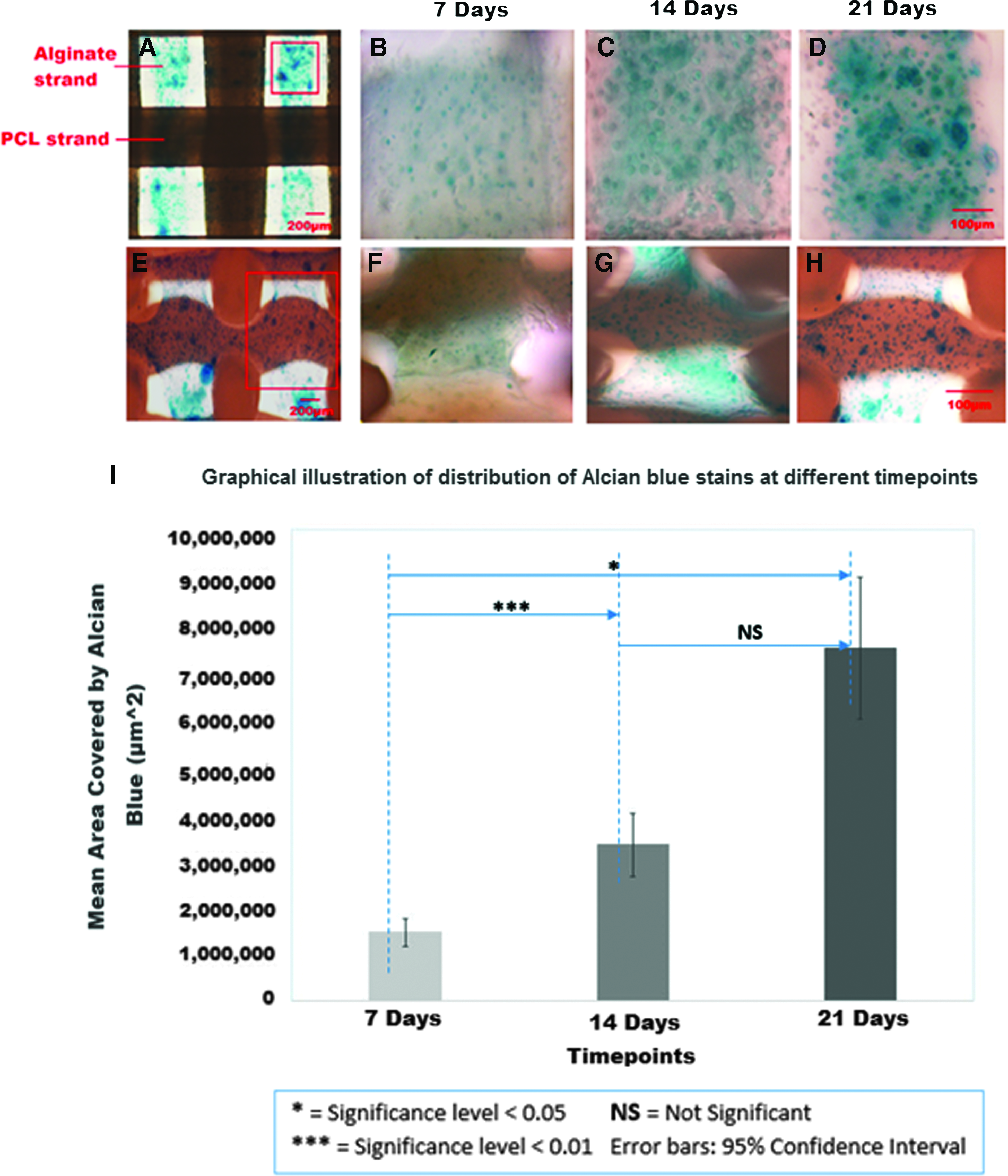
Alcian blue-positive matrix increased over time in 3D-printed hybrid cartilage constructs implanted subcutaneously into the backs of nude mice.
Col2 immunostaining suggested that Col2 matrix was progressively secreted in the 3D-printed hybrid cartilage constructs over the 21-day period postimplantation in mice (Fig. 4A–D). This apparent increase in Col2-positive matrix occurred around cells at both the center and periphery of the alginate strands (Fig. 4F–H). Quantitative analyses of the area covered by Col2-stained matrix confirmed progressive secretion of collagen matrix in the excised hybrid constructs (Fig. 4I). Specifically, the mean area covered by Col2 staining in the alginate strands was significantly different among time points (F (2, 14) = 57.624, p < 0.001; Fig. 4). Post hoc tests revealed that Col2 secretion increased significantly between 7 and 21 days (p < 0.001) and between 14 and 21 days (p < 0.001).
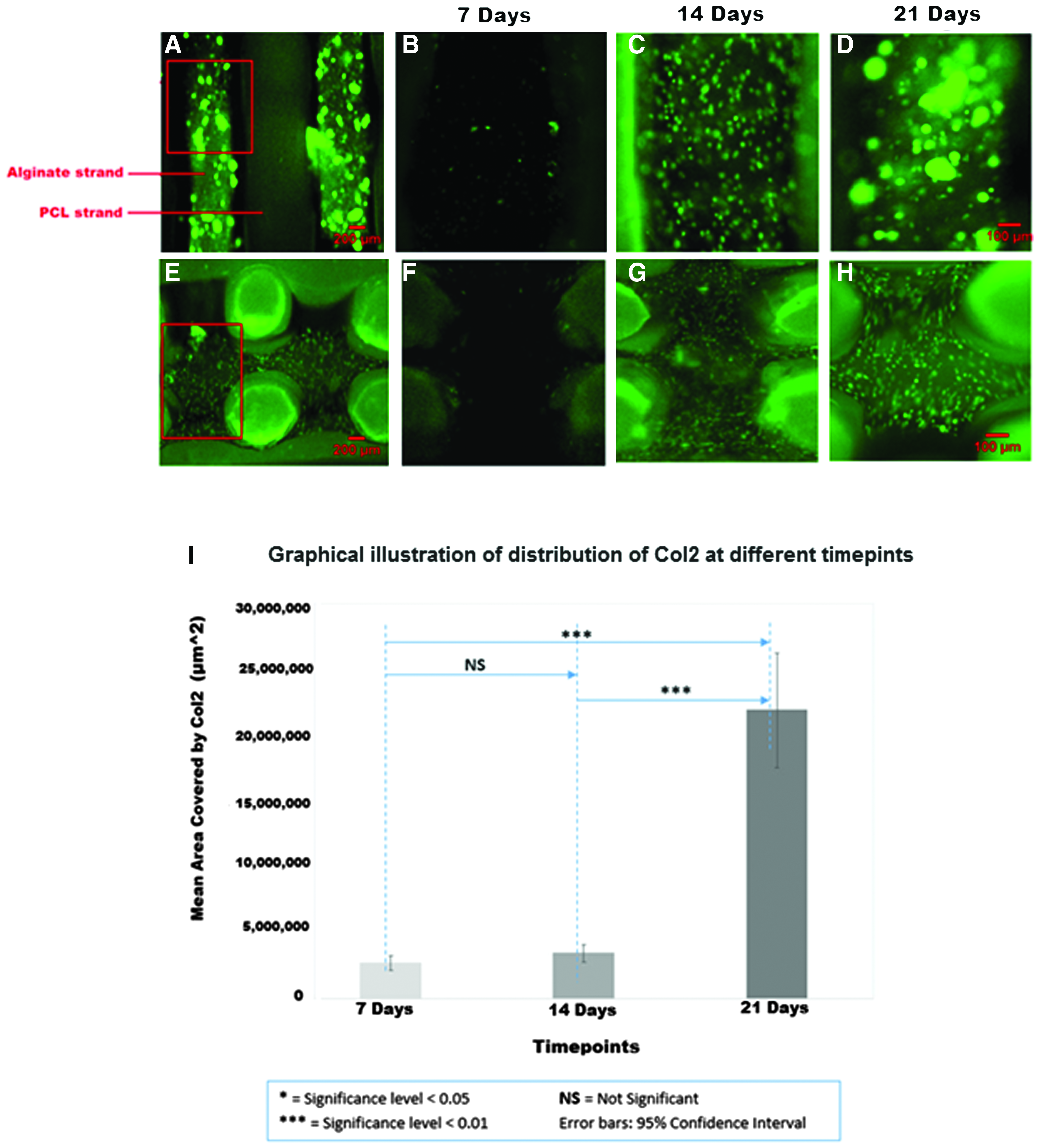
Col2 staining increased over time in 3D-printed hybrid cartilage constructs implanted subcutaneously into the backs of nude mice.
SR-inline-PCI-CT enabled noninvasive visualization of the individual components within the hybrid cartilage constructs and surrounding host tissues
To visualize intact 3D-printed hybrid cartilage constructs noninvasively, SR-inline-PCI-CT was conducted using parameters established in our previous in vitro study of the 3D-printed hybrid cartilage constructs. 7 Mice were sacrificed at 7 and 21 days postimplantation, and the SR-inline-PCI-CT images were acquired. Reconstructed images of SR-inline-PCI-CT projection data clearly showed various soft and hard tissues, such as skin, bone, lung, intestine, and internal soft tissues, of the host mouse (Fig. 5A). Importantly, the reconstructed slices appeared to show the individual alginate and PCL strands within the hybrid cartilage constructs in intact conditions within the mice at the earlier time point (Fig. 5B). In addition, 3D rendering of the reconstructed slices from SR-inline-PCI-CT data further enhanced the delineation of interfaces between the PCL and alginate strands within the hybrid cartilage constructs (Fig. 5C).
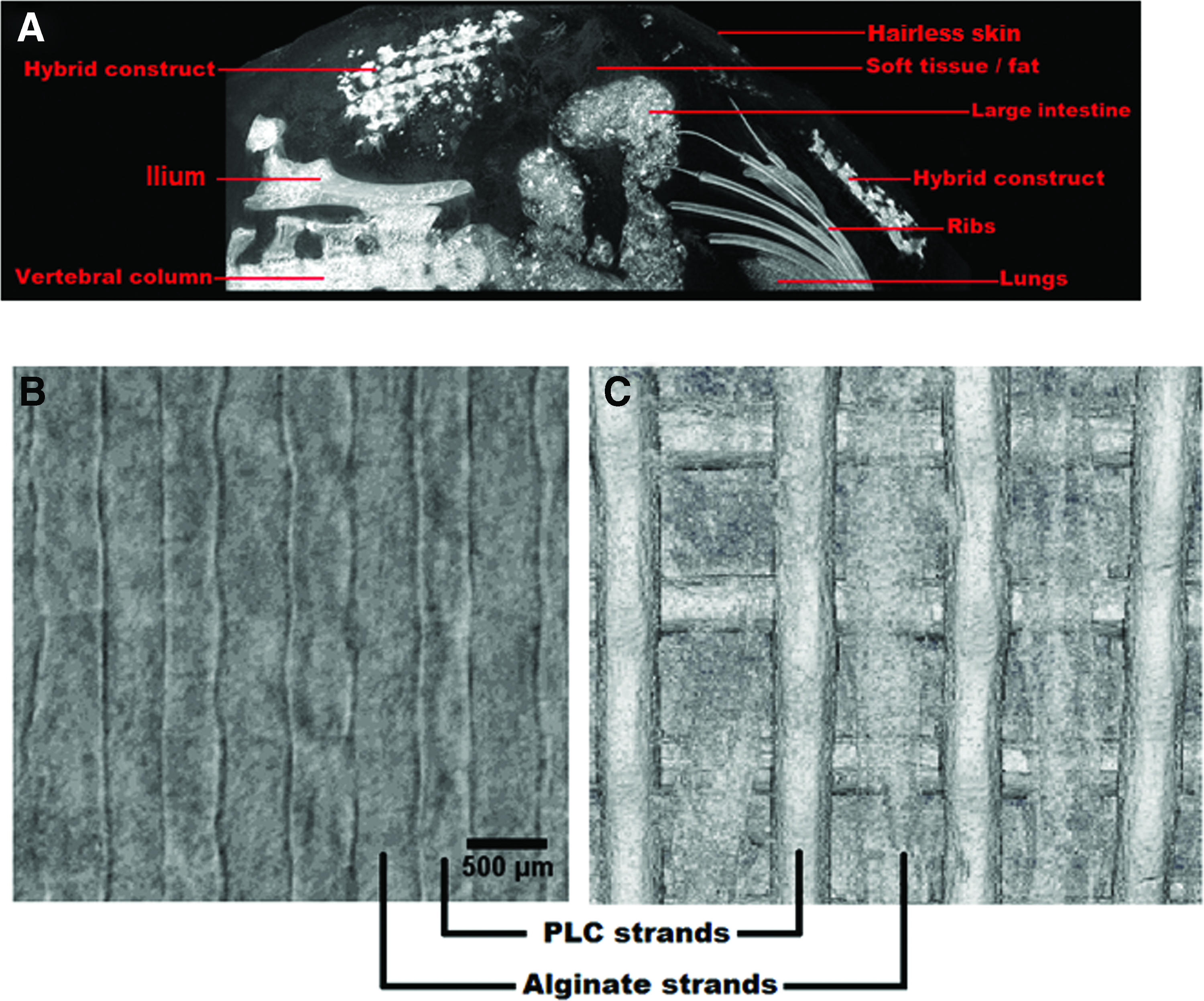
SR-inline-PCI-CT visualized 3D-printed hybrid cartilage constructs in vivo.
SR-inline-PCI-CT enabled noninvasive visualization of structural changes within hybrid cartilage constructs over 21 days postimplantation
Furthermore, structural changes within the 3D-printed hybrid cartilage constructs over the 21 days postimplantation were explored noninvasively using SR-inline-PCI-CT. Images representing equivalent regions of a stack of reconstructed slices showed visible structural changes within the cell-impregnated hybrid constructs between 7 and 21 days postimplantation (Fig. 6). At 7 and 21 days postimplantation, representative alginate and PCL strands were apparent (Fig. 6B, C). Careful analyses of image pixel intensities more clearly demonstrated the capabilities of SR-inline-PCI-CT to distinguish components of the hybrid cartilage constructs. Very large gray-value peaks reflecting high phase contrast correlated with the edges of the PCL strands and the interfaces between the alginate and PCL strands (Fig. 6E–J; see regions 1 and 3). Much lower gray-value peaks correlated with changes apparent within the alginate strands (Fig. 6E–J; see region 2). Comparison of the cell-laden alginate strands at 7 and 21 days postimplantation showed time-dependent structural changes. Recorded gray values tend to decrease overall at 21 days postimplantation, but visible structural changes in the alginate strands between 7 and 21 days postimplantation were associated with increased peak correlating to gray values (Fig. 6E–G, H–J; see region 2). These time-dependent changes within alginate strands revealed by SR-inline-PCI-CT may be, in part, dependent on the presence of cells within the alginate strands. This is because cell-free alginate strands looked very different from these cell-impregnated alginate strands at 21 days postimplantation (Fig. 6C, D). In some regions, the cell-free alginate strands have varying phase contrast and edge enhancement characteristics (Fig. 6D, upper left region). This characteristic was apparent after harvesting the constructs from the mice, as regions similar to those analyzed in the SR-inline-PCI-CT for the cell-free constructs showed clear and opaque alginate strands in between the PCL strands (data not shown). In regions showing differences between the PCL and alginate strands, there was much less phase contrast information over the width of the alginate strand (Fig. 6J, region 2). In summary, noninvasive SR-inline-PCI-CT revealed time-dependent structural changes with the 3D-printed hybrid cartilage constructs.
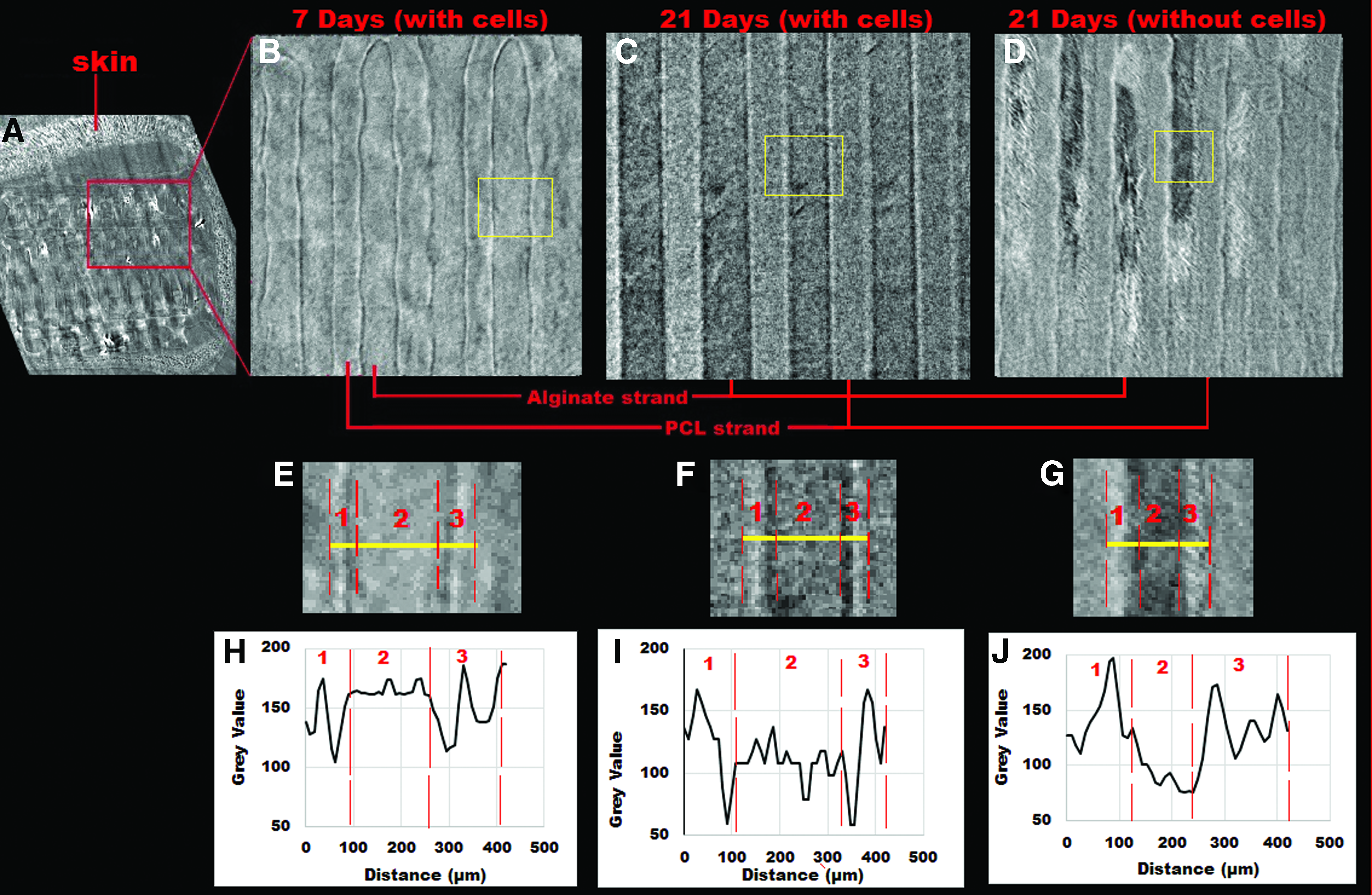
SR-inline-PCI-CT reflected time-dependent structural changes to 3D-printed hybrid cartilage constructs in nude mice.
Discussion
Two of the main problems facing CTE today are: (1) fabrication of constructs that mimic the native human articular cartilage and (2) noninvasive imaging of the progression of functionality of these cartilage constructs postimplantation in either animal models or human subjects. In recent years, rapid-prototyping-based additive manufacturing (i.e., 3D-printing) techniques are being investigated for fabrication of hybrid cartilage constructs. Specifically, 3D-printed cartilage constructs fabricated from PCL and cell-impregnated alginate mimic the biphasic nature of native articular cartilage and are attracting attention from researchers around the globe. A few in vitro studies have shown that cells impregnated within these 3D-printed hybrid cartilage constructs stayed viable and progressively secreted cartilage-specific ECM over time.4–7 Furthermore, secretion of Col2 by cells within the hybrid cartilage constructs has been demonstrated in vivo. 5 However, the in vivo performance of these constructs needs further investigation and extensive study to transition this approach to commercialization or clinical application. Regarding noninvasive assessments of construct performance, it is currently a challenge for clinically available techniques (e.g., MRI, radiography, or ultrasound) to simultaneously delineate samples with a combination of hydrophilic and hydrophobic properties such as those found in these cartilage constructs.24–27,31 To address this knowledge gap, 3D-printed hybrid cartilage constructs were implanted subcutaneously in the backs of nude mice, and their performance was evaluated over a 21-day period using both traditional invasive histological and synchrotron-based noninvasive assessments.
The suitable in vivo performance of the 3D-printed hybrid cartilage constructs demonstrated in this study supports their continued development for clinical CTE applications. Cell viability within the hybrid cartilage constructs remained at least as high as 70% over the 21-day implantation period in nude mice (Fig. 2). Although, a cell viability of 70% is still sufficient for ECM secretion, the in vivo results indicated a 10% drop in cell viability in the constructs at 14 days postimplantation in mice. The change from in vitro to in vivo conditions, along with other unknown physiological conditions, is speculated to have caused the drop in cell viability within these constructs. Interestingly, a slight increase in cell viability was observed at 21 days postimplantation in mice, that is, the cell viability increased from 70% ± 6.6% to 73% ± 7.9%. This trend is similar to what we reported in our previously reported in vitro study, where cell viability also decreased at 14 days, but increased at 28 and 42 days. 7
Meanwhile, cells in these 3D-printed cartilage constructs progressively secreted cartilage-specific ECM (Col2 and sulfated GAGs) that are essential to maintain both structural and biological functionality of the hyaline cartilage44–46 (Figs. 3 and 4). The secretion of these cartilage-specific ECM is speculated to have the potential to continue and eventually generate articular cartilage neotissue at more distant time points. However, questions such as length of time required for generation of articular cartilage, how to increase the amount of cartilage-specific ECM secreted over time to hasten articular cartilage formation, whether secretion of articular cartilage-specific ECM will be influenced by mechanical stimulation when these cartilage constructs are implanted in the stifle joint of animal models, and so on, require answers. While cartilage-specific ECM is secreted within these hybrid cartilage constructs, the PCL framework ensured their structural integrity in a manner not possible in hydrogel-only constructs. Because complete degradation of PCL is expected within ∼2 years, 47 the PCL strands did not exhibit any apparent degradation over the 21-day implantation period. In the future, long-term in vivo studies are needed to examine the rate of degradation of the PCL strands and to determine how the degradation of PCL affects the structural integrity of the neotissues formed within hybrid cartilage constructs.
In summary, the favorable biological performance of the 3D-printed hybrid cartilage constructs presented in this study supports the conclusions of previous in vitro studies,4–7 which add to the considerably limited in vivo data published 5 and indeed demonstrates the potential of the 3D-printed hybrid cartilage constructs for generation of articular cartilage. Looking forward, hybrid cartilage constructs might solve a few major challenges currently facing clinical CTE applications. First, the mechanical and biological roles of native articular cartilage can be mimicked by the 3D-printed cell-impregnated hybrid constructs in this hybrid biofabrication approach. Second, articular cartilage has four basic zones with different mechanical and biological properties. This zonal structure can be mimicked during the layer-by-layer deposition of the 3D-printing technique by adjusting materials and printing parameters. 48 Finally, patient-specific parameters that correspond to the shape and size of the defect to be repaired can be incorporated directly into the 3D CAD model used for fabrication of the hybrid cartilage constructs in the future.
Noninvasive imaging techniques to examine the performance of soft-tissue engineered constructs (from multiple biomaterials with different material properties) and their time-dependent structural changes at high spatial resolution are hard to come by. We previously investigated how changing the sample-to-detector distance of SR-inline-PCI-CT affected phase contrast, edge enhancement, and consequent image quality of the 3D-printed hybrid cartilage constructs in vitro. 7 To build of the findings of the in vitro study, in this study we presented the first attempt to use SR-inline-PCI-CT to noninvasively characterize (in situ) the different components of 3D-printed hybrid cartilage constructs. Importantly, the phase contrast and edge-enhancement fringes of SR-inline-PCI-CT enabled characterization of both the hydrophobic (i.e., PCL) and the hydrophilic (i.e., cell-laden or cell-free alginate) components of the hybrid cartilage constructs with different densities and refractive indices (Figs. 5 and 6). In addition, hard and soft tissues around hybrid constructs were clearly apparent in the SR-inline-PCI-CT images (Fig. 5), allowing evaluation of a critical clinical parameter: integration of the construct with surrounding host tissues. Not only could SR-inline-PCI-CT visualize the multiple components of the hybrid cartilage constructs and their surrounding host tissues but also it does this with a relatively high spatial resolution compared to conventional clinical imaging techniques. The spatial resolution of PCI is hard to specifically calculate because it depends upon many factors, including the exact hardware and samples used, but the images presented in this study clearly appear to exceed the resolution achievable by MRI or absorption-based radiography. Although these findings are phenomenal, imaging parameters such as detector pixel size, imaging energy, and number of projections need to be optimized to lower the absorbed radiation dose without affecting the image quality for prospective future live animal studies. 30
Apart from characterization of the overall macrostructural details of the implanted construct, SR-inline-PCI-CT also enabled evaluation of time-dependent structural changes to the biomaterials within the constructs. Structural changes were evident in the alginate strands at 21 days compared to 7 days postimplantation (Fig. 6). Whether the time-dependent changes observed in images of alginate strands reflect new ECM secretion by the impregnated cells is unclear. On the one hand, the timing of these changes correlated with the timing of ECM secretion in parallel invasive experiments (Figs. 2–4). An increase in phase contrast was associated with secretion of ECM by cells within constructs in reported in vitro and in vivo studies.7,10,39,49 On the other hand, the cell-free hybrid scaffolds also showed structural changes, especially within the alginate strands (Fig. 6). Loss of phase contrast might be due to gradual degradation of the alginate hydrogel strands as previously concluded.50,51 In fact, phase contrast decreased in alginate strands in some regions of the cell-free scaffolds, while it increased in other regions of the alginate strands. This might be due to dryness experienced within the cell-free alginate strands, because similar regions were observed to be opaque (white) upon harvesting, and these changes were not seen in cell-impregnated alginate strands. More distant time points are required to resolve what the change in phase contrast within the hybrid cartilage constructs represents, because thicker cartilage should be easily visualized by SR-inline-PCI-CT. In summary, this study supports the conclusion that phase contrast information provided by optimized SR-inline-PCI-CT has potentials for noninvasive characterization of soft tissues, hybrid tissue-engineered constructs, and their associated time-dependent structural changes.7,40,41
Conclusions
In summary, these results not only detail the in vivo functionality of 3D-printed hybrid cartilage constructs for CTE applications but also demonstrate the potentials of SR-inline-PCI-CT for noninvasive assessments of these multimaterial cartilage constructs and their associated time-dependent structural changes in situ. Over a 21-day period, hybrid cartilage constructs (consisting of strands of PCL and cell-impregnated alginate hydrogel) subcutaneously implanted in the backs of nude mice demonstrated good biological performance. Cell viability was always above 70% and cartilage-specific ECM secretion significantly increased over time. As such, this study presents a breakthrough in the implementation of 3D-printed hybrid cartilage constructs for CTE applications. Furthermore, SR-inline-PCI-CT enabled characterization of the individual components of the hybrid cartilage constructs, their time-dependent structural changes, interfaces, and the surrounding host tissues (all with different refractive indices) in situ. Therefore, SR-inline-PCI-CT offers great potential for noninvasive 3D visualization of fine features in weakly absorbing materials and tissues commonly encountered in soft-tissue engineering applications.
Footnotes
Acknowledgments
The authors acknowledge funding from the Saskatchewan Health Research Fund (SHRF), Canadian Institutes of Health Research (CIHR), and the Natural Sciences and Engineering Research Council of Canada (NSERC). Images presented in this article were produced from data captured at the Canadian Light Source (CLS), which is supported by the Canada Foundation for Innovation (CFI), NSERC, the University of Saskatchewan, the Government of Saskatchewan, Western Economic Diversification (WED) Canada, and CIHR.
Disclosure Statement
No competing financial interests exist.