
Abstract
Natural extracellular matrix-derived biomaterials from decellularized allogenic tissues are of increasing interest for tissue engineering because their structure and composition provide a complexity that is not achievable with current manufacturing techniques. The prerequisite to bring allogenic tissue from bench to bedside as a functional biomaterial is the full removal of cells while preserving most of its native characteristics such as structure and composition. The exceptionally dense structure of articular cartilage, however, poses a special challenge for decellularization, scaffold preparation, and reseeding. Therefore, we tested 24 different protocols aiming to remove cells and glycosaminoglycans (GAG) while preserving the collagen backbone and ultrastructure. The resulting matrices were analyzed for cell removal (DNA quantification, haematoxylin and eosin staining), GAG content (dimethyl methylene blue assay, Alcian blue staining and micro-computed tomography), collagen integrity (immunohistochemistry and ultrastructure), and biomechanics (compression test). Furthermore, seeding tests were conducted to evaluate cell viability and attachment to the scaffolds. Sodium dodecyl sulfate-based protocols yielded satisfactory reduction of DNA content, yet had negative effects on cell viability and attachment. Hydrochloric acid efficiently decellularized the scaffold and pepsin emerged as best option for GAG depletion. Combining these two reagents led to our final protocol, most efficient in DNA and GAG depletion while preserving the collagen architecture. The compressive modulus decreased in the absence of GAG to ∼1/3 of native cartilage, which is significantly higher than that by commercially available scaffolds tested as a reference (ranging from 1/25 to 1/100 of native cartilage). Cytocompatibility tests showed that human adipose-derived stromal cells readily adhered to the scaffold. In this study, we established a protocol combining freeze–thaw cycles, osmotic shock, and treatment with hydrochloric acid followed by a pepsin digestion step, achieving successful decellularization and GAG depletion within 1 week, resulting in a cytocompatible material with intact collagen structure. The protocol provides a basis for the generation of allogeneic scaffolds, potentially substituting manufactured scaffolds currently used in clinical articular cartilage treatment.
Introduction
T
The use of allogenic scaffolds derived from decellularized tissues and organs has come into focus for reconstructive surgery and regenerative medicine. Their advantage over manufactured biomaterials is that decellularized tissues are homologous to their destination. They provide a specific extracellular matrix (ECM) composition, growth factors, and spatial organization, guiding and influencing seeded or invading cells in a tissue-specific manner at the regenerative site.3–6
Requirements for the use of allogenic material are the full removal of cellular components to avoid immune-mediated graft rejection, while the overall ECM structure should be maintained. Decellularization approaches typically involve a variety of steps, which can be grouped into three categories: physical, chemical, and enzymatic.6–8 Physical approaches include freezing and thawing the tissues to provoke cell burst by intracellular crystal formation, but also compression or application of ultrasound treatment. Hypotonic buffers that cause cells to burst as well as detergents and acids or bases that break down cellular and/or matrix components fall under the category of chemical approaches. Finally, enzymes such as nucleases or lipases are used as biological approaches for degradation of specific target components. The protocols aim for complete removal of the resident cells, inactivation of pathogens, and elimination of harmful residues of reagents used in the process while preserving the ECM (e.g., collagen) structure and tissue architecture.
Decellularized products of various tissue types have already been successfully implemented into clinics and are commercially available. 9 In this regard, also some types of decellularized native cartilage are about to experience transition to clinics. Schwarz et al. published and patented the successful decellularization of nasal septum that could then be infiltrated superficially by seeded chondrocytes. 10 For decellularization of articular cartilage, several protocols have been reported, mostly based on sodium dodecyl sulfate (SDS).11–14 A protocol combining thermal, chemical, and enzymatic decellularization techniques was developed for menisci and later also tested on articular cartilage, showing superficial reseeding of the fully decellularized and partially glycosaminoglycan (GAG)-depleted xenograft.12,13 Also decellularized articular cartilage in the form of minced particles or homogenized slurry with embedded cells has been examined recently.15,16 The specific architecture of the native tissue, however, is lost in these cases.
The exceptionally dense structure of articular cartilage of narrowly arranged collagen fibrils filled up with GAG in their interfibrillar space makes it necessary to not only decellularize but also to remove GAG from the cartilage tissue to some extent to increase the scaffold's porosity. The balance of allowing recellularization through matrix depletion and simultaneously retaining most of the relevant tissue-specific properties is therefore particularly challenging.
The aim of this study is to achieve an optimal decellularization and GAG removal protocol for human articular cartilage by further developing, optimizing, and combining previously established methods,10–13,17–22 with a focus on retaining the complex collagen architecture present in native cartilage. For this we have systematically compared the resulting cartilage scaffolds for content, composition, structure, and biomechanics as well as their impact on vitality and adherence of therapeutically relevant cells (adipose-derived stromal/stem cells [ASCs]). The selected protocol provides a basis for the generation of allogenic or potentially xenogenic scaffolds, substituting the synthetic scaffolds currently used in articular cartilage treatment.
Materials and Methods
A more detailed version of our materials and methods section is provided as Supplementary Data (Supplementary Data are available online at
Sample harvest
Macroscopically intact human articular cartilage was harvested from femoral heads of donors undergoing hip replacement, with patient's consent and the approval of the local ethical board. Full thickness noncalcified cartilage was separated from the subchondral bone and biopsies prepared with a biopsy punch of 8 mm in diameter and washed with phosphate-buffered saline (PBS) plus penicillin and streptomycin.
Decellularization protocol workflow
Before each decellularization protocol, four freeze–thaw cycles were performed and each protocol was concluded by a decontamination step in peracetic acid followed by two 30-min wash steps and incubation in PBS overnight. During the decellularization cycles, which formed the core of the protocol, samples were incubated consecutively in hypotonic buffer (Tris-base, pH 8), the respective decellularizing agent, and PBS for 24 h. Additional enzyme steps were incorporated in some protocols before the decontamination step in an attempt to reduce the GAG content.
Protocols in the SDS-based, SDS-free, and part of the pepsin-based protocol groups follow this workflow, whereas during enzyme screening, only the respective enzyme was tested (Table 1).
Shaded section I indicates SDS-based protocol; section II, protocols from the SDS-alternative group; shaded section III, the enzyme screening; section IV, protocols involving pepsin treatment; and shaded section V, the final protocol. Stars indicate the protocols selected for further analysis with SEM, TEM, μCT, cytotoxicity, and indentation testing. The experiments were repeated on at least six different donors.
μCT, micro-computed tomography; SDS, sodium dodecyl sulfate; SEM, scanning electron microscopy; TEM, transmission electron microscopy.
SDS-based protocols
In all these protocols, SDS was used as decellularizing agent. The SDS+nucleases protocol included treatment with 50 U/mL DNAse and 1 U/mL RNAse directly following only one single decellularization cycle. In the nuclease-free SDS protocol, the nuclease step was replaced by two extra decellularization cycles (to a total of three). The SDS+trypsin protocol and SDS+elastase incorporated an enzyme step after three decellularization cycles.
SDS-free protocols
The decellularizing agents Triton X-100, Triton QS-15, hydrogen peroxide, sodium deoxycholate, CHAPS buffer, NaOH, and HCl were tested in three decellularization cycles for their effect on tissue decellularization and GAG depletion.
Enzyme screening
To achieve a more targeted depletion of matrix components, enzymes were chosen for their selective mode of action. A cell isolation protocol combining 0.1% hyaluronidase, 0.5% pronase, and 0.2% collagenase was tested, followed by multiple PBS wash steps. To assess their individual effect, the enzymes were also tested separately and others were included in the screening: 0.2% hyaluronidase, 1% pronase, 0.4% collagenase, 0.1% pepsin, 0.25% trypsin, and 0.03 U elastase. Samples were incubated in the respective enzyme for 24 h, followed by multiple wash steps in PBS.
Pepsin-based protocols
After freeze–thaw cycles, samples were incubated in either 0.4% collagenase, 0.1 M NaOH, or 0.1 M HCl for 24 h, before a 24 h 1 g/L pepsin treatment.
Finally pepsin incorporated in the HCl-based decellularization protocol was established as our optimized protocol: freeze–thaw cycles, three decellularization cycles consisting of 24 h in hypotonic buffer, 24 h in HCl and multiple wash steps overnight, and 24 h pepsin treatment after the last cycle, and concluded by decontamination and final extensive washing. It was also tested whether reducing the number of decellularization cycles to one single cycle would be equally efficient with a greatly reduced treatment time.
Biochemical assays
Three samples per treatment and donor were digested with papain according to a protocol adapted from Kim et al. 23 DNA content was quantified using the CyQUANT® Cell Proliferation Kit and GAG content using the dimethyl methylene blue (DMMB) assay described by Farndale et al. 24
Histological examination
Samples were fixed in 4% neutral buffered formalin, washed in PBS, and dehydrated with a graded series of alcohol. Then, samples were embedded in paraffin and sectioned with 3 to 4 μm thickness with a rotatory microtome. Sections were either stained with hematoxylin and eosin (H&E) for observation of cells and cell residuals, Alcian blue with Sirius red counterstaining for GAG detection, or immunolabeled against collagen type II.
Scanning and transmission electron microscopy
For scanning electron microscopy (SEM), cartilage chip samples were fixed in 4% neutral buffered formalin overnight, washed in PBS, dehydrated in a graded series of alcohol, and chemically dried with hexamethyldisilazane. Samples were sputter coated using a Quorum Q150R ES (Quorum Technologies, UK) with gold, and imaging was performed with a Jeol JSM-6510 scanning electron microscope (JEOL Ltd., Japan).
Samples for transmission electron microscopy (TEM) were fixed in 2.5% glutaraldehyde in 0.1 M sodium cacodylate buffer overnight at 4°C, rinsed, dehydrated in a graded series of alcohol, and embedded in low-viscosity resin. Ultrathin sections were stained with gadolinium and lead citrate and imaged using a TEM Zeiss 902 (Carl Zeiss AG, Germany) microscope.
Micro-computed tomography imaging
For 3D observation of GAG removal, 25 samples were fixed in 2.5% glutaraldehyde in 0.5 M sodium cacodylate containing 0.07% ruthenium hexamine trichloride (RHT), rinsed in 0.1 M sodium cacodylate buffer, and post fixed in 1% osmium tetroxide and 0.07% RHT. Then samples were rinsed and dehydrated in a graded series of alcohol and dried with hexamethyldisilazane, as used for SEM. CT scans of the stained cartilage samples were performed using a SCANCO micro-computed tomography (μCT) 50 (SCANCO Medical AG, Switzerland).
Cytotoxicity screening
ASCs were harvested from liposuction material with patient's consent and the approval of the local ethical board. Scaffolds were placed in 1.5 mL screw cap vials, and 200,000 cells (ASCs) in endothelial growth medium (EGM-2) were seeded into each vial. The vials were then placed on a device performing continuous rotation of the vials on a Stuart Roller Mixer SRT1 (Bibby Sterilin Ltd., UK) at 37°C. After 3 days, samples were transferred to a 48-well plate and subjected to Calcein AM staining.
In case of negative influence on cell attachment, all reagents used in the respective protocol were tested separately for the release of cytotoxic components. Samples were incubated in these reagents for the same time span as in the protocol and washed overnight in PBS. Samples were then placed in a 24-well plate and ASCs (50,000 cells per scaffold in EGM-2) were seeded directly onto the surface. After 24 h, cell viability and attachment on the scaffolds and in the well were observed.
Mechanical compression test
Samples were mechanically tested using a custom-made unrestrained compression set fitted on a Zwick BZ2.5/TN1S uniaxial testing machine (Zwick GmbH & Co. KG, Germany) equipped with a 50 N load cell. Before testing, all specimens were equilibrated in PBS for at least 24 h. After achieving a preload of 50 mN, data were recorded and the samples were compressed until 80% deformation with a constant speed of 100 μm/min. As controls, three commercially available scaffolds, TissuFleece™ (Baxter, USA), Hyalograft® C (FIDIA Advanced Biopolymers, Italy), and HYAFF® (FIDIA Advanced Biopolymers), were tested under the same conditions.
Statistical analysis
Quantitative data (Figs. 1, 2, and 10; Supplementary Fig. S1) were evaluated using one way analysis of variance (ANOVA) with a 95% confidence interval.

DNA content as an index for decellularization efficiency, measured using the CyQuant DNA dye. Shown as percentage (calculated from w/w) of the content in untreated cartilage, mean ± SD, n = 6; red bars indicate SDS-based protocol; green bars, protocols from the SDS-alternative group; yellow bars, the enzyme screening; purple bars, protocols involving pepsin treatment; blue bars, the final protocol; and full bars indicate the protocols selected for further analysis; one way ANOVA (p ≤ 0.05) shows all of them (*) to differ significantly from untreated tissue; DNA contents of NaOH HCl and HCl (1 cycle)+pepsin are significantly lower (*, #) than the other two (* only) and do not differ significantly from each other (or from the SDS+nucleases protocol). ANOVA, analysis of variance; SD, standard deviation; SDS, sodium dodecyl sulfate. Color images available online at
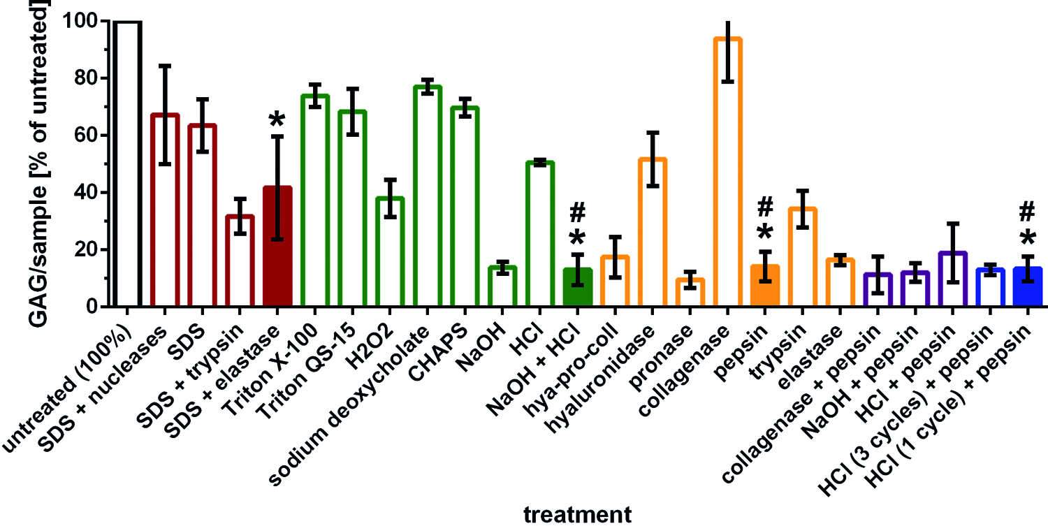
GAG content as measured using the dimethyl methylene blue assay
Results
In total, 24 protocols were tested for their effect on decellularization and/or selective matrix depletion (Table 1). Decellularization protocols (SDS-base, SDS-free, and part of the pepsin-based group) followed a basic workflow of freeze–thaw cycles, decellularization cycle(s), an optional enzyme treatment, and final decontamination and washing. Decellularization cycles, consisting of hypotonic buffer followed by the respective decellularizing agent and washing, formed the core of every protocol and were repeated three times unless mentioned otherwise.
SDS-based protocols
Within the four tested SDS-based protocols, SDS+nucleases most efficiently reduced the DNA content to 2.7% (±2.6%) of the amount in native cartilage (Fig. 1). Without nucleases, the DNA content could be diminished with every decellularization cycle (Supplementary Fig. S1), resulting in 23.1% (±8.6%) of the original content after 3 cycles (Fig. 1). Both protocols had moderate effects on the GAG content (Fig. 2; Supplementary Fig. S2).
Addition of trypsin treatment to the nuclease-free protocol led to a reduction of the DNA content to 6.4% (±3.7%), and depleted the GAG to 31.8% (±6.1%). The SDS+elastase protocol resulted in a DNA content of 7.7% (±6.9%) and a GAG content of 41.7% (±18.0%) (Figs. 1–3) while the collagen matrix was left unaltered (Fig. 4).
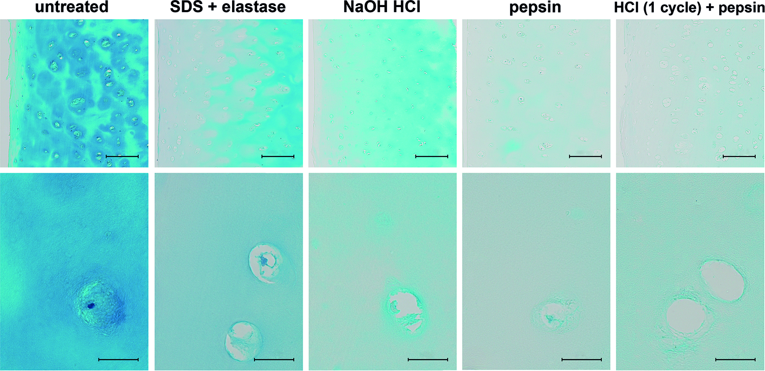
Alcian blue staining of cartilage, untreated and treated with selected protocols. The absence of staining shows efficient GAG depletion in all conditions, but most noticeably so when pepsin treatment was involved; scale bars = 200 μm (upper row) and 25 μm (lower row). Color images available online at

Collagen type II immunostaining of cartilage, untreated and treated with selected protocols. Staining shows preservation of the collagen type II in all conditions except after NaOH HCl where the outermost layer of the scaffold is rendered transparent (black arrow points at the outer edge), indicating a change of collagen type II (disruption of the integrity or epitope); scale bars = 200 μm (upper row) and 25 μm (lower row). Color images available online at
SDS-free protocols
In the screening for alternative decellularizing agents, HCl was most promising, reducing the DNA content to 2.9% (±1.9%), a level otherwise only achieved by the use of highly concentrated nucleases (Fig. 1). With 50.6% (±1.0%), the GAG content remained high. NaOH significantly reduced the DNA content to 11.9% (±5.8%) and was most effective in the removal of GAG (Figs. 2 and 3), reducing the content to 13.8% (±2.1%). Results of those two treatments were at similar levels for 30°C and 45°C during the decellularization cycle (data shown for 30°C). Therefore, the lower temperature was chosen for further studies to prevent thermal decomposition of the matrix components. The protocols did not require the application of multiple decellularization cycles, because their effect on DNA and GAG content was similar after 1 or 3 cycles (Supplementary Fig. S1). Because of their individual advantages in either DNA or GAG reduction, HCl and NaOH were both incorporated into the established decellularization protocol (freeze–thaw, hypotonic burst, NaOH treatment, washing, HCl treatment, washing, decontamination, and further washing).
After NaOH treatment, the collagen type II staining was hampered in the outer layer up to 100 μm, indicating serious changes of the collagen type II fibrils (Fig. 4; Supplementary Fig. S3).
Enzyme screening and pepsin-based protocols
Enzyme treatment alone had limited impact on scaffold decellularization (Fig. 1). The combination of hyaluronidase, pronase, and collagenase resulted in effective GAG depletion to 17.4% (±7.1%). The individual enzymes alone changed the matrix composition according to the expected effect based on their mode of action (Fig. 2; Supplementary Fig. S2). In terms of GAG depletion, pronase and pepsin performed best in this screening, with a GAG content of 9.5% (±2.8%) and 14.2% (±5.1%), respectively. Pronase comprises several peptidases, whereas pepsin is a clearly defined enzyme less likely to attack collagen fibers. Therefore, pepsin was chosen in further experiments because of its more specific mode of action.
Various combinations of pepsin with the other reagent steps were tested, aiming to obtain higher matrix porosity by removing more GAG, yet they led to no further reduction of the GAG content compared with pepsin treatment alone (Fig. 2). The combination of pepsin with HCl was deemed useful, as HCl led to most efficient decellularization, whereas pepsin treatment resulted in highly efficient GAG removal (Figs. 1–3).
Incorporation of a pepsin step into the decellularization protocol with HCl as main decellularizing agent resulted in a DNA content of 3.9% (±1.4%) and GAG content of 12.9% (±1.9%) compared with untreated cartilage. Reduction of the decellularization cycles from three to one did not significantly change this outcome, but reduced the total time of treatment to 1 week (Figs. 1 and 2).
Characterization of most promising candidate per screening group
Histology
Alcian blue staining confirmed the results of the biochemical assays on GAG (Fig. 3). Collagen type II retained its immunoreactivity with the specific antibody in all samples except that ones treated with the NaOH HCl protocol, in which the tissue in the outer 30 μm remained unstained (Fig. 4). H&E staining of the samples treated with the pepsin protocol still showed intact nuclei, whereas samples treated with the other protocols were devoid of nuclei and showed few cell remnants (Fig. 5).
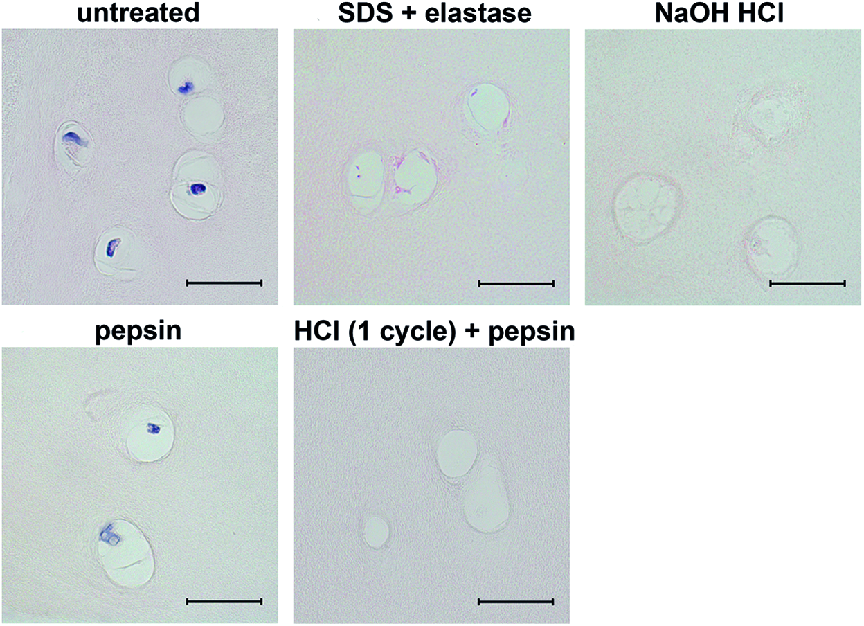
Hematoxylin & eosin staining of cartilage, untreated and treated with selected protocols. Intact nuclei (dark purple) are shown in untreated and pepsin-treated cartilage and the absence thereof in the samples treated with the SDS+elastase, NaOH HCl, or HCl (1 cycle)+pepsin protocol; scale bars = 25 μm. Color images available online at
SEM and TEM
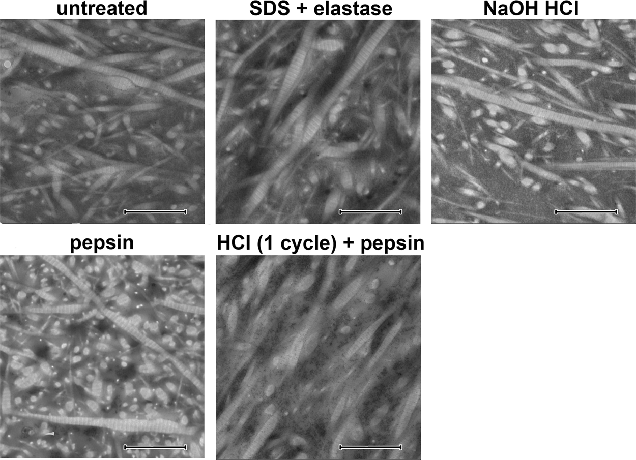
The collagen fibers in all samples appeared intact in SEM after treatment and aligned in the same way as in untreated cartilage. In samples treated with the NaOH HCl protocol, the pepsin protocol, or the HCl (1 cycle)+pepsin protocol, the fiber network was more clearly visible and looser, without homogeneous material in between the fibrils as in the untreated samples (Fig. 6). The collagen fibrils in TEM had retained their ultrastructure after treatment with each protocol, displaying the characteristic banding pattern without fraying or other signs of degradation (Fig. 7).
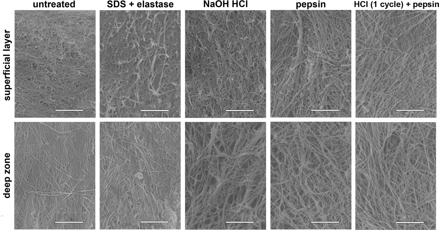
Scanning electron microscopy images (SE mode) of cartilage, untreated and treated with selected protocols. Images show the collagen fibers to be still intact and more clearly visible after NaOH HCl or protocols containing pepsin treatment due to the matrix being less dense because of the absence of GAG; scale bars = 5 μm.
Transmission electron microscopy images of cartilage, untreated and treated with selected protocols. Collagen fibrils are observed in longitudinal section as well as cross section; in all conditions, fibrils retained their characteristic 60 nm banding pattern, indicating their intactness; scale bars = 1 μm.
μCT imaging
Although untreated cartilage appeared homogeneously bright on μCT images, indicating an even distribution of GAG, cartilage treated with the pepsin or HCl (1 cycle)+pepsin protocol displayed darker areas, where the contrast agent did not bind because of the absence of GAG (Fig. 8).

μCT images of cartilage, untreated and treated with selected protocols. Samples were treated with highly positive ruthenium hexamine trichloride binding to the negatively charged GAG. Therefore, the tissue appears brighter the more GAG are present in that area. SDS+elastase- and NaOH HCl-treated samples become uniformly darker, whereas samples treated with pepsin-containing protocols show heterogeneous staining with darker areas around the lacunae; scale bars = 200 μm.
Cytotoxicity screening
Human ASCs were vital (Calcein AM metabolism) and attached well to the treated scaffolds, demonstrating a typical spindle shape morphology and forming a homogeneous monolayer spanning the entire surface of the sample (Fig. 9). The only exceptions were SDS+elastase-treated scaffolds, in which most of the surface was completely cell free and the few cells that were present had a round morphology with minimal adhesion to the sample (Fig. 9). In a subsequent cytotoxicity screening of all reagents used in the protocol (hypotonic buffer, SDS-based ionic detergent, and peracetic acid), SDS was identified as the cause for cell death (data not shown). Furthermore, in culture dishes with SDS-treated scaffolds, also cells in proximity to the material were nonvital. This indicated that SDS could not be removed from the tissue reliably.
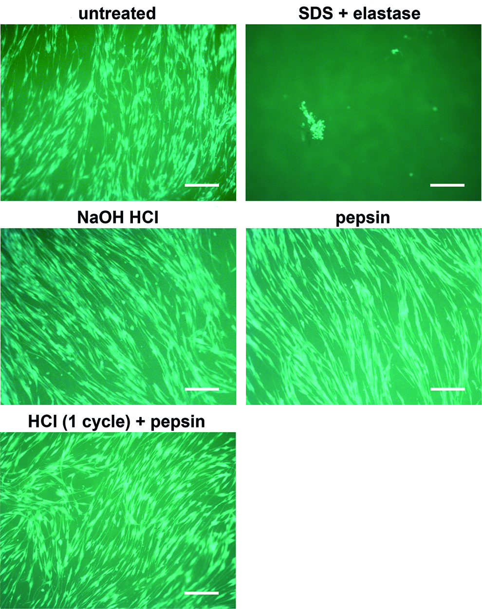
Calcein AM staining of adipose-derived stromal/stem cells seeded on cartilage, untreated and treated with selected protocols. Samples were cultivated under dynamic conditions for 3 days. Staining shows good cell viability and attachment in all conditions except for the SDS+elastase-treated scaffolds, in which attachment was very low and hardly any cells were present (picture shows the only viable cells on all three tested samples); scale bars = 100 μm. Color images available online at
Mechanical compression test
There were two phases in the strain–stress curve of untreated, native cartilage: an initial phase of low stiffness in which the material was deformed without an increase in stress value, followed by a phase of highly increasing stiffness. The treated samples lacked the initial phase and the indentation causes stress levels to rise from the beginning, indicating a loss of elasticity (Fig. 10). Compressive modulus was highest for untreated cartilage, the NaOH HCl and SDS+elastase protocols retained about half of their native compressive modulus, the protocols involving pepsin treatment about one-third. However, the values measured for the reference products ranged from about 1/25 for Hyalograft to 1/100 for TissuFleece. The compressive modulus of HCl (1 cycle)+pepsin-treated cartilage was eight times higher than any of the commercially available scaffolds measured (515.25 [±33.4] kPa vs. the highest value 65.86 kPa for Hyalograft) (Fig. 11).

Compressive stress in relation to sample deformation, untreated and treated with selected protocols. Only untreated cartilage shows an initial elastic phase, in which stress levels do not increase during compression; n = 4, colors represent the different donors. Color images available online at
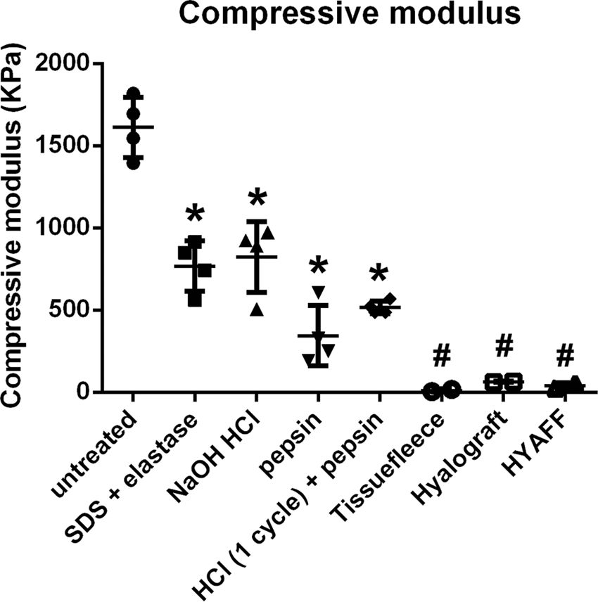
Compressive modulus of cartilage, untreated and treated with a selection of protocols, as well as several commonly used scaffolds. Modulus was calculated between 60% and 70% deformation; data shown as mean ± SD, n = 4; although compressive modulus of treated cartilage is significantly lower than that of untreated tissue, one way ANOVA (p ≤ 0.05) confirms significant difference between all treated cartilage scaffolds (*, ranging from 1/2 to 1/3 of untreated cartilage) and the commercially available controls (#, 1/25 to 1/100 of untreated cartilage).
Discussion
We systematically compared a variety of reagents and combinations thereof, which have been described in the literature for their use in decellularization and selective matrix digestion. The final protocol guarantees full devitalization (the absence of living cells), sufficient decellularization (removal of cells and cellular debris), and decontamination as well as excludes the risk of harmful residues from reagents used in the process, all while preserving the structure of the articular cartilage. The treated cartilage was evaluated not only for the absence of cellular material but also for removal of GAG, mechanical properties, and influence on cell behavior.
Efficient decellularization of allografts is crucial to exclude the possibility of immune reactions to cellular antigens in the patient. 26 SDS is the widest used reagent in decellularization protocols that acts by solubilizing cell and nucleic membranes.6,20 However, adverse effects of SDS-treated scaffolds on recellularization have been described6,27–30 and have been suggested to be caused by residues of the detergent in the tissue or matrix alterations because of the treatment. 31 In our study, a negative influence of SDS treatment on cell viability was observed restricted to not only the treated tissue itself but also visible in a radius of the material, indicating the release of a cytotoxic substance from the tissue. Although SDS works well in decellularization protocols for other tissues,32–37 articular cartilage seems to retain SDS to a higher extent. Therefore, we suggest replacement of SDS for cartilage preparation.
We found HCl to be the most efficient reagent tested for decellularization while having no adverse effects on cell viability and attachment. In our study, similar decellularization efficiency was otherwise only achieved by using SDS in combination with highly concentrated nucleases, which are far more cost intensive and more complex in terms of approval by regulatory authorities. The commonly acknowledged goal that needs to be met for a scaffold to be termed decellularized is an amount of double-stranded DNA lower than 50 ng per milligram dry ECM, a threshold associated with the absence of adverse host responses, alongside the lack of visible nuclear material in haematoxylin and eosin staining. 6 This aim was met by our final protocol, achieving a DNA content lower than 10 ng per milligram wet ECM, corresponding to a calculated 33 ng per milligram dry sample weight.
HCl is known to denature proteins by disrupting intramolecular bonds held together by ionic charges. In high concentrations, this can lead to deterioration of matrix components. However, tissue slices stained with a collagen type II antibody show no deterioration of the epitope, and SEM and TEM confirm that both the alignment and the ultrastructure of the collagen type II fibrils remained unaltered by the treatment. This leads to the conclusion that the tested concentration is low enough not to harm the collagen fibers, but sufficient for decellularization, acting through solubilization of cell components and disruption of nucleic acids. 7
The biggest challenge of the process, also considering future reseeding of the decellularized scaffold, is posed by the exceptionally dense ECM of articular cartilage. Therefore, we consider depletion of GAG content necessary to open up the matrix, enhance perfusion with reagents, facilitate the removal of unwanted remnants, and make the scaffold more accessible to cells.
Among the chemical reagents, we found NaOH to most successfully decrease the GAG content, an effect that has been previously described in the literature, 38 but histological staining using collagen type II-specific antibodies demonstrated a loss of immunorecognition at the edges of the sample, suggesting changes of the nature of the collagen fibrils by NaOH treatment.
Regarding enzymes, elastase and trypsin have been tested in combination with the SDS-based protocols. With trypsin potentially having adverse effects on the collagen matrix, addition of elastase was regarded as a more gentle option. It tackles not only elastic fibers but also GAG, rendering it useful although no elastic fibers are present in articular cartilage. The use of elastase has been described in the literature to be effective even for bovine articular cartilage, 39 yet equally strong effects could not be reproduced in our study.
While screening for targeted GAG depletion by using enzymes, pepsin emerged as most suitable. Its effectiveness in reducing the GAG content was quantified through DMMB assay and observed through Alcian blue staining, electron microscopy, and μCT imaging. Its mode of action is very specific, also compared with enzyme cocktails such as commonly used pronase or collagenase products. Pepsin preferentially cleaves after aromatic amino acids such as tyrosine or tryptophan, which are hardly present in collagen fibers.40,41 It has been used in the past, at high concentrations or after chemical GAG depletion, to solubilize collagen molecules from bone or cartilage matrices38,42,43; however, no damage of the collagen structure has been observed for our application. Pepsin can be obtained in highly purified form (or as recombinant protein) also in good manufacturing practice grade, and is a relatively cheap enzyme, thus meeting the criteria for its use in a final cost-effective and scale-up possible protocol.
Mechanical properties are of crucial importance especially in the load-bearing zones within the joint. As opposed to synthetic scaffolds commonly used in MACT, the scaffold obtained by our procedure is stable and still retaining a significant amount of its initial high stiffness, giving it a considerably higher mechanical load capacity. Therefore, the decellularized cartilage retains its shape throughout the whole process and reseeding, without shrinkage or contraction that often appears when using synthetic scaffolds. 44 The strain–stress curve of native cartilage has two phases, which can be explained by the presence of GAG that creates a highly hydrated space and that upon mechanical load releases water. 45 Once the majority of the water is excluded from the matrix, the positively charged side chains repel one another, resulting in highly increasing stiffness. This typical mechanical behavior was lost in nearly all treatment groups in our experiment because the majority of the GAG had been removed. Although compressive stiffness decreased especially in pepsin-treated samples, it remained several times higher than values measured for commercially available biomaterials.
Loss of some initial elasticity and overall compressive stiffness as a result of the decellularization process is accepted in favor of higher scaffold porosity. 10 The lack of GAG is unproblematic for application of the scaffold for cartilage regeneration, because once the cells are seeded and migrated into the materials, they are expected to reproduce GAG over time. Chondroprogenitor cells and chondrocytes constantly synthesize GAG, especially in a 3D environment that has in multiple occasions been shown to keep the cells in the chondrogenic lineage and counteract dedifferentiation.46–51 In dedifferentiated chondrocytes that had ceased chondrogenic matrix production because of extensive 2D culture, the presence of collagen type II triggers the production of GAG, activating the corresponding pathways within minutes. 52 GAG synthesis by ASCs has been shown within days after seeding in 3D environment in the presence of transforming growth factor beta. 53 In a recent study, a decellularized GAG-depleted scaffold has been reported to show GAG content and histological staining similar to untreated tissue within 5 weeks as well as fully regained biomechanical properties through the matrix synthesis of seeded cells. 21
In this study, peracetic acid was used for disinfection, as it has been shown to effectively inactivate viruses while having only a minimal effect on the ECM composition and structure.19,54 To ensure sterility including virus inactivation also after packaging, most commercially available allografts are subjected to low-dose gamma irradiation (e.g., in course of the Tutplast® process by Tutogen Medical Gmbh, Germany, or the Allowash® process by LifeNet Health, USA), which would also be an option for the scaffold proposed in this study. 55
With the final protocol presented in this article (HCl [1 cycle]+pepsin), it is possible to obtain an acellular scaffold from human articular cartilage that is suitable for recellularization, by making use of clinical waste material from patients undergoing joint replacement surgery after hip fracture or from cadaverous donors. The advantage of human tissue over xenografts is the a priori absence of the alpha-Gal epitope that is expressed in all mammals except humans and primates and leads to adverse host immune reactions if not fully depleted.56,57 Our scaffold has the potential to become a so-called off the shelf product that does not require any preparatory steps before surgery. Its mechanical properties outperform those of scaffold materials currently used in MACT. Increased porosity through GAG depletion could be confirmed on SEM images, in which the collagen fibers appear looser with open space in between that is usually occupied by GAG. Although attachment and viability of primary human mesenchymal stem cells on the scaffold have been shown in course of the cytotoxicity testing, cultivation for 3 days did not allow the cells to migrate into the matrix. As cell infiltration is desired regarding homogeneous distribution of newly synthesized matrix, further studies will focus on long-term recellularization and functionality of the scaffold. In this study, cytocompatibility has been shown by reseeding experiments, and the scaffold's preserved natural collagen structure gives reason to expect chondroinductive properties.
Footnotes
Acknowledgments
This work was supported by funding from the Austrian Research Promotion Agency FFG (project CartiScaff, No. 842455) as well as the Lorenz Böhler Fonds (16/13). The authors wish to thank the University of Applied Sciences Technikum Wien for providing the scanning electron microscopic equipment and Barbara Schädl, MSc, for electron microscopic imaging and preparation of the samples for μCT.
Disclosure Statement
No competing financial interests exist.
References
Supplementary Material
Please find the following supplemental material available below.
For Open Access articles published under a Creative Commons License, all supplemental material carries the same license as the article it is associated with.
For non-Open Access articles published, all supplemental material carries a non-exclusive license, and permission requests for re-use of supplemental material or any part of supplemental material shall be sent directly to the copyright owner as specified in the copyright notice associated with the article.