
Abstract
Introduction:
Labeling using iron oxide particles enables cell tracking through magnetic resonance imaging (MRI). However, the magnetic field can affect the particle-labeled cells. Here, we investigated the effects of a clinical MRI system on primary human hepatocytes labeled using micrometer-sized iron oxide particles (MPIOs).
Methods:
HuH7 tumor cells were incubated with increasing concentrations of biocompatible, silica-based, micrometer-sized iron oxide-containing particles (sMPIOs; 40–160 particles/cell). Primary human hepatocytes were incubated with 100 sMPIOs/cell. The particle-labeled cells and the native cells were imaged using a clinical 3.0 T MRI system, whereas the control groups of the labeled and unlabeled cells were kept at room temperature without exposure to a magnetic field. Viability, formation of reactive oxygen species (ROS), aspartate aminotransferase leakage, and urea and albumin synthesis were assessed for a culture period of 5 days.
Results:
The dose finding study showed no adverse effects of the sMPIOs labeling on HuH7 cells. MRI had no adverse effects on the morphology of the sMPIO-labeled primary human hepatocytes. Imaging using the T1- and T2-weighted sequences did not affect the viability, transaminase leakage, formation of ROS, or metabolic activity of the sMPIO-labeled cells or the unlabeled, primary human hepatocytes.
Conclusion:
sMPIOs did not induce adverse effects on the labeled cells under the conditions of the magnetic field of a clinical MRI system.
Introduction
C
A major hurdle that must be overcome to improve the existing cell-based therapies is to gain a better understanding of the fate of the transplanted cells because the clinically applicable imaging modalities are not suited for noninvasive cell tracking. 10 Among other approaches for cell tracking, such as radionuclide-based imaging or optical methods, magnetic resonance imaging (MRI) has shown promising results in several animal models and has reached clinical investigation.11–13 Cellular imaging using MRI is based on the use of nanometer-sized or micrometer-sized iron oxide particles (SPIOs/MPIOs) as an intracellular contrast agent. The most important concerns for these contrast agents, especially with regard to their translation to clinical use, are their biological harmlessness, safety of application, and possible cytotoxic effects. Although SPIOs can have dose-dependent, harmful effects on labeled cells, MPIOs have been shown to have no effects or only minimal effects on proliferation rate, motility, capacity for differentiation, metabolic function, or cell viability, and show better detection thresholds because of their higher iron content.9,14–19
However, previous studies did not consider the possible adverse effects of the magnetic fields of the MRI systems on MPIO-labeled cells. Bae et al. analyzed the effects of exposure to static magnetic fields (SMFs) on ferucarbotran, a nanometer-sized superparamagnetic iron oxide particle. 20 Interestingly, they found significantly reduced cell viability in the cells exposed to SPIOs and SMFs, enhanced SPIO-mediated cell death, and elevated levels of reactive oxygen species (ROS). 20 Since the authors used SPIOs, which are much smaller than MPIOs, and used a magnetic field strength of 0.4 T, which is much weaker than the field strength used in clinical MRI, their results were not directly translatable for the application of MPIOs as MRI contrast agents. Moreover, these data raised the question of whether comparable effects could be obtained when using larger MPIOs as tools for human cell tracking.
The aim of this study was to investigate the possible harmful effects of MPIOs on labeled cells using a clinical MRI strength magnetic field of 3.0 T. We used silica-based MPIOs (sMPIO) with a diameter of 1.18 μm and an average iron content of 1 pg of Fe/particle that were already shown to be nontoxic to labeled cells in previous in vitro studies that did not address the effects of the magnetic field.9,21 Primary human hepatocytes were used as a model for the cells that need to be tracked using MRI after transplantation.
Materials and Methods
Experimental design and groups
A dose-finding study was performed using HuH7 cells to investigate a possible particle dose-dependent effect of the magnetic field. Primary human hepatocytes were then used for a detailed evaluation of the possible harmful effects of sMPIOs on adult cells caused by MRI.
For the dose finding studies, HuH7 cells were divided into two groups, one of which was imaged using a clinical 3.0 T MRI system, whereas the other group did not undergo MRI. Each group contained seven subgroups: one group of native cells and six groups that were incubated with sMPIOs at labeling concentrations of 40, 60, 80, 100, 120, and 160 particles/cell. The cells of all groups were stored in warmed isolating boxes until the time of MRI to prevent cooling. After MRI, the cells were cultured for an additional 48 h, and their viability was assessed every 24 h using the CellTiter-Glo Luminescent Cell Viability assay (Promega, Madison, WI). Light microscopy (Axiovert 40CFL; Zeiss, Oberkochen, Germany) was performed to verify the particle uptake and to analyze the cellular morphology after MRI.
The primary human hepatocytes (cells labeled with 100 sMPIOs/cell and native cells) were treated using the same protocol and were then cultured for 5 days. Viability and ROS analyses were performed at 24, 72, and 120 h after MRI. Samples for the measurement of aspartate aminotransferase (AST), alanine aminotransferase (ALT), urea, and albumin were obtained after 24, 72, and 120 h.
Silica-based MPIOs
The particles used in this study were kindly provided by Microparticles GmbH (Berlin, Germany) and were manufactured according to a previously published protocol. 21 The core of the sMPIOs is formed using a silica-based polymer matrix that allows for the homogeneous distribution of superparamagnetic iron oxide particles with an average diameter of 10–15 nm. 21 The core is coated with a surface of silica-based polymer that contains carboxylic acid groups. 9 The sMPIOs have a mean size of 1.18 μm and a mean iron content of 1.13 pg of Fe per particle. 9
Cell isolation and cell culture
HuH7 cells
HuH7 cells (derived from well-differentiated human hepatocellular carcinoma) were obtained from the JCRB Cell Bank (Shinjuku, Japan; JCRB0403) and were cultured at 37°C in 95% O2, 5% CO2, and a humidified atmosphere in 75 cm2 culture flasks using Dulbecco's modified Eagle's medium (Biochrom, Berlin, Germany) supplemented with 1 mM sodium pyruvate, 4 mM
For the experiments, HuH7 cells were harvested using a trypsin 0.25%/EDTA 0.02% solution and were transferred to 96-well plates at a density of 2 × 10⁴ cells per well. The HuH7 cells were cultured overnight in the culture plates before sMPIO labeling the next day. For particle labeling, the HuH7 cells were incubated with superparamagnetic sMPIOs for 4 h and were then washed extensively with phosphate-buffered saline (PBS) and incubated with fresh medium; afterward, they received MRI stimulation. After MRI, the culture period lasted 2 days, and the medium was changed every 24 h.
Primary human hepatocytes
Primary human hepatocytes were isolated using the two-step collagenase procedure from liver specimens obtained from patients undergoing a partial hepatectomy; prior informed consent and ethical approval by the institutional review board were obtained (EA2/137/09). The detailed protocol for the isolation procedure has been previously described.22–24
The freshly isolated hepatocytes were seeded onto collagen-coated culture plates at a density of 1 × 10⁶ cells on 6-well plates, 0.5 × 10⁶ cells on 12-well plates, 0.25 × 10⁶ cells on 24-well plates, and 5 × 10⁴ cells on 96-well plates using William's E medium (Biochrom GmbH, Berlin, Germany) supplemented with 1 μM fortecortin (Merck Serono GmbH, Darmstadt, Germany), 1 μM insulin (Lilly, Indianapolis, IN), 1 mM sodium pyruvate, 100 U/mL ·100 μg penicillin·streptomycin, 10 mM HEPES, and 10% fetal calf serum (all: Biochrom). Human hepatocytes were only used for experiments once they achieved a mean viability
Magnetic resonance imaging
MRI was performed using a Siemens Magnetom Skyra 3T scanner (Siemens Healthcare, Erlangen, Germany) with a gradient strength of 45 mT/m @ 200 T/m·s. The cells were prepared in 96-, 24-, 12-, and 6-well culture flasks and were positioned in the isocenter of the scanner with the spine coil below and the body matrix coil above the cells. The dimensions of the objects measured by MRI were 20 × 30 × 20 cm. The scanning protocol consisted of the sequences shown in Table 1 to maximize the specific absorption rate, and the total scanning time was ∼20 min.
FA, flip angle; TE, echo time; TR, repetition time; TRUFI, true fast imaging with steady-state free precession; TSE, turbo spin echo; VIBE, volumetric interpolated breath-hold examination.
Cell viability and ROS formation
Cell viability was measured using the CellTiter-Glo Luminescent Cell Viability assay (Promega, Madison, WI) and the FLUOstar Optima plate reader (BMG Labtech, Ortenberg, Germany). The measurements were obtained in sextuplicate and according to the manufacturer's instructions.
ROS levels were measured using chloromethyl-2′,7′-dichlorofluorescein-diacetate (CM-H2DCFDA) ROS Detection Reagent (Invitrogen, Carlsbad, CA) and a protocol previously described by Raschzok et al. 25 In brief, CM-H2DCFDA was dissolved in 100 μL dimethyl sulfoxide and diluted with PBS to a final concentration of 10 μM. The cells were incubated with CM-H2DCFDA for 30 min at 37°C.
Afterward, the cells were incubated on ice for 5 min with lysis buffer (0.1 M potassium phosphate [pH 7.6], 0.1 mM EDTA, and 0.1% Triton-X 100).
The cell lysates were centrifuged at 4°C for 10 min at 13,800 g.
A volume of 200 μL of the supernatant was transferred to a black 96-well plate, and the relative fluorescence units were measured using a fluorescence reader (excitation, 492 nm; emission, 530 nm). The measurements were repeated six times.
Enzyme leakage and metabolic function
The enzyme activities of AST/ALT were measured using the Fluitest® GOT AST and Fluitest® GPT ALT detection kits (both Analyticon Biotechnologies AG, Lichtenfels, Germany). Analysis was performed according to the manufacturer's instructions, and the measurements were obtained in triplicate.
Albumin synthesis was quantified using the Human Albumin ELISA Quantitation Set (Bethyl Laboratories, Montgomery, TX).
Urea synthesis was analyzed using the QuantiChrom™ Urea Assay Kit (BioAssay Systems, Hayward, CA). Both assays were measured in duplicate and were performed according to the manufacturer's instructions; however, urea was measured at 450 nm instead of 430 nm that was recommended in the manufacturer's manual.
Statistical analysis
All of the data are expressed as the mean ± standard error of the mean. Statistical analysis was performed using GraphPad Prism software, version 6.0 (GraphPad Software, Inc., La Jolla, CA). For the dose finding studies using HuH7 cells, two-way analysis of variance and, for multiple comparisons, Sidak's multiple comparisons test were used. For studies using primary human hepatocytes, the nonparametric Kruskal–Wallis test was used to analyze the parameters with non-Gaussian distribution, and Dunn's multiple comparisons test was used for multiple comparisons. A p value ≤0.05 was considered significant.
Results
Treatment of the sMPIO-labeled cells with MRI
The sMPIO-labeled cells incubated with a labeling dose of 100 particles/cell induced signal voids on the T2-weighted sequence, whereas no signal changes were detectable using the native cells (Fig. 1). Light microscopy showed no changes in the particle orientation or cell morphology after MRI (Fig. 1).
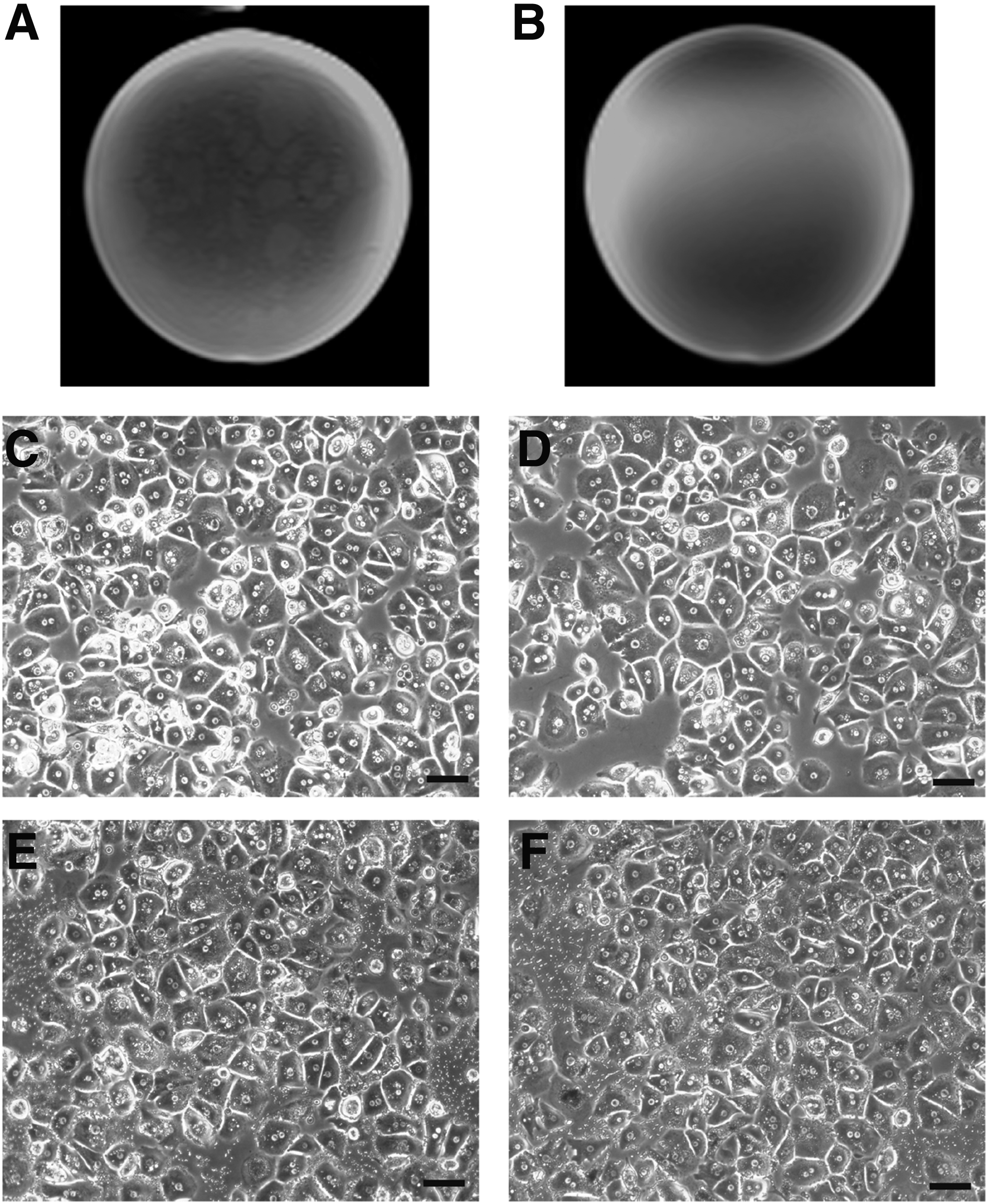
Cell morphology and MRI signal of the sMPIO-labeled cells. Labeling dose was 100 particles/cell. Scale bars: 10 μm.
Dose-dependent viability of the MPIO-labeled HuH7 cells after MRI
The analysis showed no interaction between the MRI treatment and the labeling dose at 24 or 48 h (Fig. 2). The particle concentration had no influence on viability (Fig. 2), and there were no differences in the multiple comparisons at any time point. Multiple comparisons of the influence of MRI treatment showed no differences between the groups at the 24-h time point, but there was a difference at 48 h after MRI treatment (Fig. 2). However, this effect could not be verified in the subsequently performed multiple comparisons.

Cell viability of the HuH7 cells depends on the particle load. Bars and whiskers indicate the means and standard error of the means, respectively. p values were calculated using two-way analysis of variance.
MRI does not alter the viability of the sMPIO-labeled primary human hepatocytes
The relative luminescence units (RLU) of the human hepatocytes incubated with the sMPIOs (100 particles/cell) was not changed by MRI (24 h, p > 0.999; 72 h, p > 0.999; 120 h, p > 0.999). Both groups that received the sMPIOs had slightly lower RLUs than the groups without the sMPIOs, but the difference was not statistically significant at any time point. In particular, the native controls without MRI stimulation with mean RLUs of 498912 ± 64106 at 24 h, 626515 ± 41103 at 72 h, and 671.348 ± 80687 at 120 h were not significantly higher than the corresponding RLUs of the particle-labeled hepatocytes that underwent MRI (24 h, p > 0.999; 72 h, p = 0.895; 120 h, p > 0.999) (Fig. 3).

Cell viability and ROS level of the human hepatocytes. Box plots display the 25th to 75th percentiles; whiskers indicate the smallest and largest values. p values were calculated using the Kruskal–Wallis test. Viability of the sMPIO-labeled and native human hepatocytes with or without MRI treatment after 24 h
MRI has no effect on ROS formation in the sMPIO-labeled cells
The relative fluorescent units (RFLs) indicating the formation of ROS remained stable for the entire culture period of all of the experimental groups.
Both groups that received the sMPIOs had slightly increased ROS levels at 24, 72, and 120 h after MRI. However, these differences did not reach statistical significance (24 h, p = 0.388; 72 h, p = 0.390; 120 h, p = 0.373). MRI did not induce changes in the RFLs of the MPIO-labeled or native cells (Fig. 3).
Neither MRI nor the sMPIOs cause alterations in the enzyme leakage of primary human hepatocytes
The levels of AST and ALT remained stable throughout the entire culture period until 120 h after MRI. There were no statistically significant differences between the native cells with or without MRI or between the sMPIO-labeled cells with or without MRI at any time point (24 h/AST, p = 0.263; 72 h/AST, p = 0.829; 120 h/AST, p = 0.652; 24 h/ALT, p = 0.958; 72 h/ALT, p = 0.679; 120 h/ALT, p = 0.949) (Fig. 4).

Enzyme leakage. Box plots display the 25th to 75th percentiles; whiskers indicate the smallest and largest values. p values were calculated using the Kruskal–Wallis test. Aspartate aminotransferase leakage of the sMPIO-labeled and native human hepatocytes with or without MRI after 24 h
Metabolic function is unaffected by MRI and the sMPIOs
Albumin synthesis was lowest at 24 h after MRI, and it increased in all of the groups for the entire culture period until 120 h after MRI. The albumin synthesis did not differ significantly at any time point among any of the experimental groups (24 h, p = 0.862; 72 h, p = 0.399; 120 h, p = 0.676) (Fig. 5). Urea synthesis remained stable throughout the entire culture period and did not show any statistically significant differences among any of the groups (24 h, p = 0.930; 72 h, p = 0.964; 120 h, p = 0.640) (Fig. 5).

Albumin and urea synthesis. Box plots display the 25th to 75th percentiles; whiskers indicate the smallest and largest values. p values were calculated using the Kruskal–Wallis test. Albumin synthesis of the sMPIO-labeled and native human hepatocytes with or without MRI after 24 h
Discussion
The key for improving the efficiency of cell-based therapies is to understand the way in which transplanted cells are distributed in the recipient organism and how they implant in the targeted tissue or organ. Iron oxide particles, SPIOs and MPIOs, could be a useful tool used to gain knowledge of the mechanisms of cell retention, survival, migration, and integration. 11 Several authors have described the potential advantages of this approach, and initial clinical studies have been performed; there are still major limitations, however, for the application of iron oxide particles for MRI-based cell tracking in humans. In brief, these are the challenge of distinguishing labeled cells and cells of the phagocytotic system that have taken up free particles,26–28 unclear long-term effects of the iron oxide particles inside the human body,10,14–16,18,19,21,28 and possible harmful unwanted effects provoked by iron oxide particles in combination with magnetic fields. 20
Bae et al. reported an early study that questioned the possible side effects arising from magnetic fields. The authors of this study used an SMF with a relatively low field strength of 0.4 T to prepare ferucarbotran aggregates, which were then added to a set of cells. They reported adverse effects, such as reduced viability, apoptosis, and cell cycle aberrations—even at clinical doses of SPIOs. 20 To gain a resolution enabling single cell discrimination, as demonstrated by previous studies,14,17,28 higher field strength is inevitable. We attempted here to establish experimental conditions closer to the clinical setting of the transplantation of MPIO-labeled cells. The main differences from the previous study are as follows. (1) Regarding the different particle types used, Bae et al. used ferucarbotran (Resovist; Bayer Healthcare, Berlin, Germany), which has a core composed of SPIO crystals with a carbodextran coating and a diameter between 45 and 60 nm. 29 In contrast, the iron oxide particles used in this study have an average diameter of 10–15 nm and are embedded in a silica-based polymer matrix. A possible mechanism of deterioration of the iron oxide particles is that the nanometer-sized iron cores oscillate in the alternating magnetic field of MRI, which might lead to damage by mechanical magnetic pull. Induction of heat in the magnetic field and, therefore, thermal damage of the cell would be another possible mechanism. Since the iron oxide particles in the sMPIOs that we used here are embedded in a polymer matrix, it is supposed that this effect would occur only locally and in an attenuated form. (2) A stronger magnetic field was applied using a clinical 3 T MRI, which combines SMF and dynamic magnetic field. (3) The cells were incubated with the MPIOs before they underwent MRI, as it would be when cells are used for cell tracking in hepatocyte transplantation. (4) Bae et al. used a murine epithelial cell line (NCTC 1469), whereas we used primary human hepatocytes to evaluate the applicability for cell tracking for LCT. SPIOs, such as Resovist, have already been used for the labeling of human hepatocytes.30,31 Although we would have preferred to compare directly the effects of MRI on the SPIO- and sMPIO-labeled cells, we were not able to use a control group with equivalent SPIOs because Resovist and other similar SPIOs are no longer available on the market.32,33
The results of our dose-finding studies using HuH7 cells showed no reduction of viability after 24 or 48 h, by MRI alone, by sMPIOs alone, or by a combination of both. Moreover, the experiments with the HuH7 cells showed no dose-dependent reduction in viability up to a labeling dose of 160 particles/cell. These results prompted us to perform an in-depth MRI toxicity assessment using primary human hepatocytes at a labeling dose of 100 particles/cell.
We could not observe any significant effects of the magnetic field of MRI on cellular viability in any experimental group at any time point. These findings were supported by the transaminase levels measured for 120 h, which peaked at 24 h and slightly decreased for the culture period but were the same levels in all groups. Urea and albumin synthesis was slightly higher in the unlabeled groups but did not differ significantly among any of the groups and increased for the culture period, allowing us to assume that metabolic function was not severely affected by sMPIO labeling, regardless of additional MRI stimulation. ROS levels showed inverse profiles compared with cell viability, with slightly higher relative fluorescence in the sMPIO-labeled groups. Oxidative stress induced by iron oxide particles might be an explanation for our other findings, namely the slightly reduced viability and albumin and urea synthesis in the particle-labeled cells. However, it is important to emphasize that stimulation by the magnetic field of MRI neither influenced these effects nor caused any additional adverse effects.
The biocompatible sMPIOs that we used in this study have already been shown to be nontoxic 9 at a labeling concentration of 60 particles/cell. We decided to increase the labeling dose to 100 particles/cell to detect even small effects provoked by MRI. This increased labeling dose probably accounted for the slightly elevated ROS levels and decreased viability in both particle-labeled groups.
In contrast to the observations reported by Bae et al., we could not detect any harmful effects caused by MRI stimulation in combination with the sMPIOs in this in vivo analysis. Therefore, sMPIOs continue to appear to be a safe contrast agent for MRI-based cell tracking and provide two important advantages toward SPIOs. MPIOs are larger and have a higher iron content and, therefore, the necessary labeling doses under-run those of SPIOs by far while signal strength in MRI remains equivalent. 14 Bae et al. already pointed out in their study that particle aggregation in the presence of magnetic fields might be a key factor for particle-provoked cell damage. Furthermore, they suggested that different surface fabrication could prevent particle aggregation and solve related cytotoxic effects. 20 The particles used in our study are coated with a silica-based polymer surface and only the particle core contains iron oxide particles. We could not observe any particle aggregation, neither under the influence of magnetic fields nor without. Nevertheless, with regard to obstacles such as donor–host cell distinction and their unclear long-term effects in the recipient organism, further studies are needed to optimize MRI-based cell tracking using iron oxide particles.
Footnotes
Acknowledgments
Dr. Nathanael Raschok is participant of the BIH Charité Clinician Scientist Program funded by the Charité – Universitätsmedizin Berlin and the Berlin Institute of Health (BIH).
Disclosure Statement
No competing financial interests exist.