
Abstract
Herein, we report the fabrication of orientation-controlled tissues similar to heart and nerve tissues using a cell accumulation and three-dimensional (3D) printing technique. We first evaluated the 3D shaping ability of hydroxybutyl chitosan (HBC), a thermoresponsive polymer, by using a robotic dispensing 3D printer. HBC polymer could be laminated to a height of 1124 ± 14 μm. Based on this result, we fabricated 3D gel frames of various shapes, such as square, triangular, rectangular, and circular, for shape control of 3D tissue and then normal human cardiac fibroblasts (NHCFs) coated with extracellular matrix nanofilms were seeded in the frames. Observation of shape-controlled tissues after 1 day of cultivation showed that the orientation of fibroblasts was in one direction when a short-sided, thin, rectangular-shaped frame was used. Next, we tried to fabricate orientation-controlled tissue with a vascular network by coculturing NHCF and normal human cardiac microvascular endothelial cells. As a consequence of cultivation for 4 days, observation of cocultured tissue confirmed aligned cells and blood capillaries in orientation-controlled tissue. Our results clearly demonstrated that it would be possible to control the cell orientation by controlling the shape of the tissues by combining a cell accumulation technique and a 3D printing system. The results of this study suggest promising strategies for the fabrication of oriented 3D tissues in vitro. These tissues, mimicking native organ structures, such as muscle and nerve tissue with a cell alignment structure, would be useful for tissue engineering, regenerative medicine, and pharmaceutical applications.
Introduction
R
Based on these facts, several research groups have actively researched cell orientation control by using a variety of techniques in the study of 3D tissue structure with native organ-like structure. Cimetta et al. have reported that cell adhesive protein was printed on a substrate in a thin straight line by using microcontact printing technology. As a result of adhesion of cells on the line, orientation control of the cells was achieved. 10 Nakamura et al. reported that a nonadhesive polymer was printed on an adhesive substrate by using an inkjet printing technique. As a consequence of cell adhesion on the thin line, they achieved fabrication of orientation-controlled tissue. 11 Bian et al. have reported a mold with a micropattern like a meshwork by using photolithography. As a result of cultivation of cell-containing gel solution by using the mold with meshwork pattern, the 3D tissue with cell orientation along the meshwork was fabricated. 12 Thus, orientation control of cells has been achieved by controlling the arrangement of cells using microfabrication technology. On the other hand, research on construction using 3D printer technology has also been actively conducted to obtain tissues with a predetermined cell arrangement and shape. For example, many kinds of bioprinting systems have been developed, such as robotic dispensing, inkjet printing, laser-assisted printing, and stereolithography techniques.13,14 Furthermore, construction of a 3D structure, including cells has been achieved using these techniques.15–18 However, the fabrication of orientation-controlled 3D tissue in which predetermined shape control is performed by using a 3D printing technique has not yet been achieved.
In a previous study, we developed a cell-accumulation technique, which was able to fabricate 3D multilayered tissues by coating ECM nanofilms onto a cell membrane using a layer-by-layer (LbL) technique (Fig. 1a).19,20 The ECM nanofilms were about 10 nm thick (nine layers) with fibronectin and gelatin (FN-G), which induced cell adhesion protein. We have achieved the fabrication of various kinds of tissues, such as blood vessel models and cardiac tissue models, by using human induced pluripotent stem cell-derived cardiomyocytes.21–25 In addition, we have fabricated micrometer-sized multilayered tissue for microarray using an inkjet technique. As a result, cells and ECM were alternately ejected and laminated to obtain functional 3D multilayered tissue. 26 The 3D multilayered structure made by the LbL technique had a high cell density because it consisted of only cell and ECM nanofilms. It is advantageous to fabricate cell–cell interactions such as those occurring at the tight and gap junction. Furthermore, these cells coated with ECM nanofilms could be expected to exhibit cell–ECM interaction. With this technique, we could construct biomimetic structures, such as 3D skin tissues, which have a layered structure.27,28 In addition, evaluation of drug response in myocardial tissue with a vascular network has been achieved using the cell accumulation technique. Control of orientation is indispensable for further evaluation in vitro; however, construction of myocardial tissues with controlled orientation has not yet been achieved using a cell accumulation technique.

In this study, we report the fabrication of orientation-controlled 3D tissue models by combining the LbL technique and a 3D printing system. At first, we selected the thermoresponsive polymer gel, hydroxybutyl chitosan (HBC) as the printing material because it has the ability of sol–gel transition dependent on the temperature. 29 In the next experimental step, we evaluated the molding ability of HBC gel using a 3D printer. Next, we fabricated a HBC gel frame on a culture dish by using a 3D printer. In addition, LbL coated normal human cardiac fibroblasts (NHCF) were seeded in the HBC gel frame and culture. The cell morphologies of the shape-controlled 3D tissues were observed by using cytoskeleton staining and quantitative alignment analysis (Fig. 1b). Finally, we tried to fabricate orientation-controlled 3D tissue using cocultured NHCF and human cardiac microvascular endothelial cells (HMVEC).
Materials and Methods
Materials
All of the chemical reagents were used without further purification. Chitosan was kindly donated by Dainichiseika Color & Chemicals Mfg. Co. Ltd. Fibronectin (FN) from human plasma, Triton X-100, and bovine serum albumin were purchased from Sigma-Aldrich. Gelatin (G) and 4% paraformaldehyde (PFA)/PBS were purchased from Wako Pure Chemical Industries. 4′6-diamidino-2-phenylindole, dihydrochloride (DAPI) and Alexa Fluor 546-conjugated goat anti-rabbit IgG cross-absorbed secondary antibody were purchased from Thermo Fisher Scientific. Cell culture inserts with a 0.4 μm pore membrane (polyester) and cell culture inserts with a 3 μm pore membrane (polycarbonate) were purchased from Corning. NHCFs, human cardiac microvascular endothelial cells (HMVEC-C), Fibroblast Basal Medium (FGM-3), and endothelial growth medium (EGM-2 MV) were purchased from Lonza. Acti-stain 488 fluorescent phalloidin was purchased from Cytoskeleton, Inc. Cellmatrix Type I-C was purchased from Nitta-Gelatin. Rabbit polyclonal anti-CD31 antibody was purchased from Abcam.
Synthesis of thermoresponsive polymer
In this study, HBC was used as a thermoresponsive polymer (Fig. 2). HBC has the ability of sol–gel transition dependent on the temperature and it was synthesized based on a previously reported method. 29 Chitosan (1 g) was dissolved in 60 mL of 0.1 M hydrogen chloride (HCl) at an ambient temperature with stirring. Sodium hydroxide (5 M) was then added in a dropwise manner to adjust the pH to 8. After the solution was heated to 85°C, 10 mL of 1,2-butylene oxide (BO) was added and mixed at 85°C for 3 h. During the reaction, some precipitates of HBC appeared. To resolve these precipitates, 5 M HCl was added until all precipitates were dissolved. Then, a further 10 mL of BO was added and the reaction was allowed to continue for 24 h at 85°C. The product was then thoroughly washed with pure water by centrifugation, and lyophilized for 4 days. The degree of substitution (DS) was evaluated by elemental analysis according to a previous report. 30 The DS was calculated by the following equation: DS = {(C/N)m–(C/N)0}/4, where (C/N)m was C/N (mole ratios) of the chitosan derivative, (C/N)0 was the C/N (mole ratios) of the chitosan. 30 The degree of substitution (DS) could be controlled by changing the feeding quantity of BO. Elemental analysis revealed that the DS of the obtained HBC was 2.1. This polymer showed a thermoresponsive reaction, whereby the solution immediately showed gelation above 37°C while rapidly dissolving below 4°C.
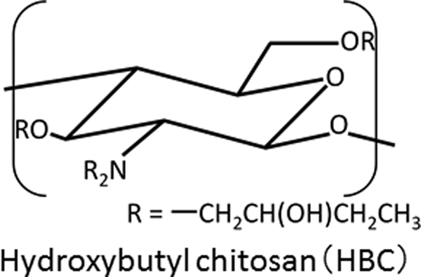
Chemical structure of HBC. HBC, hydroxybutyl chitosan.
Designing 3D frames with a thermoresponsive polymer using a 3D printer
The thermoresponsive polymer was printed by using a dispenser type 3D printer (SHOTMASTER 200DS; Musashi Engineering). Figure 3 shows a schematic of the robotic dispenser type 3D printer. It mainly consists of three modules: a syringe pump module, Y moving stage and the X-Z motors module, and a temperature control module (the nozzle was cooled to 10°C and the stage was heated up to 40°C). The HBC (25 mg/mL) solution was loaded into a 1-mL syringe with a double thread screw taper nozzle with an internal diameter of 610 μm. In this study, the syringe pump, moving speed, and nozzle-to-collector distance were fixed at 7 μL/s, 2.5 mm/s, and 250 μm/layer, respectively. The morphology of the printed HBC gels were observed from a horizontal angle from where the HBC gel height was measured using ImageJ software. 31 To prepare the cell culture dish, the HBC gel was printed onto glass bottom dishes which were coated with type-I collagen (nozzle cooled to 10°C, stage heated up to 40°C). The fabricated HBC gel frame shapes were square (10 × 10 mm), circular (11.6 mm diameter), triangular (15.2 mm sided), and rectangular (20 × 5 mm). The area of the HBC frame was designed to be unified to 1 cm2. In the case of the culture insert with a 0.4 μm pore membrane, where the HBC gel was printed onto the membrane (nozzle cooled to 10°C, stage heated up to 50°C), the fabricated HBC gel frame shape was rectangular, where the long side was 15 mm and the short side was 2 mm and 3 mm.
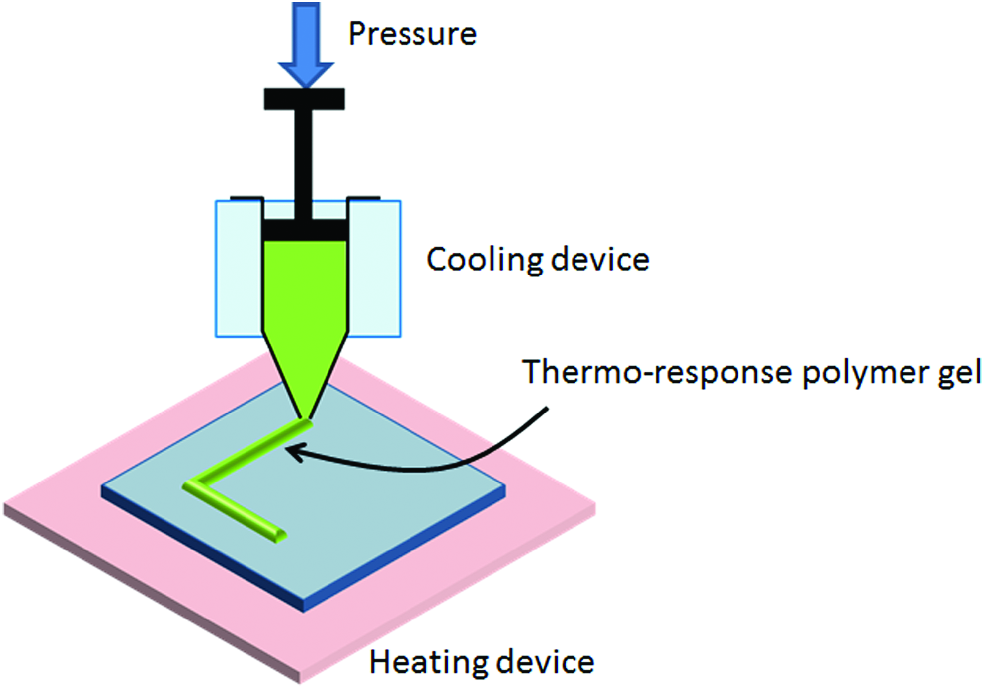
Schematic illustration of the robotic dispensing 3D printer setup. Color images available online at
Fabrication of ECM nanofilm using a filtration-LbL method
NHCFs (passage was less than 7) were cultured with FGM-3 at 37°C in an incubator at 5% CO2. We performed filtration-LbL based on a previously reported method. 23 For the filtration-LbL, 2.5 mL of 0.2 mg/mL FN and G/PBS solution and PBS were added into each well in a six-well cell culture plate. Isolated NHCFs which used 0.1% trypsin were suspended in 500 mL of PBS after centrifugation and added a six-well culture insert with a 3 μm pore membrane. The insert was set at the well containing FN/PBS and G/PBS solution and alternately soaked and agitated at 500 rpm for 1 min at room temperature using a MixMate shaker (Eppendorf) through PBS washing steps. These coating steps were executed for a total of nine steps (FN: five times, G: four times).
Fabrication of shape-controlled 3D tissues using a cell accumulation technique
After nine steps of the LbL coating process, FN-G nanofilms were fabricated on each of the cell surfaces. In the case of glass bottom dishes, these cells were suspended in a medium and seeded into the HBC gel frame (1 × 10⁶ cells per frame). After 2 h, an additional 2 mL of medium was added into each dish and incubated in 5% CO2 at 37°C. On the other hand, in the case of the culture insert, 3 mL of LbL coated cell suspension (2.4 × 10⁶ cells/mL) were seeded into the culture inserts with a HBC gel frame and 2 mL culture medium was added to the outside of the insert. After a 2-h culture, an additional 5 mL of medium was added into each well and incubated in 5% CO2 at 37°C. In the case of fabrication of tissue with a vascular network, 3 mL cell suspension mixed with ECM nanofilm-coated NHCFs (7.1 × 10⁶ cells) and HMVEC (7.1 × 10⁵ cells) were seeded into the culture inserts (4.67 cm2) with a HBC gel frame and 2 mL culture medium was added to the outside of the insert. After 2-h culture, an additional 5 mL of medium was added into each well and incubated in 5% CO2 at 37°C. After 1 day of incubation, the samples were fixed with 4% PFA for 20 min at room temperature.
Fluorescence staining of 3D tissues and histological analysis
For the histological evaluation of the 3D tissues, the samples were fixed with 4% PFA. Samples were permeabilized with 0.2% Triton X for 20 min at room temperature. After washing three times with PBS, these samples were then incubated with Acti-stain 488 fluorescent phalloidin for 60 min at room temperature in darkness. After washing three times with PBS, the samples were incubated with DAPI (10 μg/mL) for 30 min at room temperature in darkness to stain the nuclei. The endothelial cells were immunostained with an anti-CD31 antibody. Fixed tissues were permeabilized with 0.2% Triton X for 20 min at room temperature, blocked with 1% BSA/PBS for 1 h at room temperature, and incubated with the primary antibodies (1:200) overnight at 4°C. After the washing step, the secondary antibodies (1:200) were added to the tissues for 2 h at room temperature. The samples were observed by confocal laser scanning microscopy (CLSM) (FLUOVIEW FV10i; Olympus) (Cell Voyager CV1000; Yokogawa) (LSM 780; Carl Zeiss). The local orientation of the cell alignment was estimated using an ImageJ plug-in, OrientationJ. This plug-in is able to calculate the directional coherency coefficient of the F-actin fibers and CD31, a marker of blood capillaries.32,33
Results
Fabrication of HBC gel structure by 3D printer
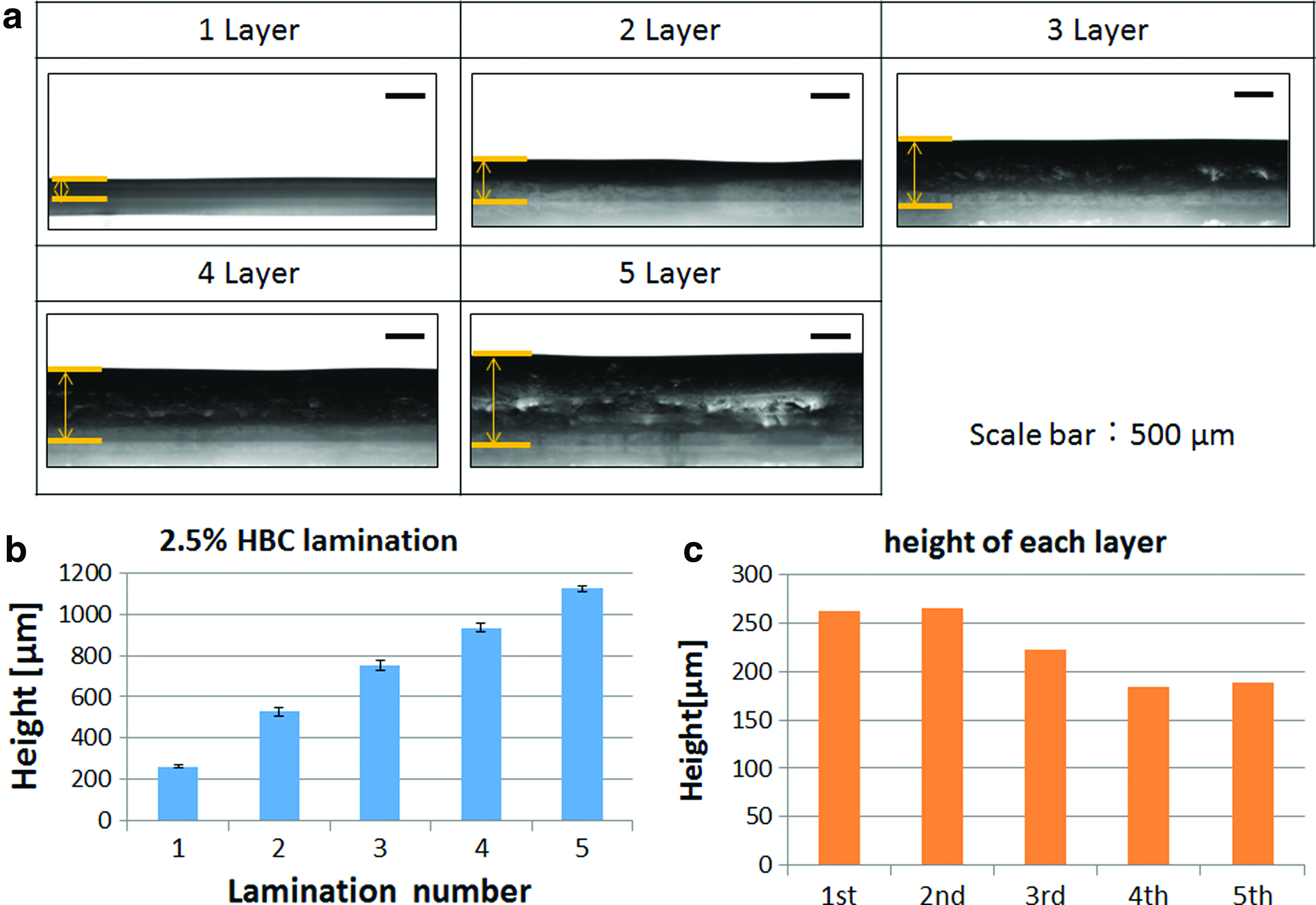
First, we assessed the shaping ability of the HBC gel using a robotic dispensing 3D printer for fabricating a gel frame. HBC gel cooled to 4°C was printed and laminated into a linear shape on the slide glass heated to 40°C. As a result, the HBC line width was about 1.4 mm and the HBC gel could be printed and laminated until 5 layers were observed from the horizontal direction by a contact angle measuring device, as shown in Figure 4a. Figure 4b shows the measured height of the laminated HBC gel. The HBC gel height gradually increased from 262 ± 4 μm to 1124 ± 14 μm as the lamination number increased from 1 to 5. Figure 4c shows the height of each layer calculated using the data from Figure 4b. These data indicate that the layer thickness of the laminated HBC gel decreased with increasing layer numbers.
Shape control of 3D tissue by HBC polymer gel
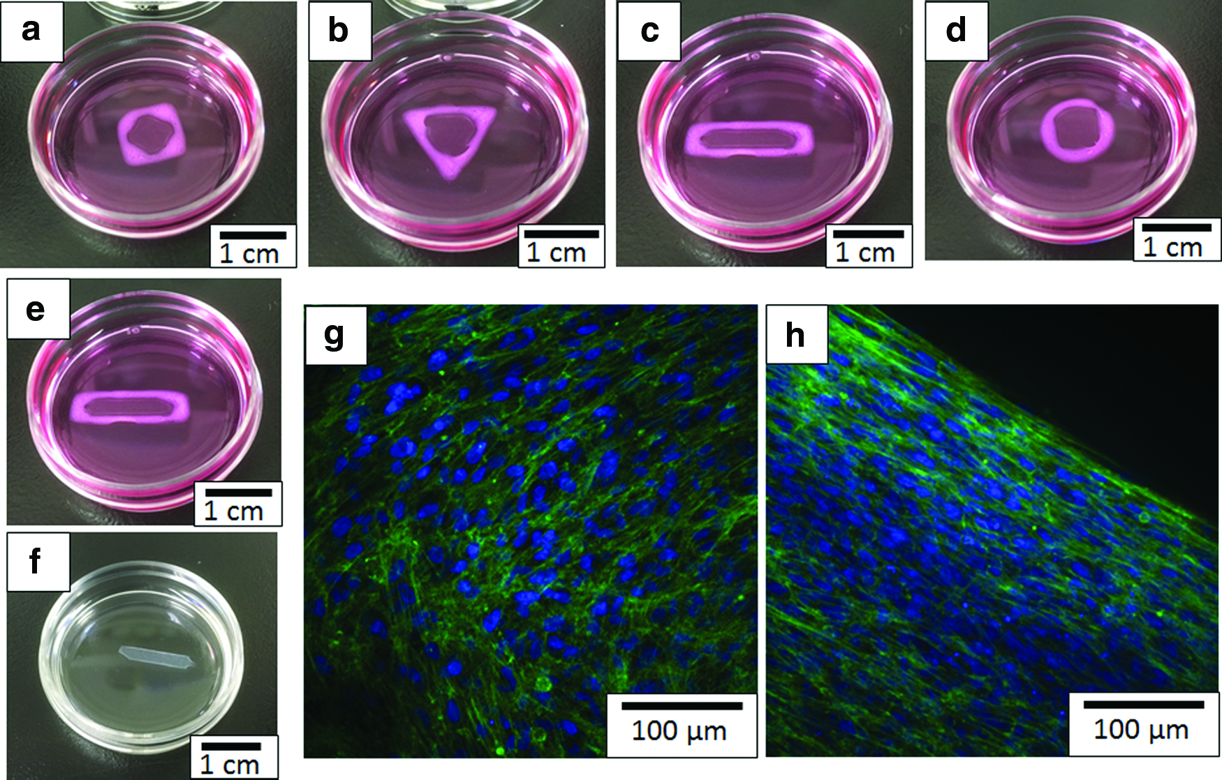
The HBC gel could be three-dimensionally laminated by using a 3D printer. Next, we tried to fabricate the shape-controlled 3D tissue by using a HBC polymer gel print and cell accumulation technique. Figure 5a–d shows that variously shaped 3D tissues, such as square, triangular, rectangular, and circular were obtained. HBC gel frame could be easily removed when it was cooled on ice. Because it is water soluble due to the hydrophilic change (sol–gel transition) at a low temperature (Fig. 5e, f). The HBC gel frames dissolved in about 5 min. Figure 5g and h shows that F-actin of NHCF in these fabricated shape-controlled 3D tissues were stained with Acti-stain 488 fluorescent phalloidin. The images indicated that the direction of cell extension in the central region of the shape-controlled 3D tissues was random (Fig. 5g). On the other hand, the images indicated that the direction of cell extension in the edges of the shape-controlled 3D tissues was aligned in one direction (Fig. 5h).
Orientation-controlled 3D tissue by HBC polymer gel
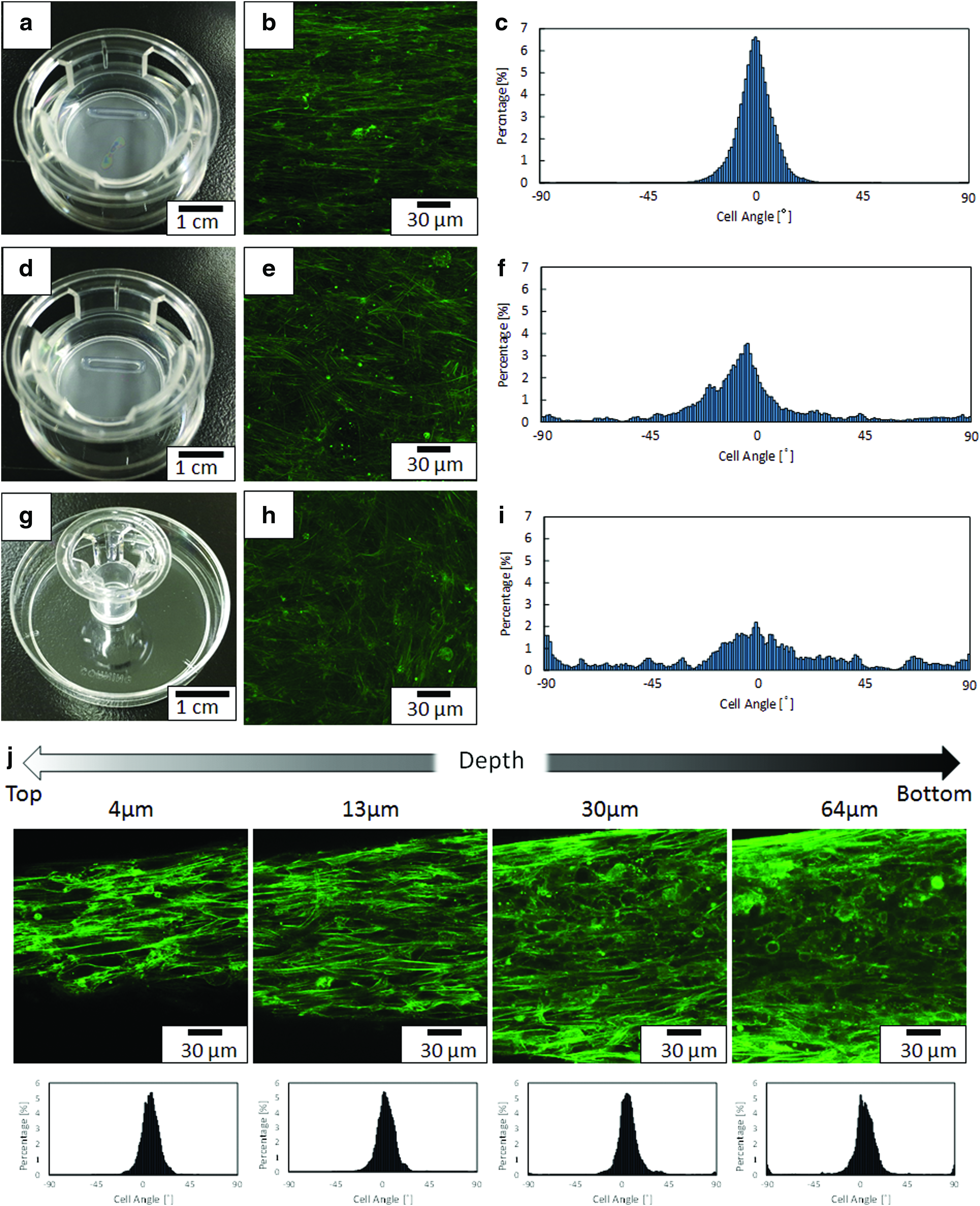
From Figure 5h, the aligned cell-orientation was observed in the edges of tissue in the frame. Therefore, we tried to fabricate orientation-controlled 3D tissue by using a shape-controlled HBC polymer gel. We focused on the rectangular-shaped frame to control the orientation of all cells in the 3D tissue. Figure 6a, d, and g shows a fabricated HBC gel frame with a 2 × 15 mm rectangle (Fig. 6a), a 3 × 15 mm rectangle (Fig. 6d), and a 24-well culture insert (Fig. 6g). The width of orientation-controlled 3D tissues constructed by using the 2 × 15 mm and 3 × 15 mm rectangle HBC gel frame were 500 μm and 1400 μm, respectively. F-actin of NHCF in these fabricated width-controlled 3D rectangular tissues was stained with Acti-stain 488 fluorescent phalloidin. In the case of the 2 mm short side rectangle, the cell orientation in the fabricated 3D tissue was in one direction (Fig. 6b). On the other hand, cell extension direction in fabricated 3D tissue with a 3 mm short side rectangle (Fig. 6e) and 24-well insert (Fig. 6h) was random. Figure 6c, f, and i shows the results of the fiber alignment measurement analyzed by ImageJ. These results are shown as a graph of F-actin fiber angles of NHCF 2 mm short side rectangle HBC gel frame (Fig. 6c), 3 mm short side rectangle HBC gel frame (Fig. 6f), and 24-well insert (Fig. 6i). These data indicate that by performing shape control, the number of cells extending in the same direction was increased in the case of the 2 × 15 mm rectangle gel frame. Figure 6j shows Z stack images of 3D tissues which were fabricated on a 2 mm short side rectangular HBC gel frame. These images indicate that it is possible to control cell orientation in 3D tissue to within 64 μm of the surface.
Vascularization of the orientation-controlled 3D tissue
Finally, we fabricated orientation-controlled 3D tissues containing vascular networks. To introduce the blood capillaries in the 3D tissue, NHCFs with FN-G nanofilms were cocultured with HMVEC using 2 × 15 mm HBC gel (5%) rectangular frame. After 4 days of culture, tissues were fixed and stained with fluorescent dyes (Fig. 7a–f). Figure 7a and d shows F-actin images of NHCF and HMVEC stained with Acti-stain 488 fluorescent phalloidin. Figure 7b and e shows immunofluorescence analyses of HMVEC for the specific endothelial marker, CD31. Figure 7c and f shows merged images of F-actin and CD31. The orientation-controlled tissues using the HBC gel frame are shown in Figure 7a–c and uncontrolled tissues are shown in Figure 7d–f. As a result of NHCF and HMVEC coculture, alignment of F-actin fibers was observed in the shape-controlled tissue on a 2 mm short side rectangular HBC gel frame (Fig. 7a). On the other hand, uncontrolled tissues did not show alignment of F-actin fibers (Fig. 7d). According to the images in Figure 7b and e, HMVEC also has orientation by shape control. Figure 7g and h shows the results of the quantitative alignment analyses of vascular networks by ImageJ. The orientation of the blood network was observed in the shape-controlled tissue, but not in the uncontrolled tissue.

Discussion
There is considerable worldwide demand for fabricated 3D tissues in regenerative medicine and tissue engineering for medical treatments and in vitro assays. In the artificial construction of 3D tissues, it should be borne in mind that tissues and organs in the human body are known to have specific structures and multiple functions, which are triggered from the ECM microenvironment, multicells, adjacent cell–cell interaction and cell–ECM interaction. However, it is difficult to make an artificial 3D tissue with native-like organ structure and functions. Our previous studies reported the fabrication of 3D tissues, which were constructed only of cells and ECM by using the LbL technique.19–28 Using this technique, it is easy to control the cell type per layer and fabricate a vascular network in the tissue. Moreover, compared with other 3D tissue fabrication techniques, such as using hydrogels, porous scaffolds, and nanofiber scaffolds, this technique can easily obtain the high cell density necessary for cell–cell interaction, because the fabricated tissues use only cells and ECM nanofilms. However, although this fabrication technique was able to construct biomimetic tissues with a layered structure such as skin, orientation, which is one of the features necessary for exerting the function of living tissue, has not yet been replicated. Thus, this fabrication technique is insufficient when attempting to reproduce biomimetic tissues for the control of cell orientation. In our previous study, drug evaluation using 3D cardiac tissue by iPS-derived cardiomyocytes has been already achieved. 23 If orientation control could be achieved by construction using the LbL technique, it is considered that this could greatly contribute to drug screening in vitro.
In this study, we focused on fabricating orientation-controlled 3D tissue through the shape control of tissue. Schell et al. reported the fabrication of alignment-controlled ECM, which depended on tissue shape in the microtissues by using micromold and self-assembly techniques. 34 From these results, we tried to fabricate orientation-controlled 3D tissue through controlled tissue shape by using a robotic dispensing 3D printer system and HBC, which is a thermoresponsive polymer gel. The reason for using a robotic dispensing 3D printer system is that it is able to easily fabricate the 3D structure of predetermined shape and can use a wide variety of materials such as those with high viscosity and temperature dependence. In addition, the ink material used for tissue construction in 3D printers possesses the required biocompatibility and biodegradability features. HBC is a biodegradable and biocompatible polymer composed of naturally derived chitosan, and has thus been variously studied for use as a scaffold in tissue engineering,35,36 coating materials for tissue deadhesion, 29 and thermosensitive drug carriers in pharmaceutical fields.37,38 We hypothesized that the rapid thermoresponsive behavior of HBC could make it useful as a 3D frame for the construction of the desired tissue shape.
At first, HBC was synthesized as a material for 3D printing ink. The synthesized HBC had properties whereby gel formation and solation occurred at 37°C and 4°C, respectively. In addition, we tried to laminate the HBC by using a robotic dispensing 3D printer. HBC gel could laminate at least five layers (Fig. 4a). The reason for this is considered to be that the sol–gel transition of HBC gel quickly occurs during the 3D printing process. By using this system, a HBC gel frame with a maximum height of 1000 μm could be formed, which has sufficient size to allow for the control of the 3D tissue shape. These results indicated that a 3D printer and HBC gel are suitable tools for fabrication of shape-controlled tissue using the LbL technique.
Next, we fabricated shape-controlled tissue by using both 3D printing of HBC gel and the LbL technique. Schell et al. reported that the orientation of cells and ECMs in shape-controlled microtissues are dependent on the shape of the tissue. 34 Hence, we investigated the cell morphologies in 3D tissues corresponding to the various tissue shapes. We observed the morphologies of NHCF which were stained with F-actin in fabricated shape-controlled tissues (Fig. 5g, h). The obtained data demonstrated that the cell extension in one direction occurred in a part of the shape-controlled tissues.
Moreover, we tried to fabricate orientation-controlled 3D tissue by using 3D printing of HBC gel and the LbL technique. Nakamura et al. reported that aligned smooth muscle cell fiber-like tissue was fabricated using a bioprinting and transfer patterning technique. Noncell adhesive polymer was printed on a substrate by drawing a linear pattern by using inkjet printer. After cell seeding, cells attached to the substrate were transferred onto a gel. 11 Cimetta et al. reported that aligned cardiac and skeletal muscle myofibers were produced by using a micropatterning technique, which used microcontact printing of ECM on a soft substrate. 10 Thus, it is considered that the 2D cell orientation can be controlled by fabricating the linear shape tissues. However, there are few reports of 3D cell orientation by scaffold-free culture method.
To fabricate the 3D cell-oriented tissue, we controlled the shape of tissue using a HBC gel when cultured using a 2 mm short side rectangular HBC gel frame. The cell orientation was confirmed in fabricated 3D tissue. Figure 6g shows that it is possible to control cell orientation to within 64 μm of the surface. This result indicates that the obtained orientation-controlled tissue is 3D. Furthermore, it is shown that orientation control of the inside of the 3D tissue was achieved. Therefore, although the orientation control in 2D was reported in the preceding example, the current method suggests that orientation can also be controlled in a 3D environment. Moreover, several research groups reported that fabrication of orientation-controlled 2D culture cells by using patterning method could be achieved until about 150 μm width.10,11,39 In this study, however, we found that cell alignment could be controlled until about 500 μm width. As a reason for this, it is considered that the tensile force generated by constructing the 3D tissue affects the cell alignment because cells attached on cell surfaces.
Finally, we tried to fabricate orientation-controlled 3D tissue with a vascular network using a 2 mm HBC gel frame. It is necessary to introduce vascular networks in 3D tissue for application in tissue engineering and pharmaceutical assays. Because oxygen diffusion was restricted to 100–150 μm depth in a tissue, blood vessels are necessary to make a thick tissue. 40 For this reason, we developed a fabrication technique for 3D tissue with a vascular network. 22 As a result of F-actin staining, shape-controlled tissue with HMVEC was shown to have an orientation similar to that of shape-controlled tissue fabricated with only NHCF (Fig. 7a, d). In addition, we observed the morphology of HMVEC-stained CD31 by immunofluorescence staining. A vascular network was formed in both shape-controlled and uncontrolled tissue (Fig. 7b, e). Moreover, as a consequence of quantitative alignment analysis by ImageJ, the vascular network was shown to be oriented by control of the shape (Fig. 7g, h). Rosenfeld et al. reported that vascular networks are oriented by tensile force in the 3D tissue. By transplanting this oriented vascular tissue in the same direction as the surrounding blood vessels, it was found that it conformed easily to surrounding tissues. 41 The obtained data suggest that orientation-controlled 3D tissue with a vascular network would be useful for transplantation therapy. This system, which is developed in this study, is useful for control of cell orientation in the construction of 3D tissues. It is important for replicating the function of organs, such as the heart, skeletal muscle, and nerve tissue, in vitro. Hence, it is conceivable that these results could contribute to regenerative medicine and pharmaceutical assays. Further studies, including fabrication of 3D cardiac tissues with blood capillaries using iPS-derived cardiomyocytes, are in progress.
Conclusions
In this study, we successfully constructed 3D tissues whose shape and cell orientation were precisely controlled about 50 μm thickness by employing LbL techniques and a 3D printing system using a thermoresponsive gel.
HBC gel, which is a temperature-responsive polymer used in this experiment, is capable of constructing 3D tissue with a height of about 1000 μm using a robotic dispenser type 3D printer. When the shape of the tissue was investigated using a frame prepared by HBC gel, it was recognized that the cells in the tissue were extended in one direction by using the shape of a 2 × 15 mm rectangle. By using this method, it is possible to fabricate 3D tissue with a specific native-like organ structure of orientation such as in heart and nerve tissues. HBC gel can be easily removed by lowering the temperature. Furthermore, the tissue assembly using the LbL technique consists only of cells and ECM components. Using this method, it is possible to obtain 3D tissue consisting of only cells and ECM.
These tissues should be useful for tissue engineering, regenerative medicine, and pharmaceutical applications.
Footnotes
Acknowledgments
This work was supported by “Development of Manufacturing Technology for Functional Tissues and Organs Employing Three-Dimensional Biofabrication” funded by the Japan Agency for Medical Research and Development (AMED)
Disclosure Statement
No competing financial interests exist.