
Abstract
Tissue growth and remodeling are essential processes that should ensure long-term functionality of tissue-engineered (TE) constructs. Even though it is widely recognized that these processes strongly depend on mechanical stimuli, the underlying mechanisms of mechanically induced growth and remodeling are only partially understood. It is generally accepted that cells sense mechanical changes and respond by altering their surroundings, by means of extracellular matrix growth and remodeling, in an attempt to return to a certain preferred mechanical homeostatic state. However, the exact mechanical cues that trigger cells to synthesize and remodel their environment remain unclear. To identify the driving mechanical stimuli of these processes, it is critical to be able to temporarily follow the mechanical state of developing tissues under physiological loading conditions. Therefore, a novel “versatile tissue growth and remodeling” (Vertigro) bioreactor was developed that is capable of tissue culture and mechanical stimulation for a prolonged time period, while simultaneously performing mechanical testing. The Vertigro's unique two-chamber design allows easy, sterile handling of circular 3D TE constructs in a dedicated culture chamber, while a separate pressure chamber facilitates a pressure-driven dynamic loading regime during culture. As a proof-of-concept, temporal changes in the mechanical state of cultured tissues were quantified using nondestructive mechanical testing by means of a classical bulge test, in which the tissue displacement was tracked using ultrasound imaging. To demonstrate the successful development of the bioreactor system, compositional, structural, and geometrical changes were qualitatively and quantitatively assessed using a series of standard analysis techniques. With this bioreactor and associated mechanical analysis technique, a powerful toolbox has been developed to quantitatively study and identify the driving mechanical stimuli of engineered tissue growth and remodeling.
Introduction
C
To unravel the mechanisms of growth and remodeling, a systematic investigation of the underlying processes and associated mechanical parameters is required. To this end, bioreactor systems are valuable for studying the response of soft tissues to mechanical stimuli at the cellular and tissue level. Initially, bioreactor systems were developed to study the effects of mechanical stimuli under static conditions.5,8–10 As cardiovascular tissues are mainly exposed to dynamic loading conditions, other bioreactors applied dynamic deformation to the tissue-engineered (TE) constructs.11–16 For example, Rubbens et al. and Gould et al. applied cyclic strain to study the remodeling of 3D TE constructs.14,16 Moreover, Engelmayr et al. tested different scaffold candidates for heart valve tissue engineering by designing a bioreactor capable of applying cyclic flexure and laminar flow. 11 In addition, several strain-driven bioreactors have been proposed applying physiological mechanical and electrical stimulation to cardiac tissues. 17 Finally, Kortsmit et al. introduced a bioreactor with a feedback system, to attain a constant maximal deformation, while culturing TE heart valves in vitro. 15 However, for cardiovascular tissues, in vivo deformation is mainly governed by hemodynamic flow and pressure, which are known to play an important role in tissue growth and remodeling. 18 To incorporate hemodynamic loading, Shaikh et al. and Hollweck et al. mimicked in vivo loading conditions by using pulsatile bioreactors that applied physiological pressures to TE constructs.19,20
The previously mentioned bioreactors lacked two main features to study mechanically driven growth and remodeling over time. First, these systems were not capable of tissue culture and mechanical stimulation for a prolonged time period, while simultaneously performing mechanical testing. Consequently, changes in the mechanical state of tissues with time could not be assessed for individual samples. Moreover, these systems did not retain a (dynamic) pressure during culture, which resembles the physiological load of many, primarily cardiovascular, tissue types and can lead to a different adaptation mechanism of these tissues compared to displacement-controlled mechanical stimulation. Particularly, when applying a constant pressure during culture, the strain will not necessarily remain constant over time. Consequently, a strain-driven bioreactor may not capture the in vivo loading conditions and hence growth and remodeling phenomena.
The goal of this study was to design and test a bioreactor that is capable of tissue culture and mechanical stimulation for a prolonged time period, while simultaneously performing mechanical testing. The bioreactor features a two-chamber design that allows easy and sterile handling of a TE construct, resembling thin cardiovascular tissues such as arteries and heart valves, in a dedicated culture chamber, while a second pressure chamber applies a physiologically relevant dynamic pressure regime, regulated by a custom-made feedback system. A novel nondestructive mechanical testing method is employed, involving a bulge test that can be performed inside the bioreactor, in which the construct's curvature is determined by ultrasound (US) imaging. This technique can be used to quantify temporal changes in tissue mechanical properties, without sacrificing the sample. Additional microscopy measurements of tissue thickness and prestretch serve to obtain the tissue geometry. Finally, confocal microscopy, histology, and biochemical assays allow for determining tissue composition and architecture.
Materials and Methods
Vertigro bioreactor
The “versatile tissue growth and remodeling” (Vertigro) bioreactor consists of two chambers: a pressure chamber (bottom) and a detachable culture chamber (top) (Fig. 1) in which a TE construct is cultured. This design was chosen to have a separate chamber for sterile tissue culture and medium changes, while the other chamber is dedicated to applying the dynamic pressure. Both chambers are made of polysulfone (PSU; Röchling) and have connection ports fitting standard luer-lock slip tips. After sealing both chambers with a silicone membrane, the culture chamber is secured on top of the pressure chamber by means of three metal clamps. In this configuration, the two silicone membranes form a double-membrane connection between the two chambers to ensure the transmission of pressure from the pressure chamber to the culture chamber.

The pressure chamber is connected to a pump containing a flexible silicone tube, which can be compressed with pressurized air. A proportional pneumatic valve (Festo) regulates the inflow of pressurized air, by opening in accordance to a programmable waveform function, supplied by a multi-IO-card using LabVIEW software (National Instruments).
The culture chamber consists of a lower and an upper part, separated by an insert that is clamping the tissue construct. Both parts of the culture chamber are filled with the culture medium. The top chamber is closed with a lid that allows free air exchange with the external environment, required to buffer the pH of the medium. Upon an increase in pressure in the pressure chamber, the membranes bulge into the culture chamber, pressurizing the tissue construct. A pressure sensor (P10EZ-1; BD|SENSORS) is connected to the lower part of the culture chamber to measure the pressure on the tissue in real time.
A feedback system was implemented in the LabVIEW framework to keep the minimal and maximal pressures at preset levels. In case of deviations from this preset level (±5% tolerance), either the offset (minimal pressure) or the amplitude (maximal pressure) of a square waveform function are changed accordingly, therefore maintaining a constant peak pressure during culture. The Vertigros, pressure sensors, and pumps can be placed in an incubator (37°C, 100% RH, and 5% CO2), while the proportional air valves, pressure modules, and other hardware remain outside.
Cell and tissue culture
Tissue constructs were engineered using vascular derived cells, previously characterized as contractile, matrix-producing myofibroblasts, 21 harvested from the human vena saphena magna according to the Dutch guidelines for secondary use of materials. Cells were expanded until passage 7 using a culture medium containing advanced Dulbecco's modified Eagle's medium (DMEM; Invitrogen), supplemented with 10% fetal bovine serum (FBS; Greiner Bio One), 1% Glutamax (Invitrogen), and 1% penicillin/streptomycin (Lonza) with medium changes twice a week.
Before seeding, rapidly degrading nonwoven polyglycolic acid (PGA) Biofelt (Biomedical Structures; 0.5 mm thickness, 70 mg/cm2 density) was cut to size to fit inside the inserts. To provide a slow-degrading fixation of the tissue on the edge of the insert, a polycaprolactone scaffold (PCL, electrospun in-house, thickness 0.25 mm) was cut to the same diameter as the PGA scaffolds, while a circular 13-mm cutout was made in the center. Next, the PGA was coated with poly-4hydroxybutyrate (P4HB; Tepha) and mounted together with the PCL rings into the bioreactor inserts. The inserts were then immersed in 70% ethanol for 30 min. After sterilization, the inserts were washed with PBS and placed in 0.25 mg/mL L-ascorbic 2-phosphate acid (Sigma-Aldrich)-supplemented standard culture medium (TE medium) overnight at 37°C and 5% CO2. The next day, the cells were seeded onto the scaffolds (15 × 106 cells/cm3) using fibrin as a cell carrier. 22 Finally, the constructs were cultured statically for 3 weeks in six-well plates in the TE medium (which was changed thrice a week). After this period, the constructs were strong enough to be cultured dynamically.
Experimental protocol
To assess bioreactor functionality and evaluate the proposed analysis techniques, a proof-of-principle experiment was performed (Fig. 2). In this experiment, 20 tissue constructs were cultured: in the first 3 weeks, these were cultured statically, taking out four samples at the end of each week. From the remaining eight constructs, four were cultured under static conditions for one additional week, while the other four samples were cultured under dynamic loading conditions (minimal pressure 0 kPa, maximal pressure 2 kPa, frequency 1 Hz).
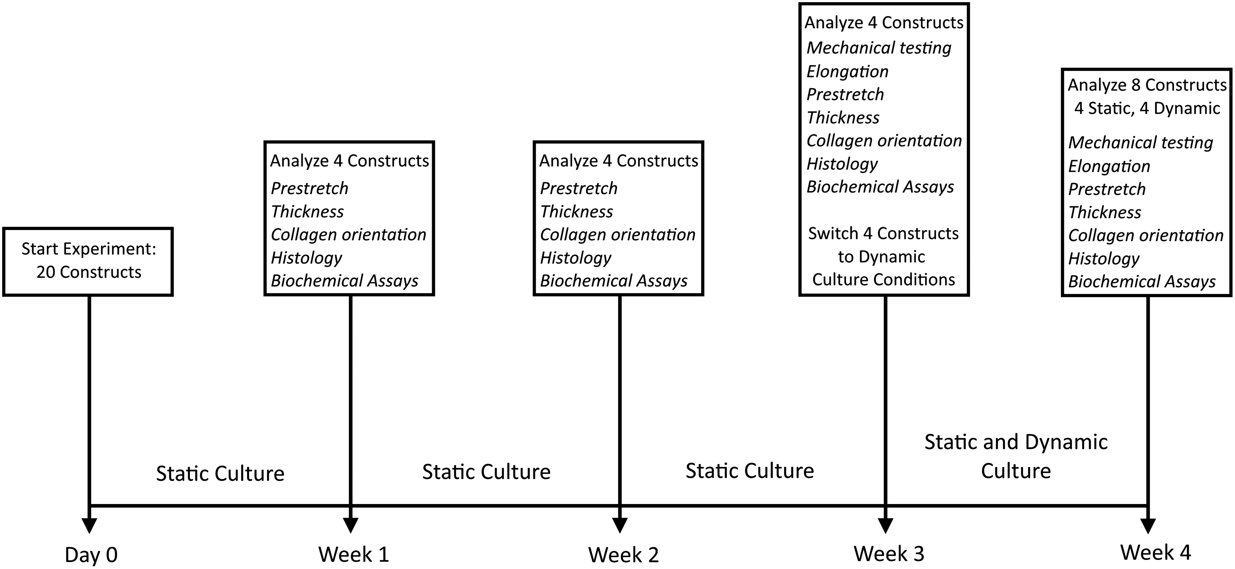
Timeline used for proof-of-concept experiment.
Analysis techniques
Mechanical testing
The bioreactor system was designed to perform mechanical testing via classical mechanical bulge tests without removing the constructs. A syringe was connected to the pressure chamber and placed in a Harvard pump (PHD2000; Harvard Apparatus) to apply a volume-controlled pressure to the samples. The volume was gradually increased until a pressure of 2 kPa was reached in the culture chamber. Subsequently, three pressure cycles were performed, of which the first two served as preconditioning cycles. A US transducer was mounted on top of the bioreactor, perpendicular to the sample surface, to nondestructively measure construct displacement as a function of pressure. Two-dimensional US imaging was performed with a MyLab70 US system (ESAOTE), equipped with a linear array (LA523, center frequency of 7.5 MHz). The data were exported in video format (AVI) at a frame rate of 20 Hz.
To quantify the mechanical behavior of the tissue, the in-plane stress was estimated as a function of stretch, assuming tissue isotropy. Tissue stretch was determined from the frames of the US movies (resolution 21.5 px/mm) using a custom MATLAB script (MathWorks). In particular, for each frame, the tissue profile was detected using MATLAB's built-in edge detection function, through which a circle was fitted in the center region. The constrained tissue length L0 at zero pressure and current tissue length L in each consecutive frame were then obtained by calculating the circle's arc length. In additon, tissue elongation, defined as the relative change of constrained length during culture time, was calculated as L0 (at 0 pressure) divided by the initial construct diameter (15.0 mm). The in-plane stretch due to bulging
To estimate the mechanical state of the tissue, the Tension T and Cauchy stress
with p the pressure,
Prestretch
Tissue prestretch was measured by calculating the change in surface area upon releasing the tissue constraints. Nine circular markers were placed in a 3 × 3 rectangular grid on each construct and positions were tracked using a high-resolution digital microscope (VHX-500; Keyence) at a resolution of 100 px/mm and 20× magnification. Subsequently, tissues were released from their constraints by dissecting them from the sample holder using a cork borer with a diameter of 13.0 mm and a second image was recorded immediately. The surface area spanned by the markers was calculated for both images using a semiautomatic custom MATLAB script. The amount of tissue prestretch was then calculated in terms of surface area change, defined as the unconstrained surface area divided by the constrained surface area. The surface area change equals the squared length change
Thickness
After the prestretch measurements, all constructs were cut in half. Three high-magnification (100 × ) images using the VHX-500 digital microscope were taken perpendicular to the cutting edge of one of the two halves, with the three positions coinciding with the three markers that were placed at the sample cutting edge for the prestretch measurements. Using the microscope's complimentary software, the thickness t0 was manually determined at five positions in each image.
Collagen orientation
The tissues were stained with CNA35 23 to enable collagen visualization. After a 1-h incubation period, constructs were visualized using a confocal laser scanning microscope (TCS SP5X; Leica Microsystems) with complimentary software (Leica Application Suite Advanced Fluorescence). A tile scan (magnification 10 × , excitation 488 nm, and emission 520 nm) was made to capture the gross collagen architecture of the entire tissue. Collagen fiber orientation was quantified by analyzing the individual images of each tile scan, using a custom MATLAB script, based on the work of Frangi et al., 24 which was previously used to quantify collagen orientation. 25 For each tile scan, a histogram containing the fiber fraction per angle was obtained.
Histology
Constructs were fixed in 3.7% formaldehyde overnight, and subsequently embedded in paraffin and sectioned into slices of 7 μm. The slices were stained for glycosaminoglycans (GAGs) and collagen using Alcian blue (AB) and Picosirius red (PR), respectively. The stained sections were imaged using a brightfield microscope (Axio Observer Z1; Zeiss) equipped with filters to provide polarized light illumination. In addition, a fluorescent costaining for collagen type 1, α-smooth muscle actin (αSMA), and cell nuclei was performed and visualized using an Axiovert 200M microscope (Zeiss).
Biochemical assays
Half of each construct was lyophilized overnight and subsequently digested using a papain digestion buffer at 60°C for 16 h. The total tissue dry weight was determined before and after digestion. Tissue GAGs were quantified using an adapted version of the protocol by Farndale et al., 26 together with a standard curve derived from chondroitin sulfate from shark cartilage (Sigma-Aldrich). Using the Hoechst dye method 27 and a standard curve of calf thymus DNA (Sigma-Aldrich), the total DNA content was quantified. Finally, the total hydroxyproline content, a measure for the amount of collagen, was determined with a modification of the assay by Huszar et al. 28 and a standard curve derived from trans-4-hydroxyproline (Sigma-Aldrich). Protein amounts were normalized with respect to the total tissue dry weight before digestion.
Results
Tissue culture in the Vertigro bioreactor
All statically cultured tissues endured the entire culture period up to 4 weeks (Fig. 3). Throughout dynamic culture, the feedback system successfully applied and maintained the preset maximal and minimal pressures to the TE constructs (Fig. 4). However, two out of four constructs that were cultured under dynamic loading conditions ruptured.


Mechanical testing
The tissue profile was successfully tracked by ultrasound imaging (Fig. 5). Also, the mechanical tests were performed for all four samples in week 3, showing similar tension–pressure relationships (Fig. 6A) and nonlinear stress–stretch behavior in all samples (Fig. 6C). During week 4, testing of two statically cultured constructs failed due to sample leakage and the presence of a fold in the tissue construct, respectively. The two successfully measured samples showed similar tension–pressure and stress–stretch behavior, featuring a higher extensibility and lower stiffness compared to week 3 (Fig. 6B, D). For 1 of the 2 dynamically cultured samples, the ultrasound signal was too weak to be processed, potentially due to the low thickness of this sample. A nonlinear stress–stretch curve was observed for the successfully measured sample (Fig. 6D).

Ultrasound B-modes images of a 3-week-old sample in the
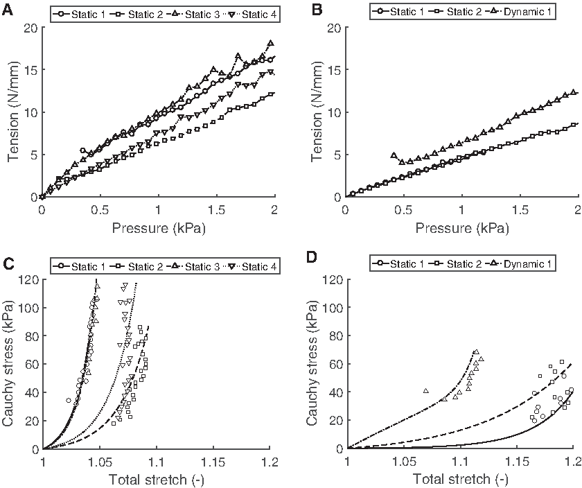
Tension–pressure relationships at
Elongation
Comparing tissue elongation (constrained tissue length at 0 pressure divided by initial construct diameter), there was a clear difference between the statically and dynamically cultured samples. Where the static sample tissue length was similar to the initial construct diameter (1.01 ± 0.01), the dynamic samples did show elongation (1.07 ± 0.00) (Fig. 7A).

Prestretch
Tissue prestretch was hardly observed after 1 week of static culture (1.01 ± 0.01), but increased with static culture time. At week 4, the area prestretch was slightly higher for the dynamically cultured samples (1.09 ± 0.01), compared to the static samples (1.07 ± 0.01) (Fig. 7B).
Thickness
From the original scaffold thickness of 500 μm, tissue thickness decreased more than 50% within the first week of culture to 226 ± 48 μm (Fig. 7C). Subsequently, tissue thickness further decreased to a value of ∼150 μm, after which only minor changes occurred.
Collagen orientation
After staining with CNA, collagen fibers could be clearly observed in the tissue samples (Fig. 8A). Analysis of the fiber architecture showed an in-plane fiber distribution with no clear preferred directionality, for all tissue samples, for the entire culture period (Fig. 8B–F).

Histology
The AB and hematoxylin (Fig. 9A–E) and PR red (Fig. 9F–J) staining show homogeneously distributed cells, GAGs, and collagen fibers in all tissues. The collagen fibers were mainly aligned in the tissue plane and, with time, collagen fibers were observed to become thicker (Fig. 9K–O). Finally, the fluorescence images (Fig. 9P–T) show vast amounts of αSMA-positive cells (green), indicating the presence of contractile cells.

Representative histology images from all weeks (week 1–4 on the first four rows, the final row represents the dynamically cultured samples at week 4).
Biochemical assays
The total tissue dry weight of the statically cultured samples appeared relatively constant with culture time (Fig. 10A). However, since the amount of PGA scaffold decreases with time, these results indicate an increase in tissue mass with time. At week 4, the dynamically cultured samples appeared to have a lower dry weight compared to the statically cultured samples.
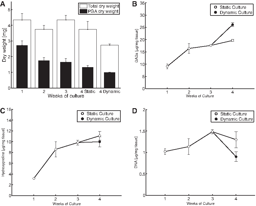
Biochemical assays.
Both the dry weight content of GAGs and hydroxyproline increased with time (Fig. 10B, C). The dynamically cultured samples contained a higher fraction of GAGs and a slightly lower fraction of hydroxyproline compared to the static samples at week 4. The concentration of DNA increased up to week 3 and marginally dropped at week 4 for the static samples, and more considerably for the dynamic samples (Fig. 10D).
Discussion
In this study, a novel bioreactor and associated analysis techniques were developed to systematically investigate the synergy between mechanics and tissue growth & remodeling. A proof-of-principle experiment was conducted to demonstrate the proposed framework's potential.
Vertigro functionality
The Vertigro is a versatile bioreactor capable of dynamically culturing planar TE constructs, and simultaneously performing mechanical testing. Its unique two-chamber design allows easy and sterile handling of the tissue construct, while the dedicated bottom pressure chamber attains the pressure application. In addition, although not treated in this study, the construct geometry can be easily changed by using removable inserts with different designs to mount tissue constructs with different geometries inside the bioreactor. The design in which currently rapidly degrading circular PGA scaffold was combined with slow-degrading PCL scaffold proved to be successful for culturing TE constructs that did not detach due to developing cell tension during static culture. Finally, the custom-made feedback system maintained the preset pressures during dynamic culturing, demonstrated in this study for a 1-week period.
Tissue composition and architecture
Histological analysis (Fig. 9) showed the presence of GAGs, collagen fibers, and αSMA-positive cells in the tissue constructs. These findings were quantitatively confirmed by biochemical assays (Fig. 10), demonstrating that tissue dry weight remained fairly constant, where the fraction PGA scaffold was gradually substituted by cell-produced matrix components such as collagen and GAGs. Finally, CNA-stained samples confirmed the presence of vast amounts of isotropically oriented collagen fibers (Fig. 8), in line with the homogeneous pressure application and isotropic boundary conditions and scaffold properties.
Tissue geometry
Besides changes in tissue composition, the geometry of the constructs underwent notable changes (Fig. 3). First, tissue thickness in week 1 was halved compared to the original scaffold thickness, gradually decreasing to a constant thickness of ∼150 μm. The large initial decrease in thickness is probably due to the high PGA porosity, which decreases immediately upon seeding. The consecutive decrease in thickness can be attributed to developing cell contractility that leads to tissue compaction in the unconstrained directions. These results are in line with the prestretch measurements that show an increase in prestretch with culture time, corresponding with previous observations 10 and the αSMA-positive cells observed by histology. Besides changes in thickness, the dynamically cultured samples were longer when constrained compared to their statically cultured counterparts. However, in contrast to the observed increase in unloaded length, the biochemical assays showed that the weight of the dynamic samples was lower compared to the static samples. This phenomenon may be explained by the fact that the amount of GAGs in the dynamic samples was higher compared to the static samples. The highly polar GAGs retain vast amounts of water and therefore increase tissue volume, which is not taken into account in the dry weight measurements.
Tissue mechanics
The bioreactor was designed to not only culture tissue samples for a prolonged time period but also to simultaneously perform mechanical testing by means of a classical mechanical bulge test.29,30 This test can be performed nondestructively as the mechanical test is performed inside the bioreactor itself, while measuring the tissue deformation nondestructively using ultrasound. The stress–stretch relationships of all samples (Fig. 6C, D) showed the typical exponential nonlinear behavior featured in collagenous soft tissues. Furthermore, the prestretch measurements are essential when analyzing the tissue mechanics: not taking the tissue prestretch into account can lead to a significant overestimation of the tissue stiffness, dependent on the degree of prestretch. 31
Limitations
The use of ultrasound to measure tissue deformation during mechanical testing provides a great advantage of nondestructive testing. In fact, the suitability of ultrasound imaging to measure in vitro material functionality was recently demonstrated. 32 Nevertheless, the limited spatial resolution of the current US system and method used resulted in inaccuracies in estimating tissue thickness, which was therefore measured using microscopy in this study. The spatial resolution can be improved by using a higher frequency ultrasound transducer and utilize the tracking of raw radio-frequency signals, allowing for nondestructive determination of tissue thickness, and high-precision tracking of the tissue. 33 Moreover, in this study, the stresses were estimated using Laplace's law, which is only valid for true membranes. To accurately estimate the mechanical properties in case of thick tissues, the use of an inverse finite element analysis method is recommended.
All the 16 statically cultures samples were successfully cultured and analyzed. However, 2 of the 4 dynamically cultured samples failed in week 4. To prevent future loss of samples during dynamic culture, a smoother pressure gradient could be implemented. Currently, a square waveform function was used, but a sinoid function would lead to a smoother pressure transition. In addition, a slow-degrading scaffold, rather than the rapidly degrading PGA, can be used to support the tissue, which could shorten the current static culture time of 3 weeks that is required before the tissue construct is strong enough to withstand dynamic pressure.
Conclusions and outlook
In conclusion, a novel versatile bioreactor was developed that offers a potent platform to unravel underlying mechanisms of tissue growth and remodeling through a systematic analysis of the influence of, among others, scaffold material, cell type, and pressure regimens on tissue (mechanical) adaptation. An improved knowledge of this process is key to deliver insights in pathologies where adverse growth and remodeling occur, and essential in the fields of regenerative medicine and tissue engineering.
This versatile framework allows for tissue culture and pressure-driven mechanical stimulation under hemodynamic loading, while simultaneously performing mechanical testing without sacrificing samples. This allows for the monitoring of tissue samples with time and as such has great potential for investigating mechanically induced growth and remodeling. Due to the versatile setup of the two-chamber bioreactor, permissive of sterile handling while maintaining dynamic pressure profiles, many different physiological situations can be studied in a well-controlled in vitro environment. This allows for a systematic investigation of how mechanical stimuli affect tissue adaptation, for example, by changing the pressure regime, degree of tissue anisotropy, scaffold material, and construct geometry. To demonstrate the successful development of the bioreactor system, a wide range of standard analysis techniques was employed to analyze the developing tissue constructs. In future studies, the current set of techniques can be extended with, for example, an inverse finite element method to accurately estimate the evolution of mechanical properties.
Footnotes
Acknowledgments
We acknowledge the support from the Netherlands Cardiovascular Research Initiative (CVON 2012-01): The Dutch Heart Foundation, Dutch Federation of University Medical Centers, the Netherlands Organization for Health Research and Development and the Royal Netherlands Academy of Sciences, and the European Union's Seventh Framework Programme (grant agreement No 604514). We also want to acknowledge the ICMS animation studio for help with the design of the graphical abstract in ![]() .
.
Disclosure Statement
No competing financial interests exist.