
Abstract
The fields of regenerative medicine and tissue engineering offer significant promise to address the urgent unmet need for therapeutic strategies in a number of debilitating conditions, diseases, and tissue needs of an aging population. Critically, the safety and efficacy of these pioneering strategies need to be assessed before clinical application, often necessitating animal research as a prerequisite. The growing number of newly developed potential treatments, together with the ethical concerns involved in the application of in vivo studies, requires the implementation of alternative models to facilitate such screening of new treatments. The present review examines the current in vitro and in vivo models of preclinical research with particular emphasis on the chorioallantoic membrane (CAM) assay as a minimally invasive, short-term in vivo alternative. Traditionally used as an angiogenic assay, the CAM of the developing chick embryo provides a noninnervated rapidly growing vascular bed, which can serve as a surrogate blood supply for organ culture, and hence a platform for biomaterial testing. This review offers an overview of the CAM assay and its applications in biomedicine as an in vivo model for organ culture and angiogenesis. Moreover, the application of imaging techniques (magnetic resonance imaging, microcomputed tomography, fluorescence labeling for tracking) will be discussed for the evaluation of biomaterials cultured on the CAM. Finally, an overview of the CAM assay methodology will be provided to facilitate the adoption of this technique across laboratories and the regenerative medicine community, and thus aid the reduction, replacement, and refinement of animal experiments in research.
Introduction to Preclinical Testing: In Vitro and In Vivo Models
G
In vitro and organotypic culture
Biomaterial preclinical testing typically involves an initial stage in vitro using cell lines and primary cells to screen and assess the biocompatibility (proliferation, cytotoxicity), characterization (gene/protein expression), and functionality of the biomaterial construct.4,5 Indeed, in vitro culture has evolved from the conventional cell monolayer culture systems and transitioned to more complex culture conditions that include three-dimensional (3D) sphere mono or cocultures of different cell types to enable the study of cell–cell interaction and cell–environment interactions, which mimic more closely their natural niche.6–8 In addition, in vitro bioreactors have been developed for the expansion and differentiation of progenitor cells by modifying their culture and mechanical conditions.9–12 Advances in bioreactor and microfluidic technologies have led to the development of lab-on-a-chip devices able to simulate the physiological conditions and responses of the human organism. These 3D in vitro biological systems have been scaled up into devices capable of culturing multiple cell types to mimic the response of whole organs (organ-on-a-chip). 13 Thus, currently, a variety of tissues and organs (skin, cartilage, bone, lung, heart, kidney, etc.) are under development across a number of groups and offer significant potential as replacement alternatives to in vivo studies.13–15
In the meantime, the in vitro culture of organs (organotypic culture/organoids) provides a closer biological approach to in vivo physiology while minimizing the need to conduct procedures on living animals. Organotypic cultures have been used to incubate a living organ in vitro at a controlled air/liquid interface while maintaining the original 3D structure of the tissue or organ, and hence the interaction between multiple cell types and extracellular matrix. Such an approach has enabled whole organs to be cultured statically 16 or using perfusion techniques.17–28
As an example of organ culture, studies conducted implementing organotypic cultures of bone have provided an in vitro system to study tissue regeneration and repair 29 as well as insights into the skeletal tissue development of the chick embryo. 30 In particular, Kanczler et al. developed a critically sized chick femur defect model for organotypic culture, which has been used to evaluate more than 14 biomaterial combinations for tissue engineering applications.30–33
Hence, the organotypic model offers a number of advantages as an in vitro model for the study of bone and cartilage repair, as well as providing a relatively high-throughput system to test biomaterials in vitro. However, while this model provides a refined alternative for animal research, the lack of a complete animal physiology system (vasculature, inflammatory, and immune system) remains an unaddressed, crucial aspect of the preclinical research-testing pipeline. In addition, the observation that graft vascularization is a crucial component in any tissue engineering strategies, further demands the use of a vascular bed for the evaluation of tissue regeneration, 34 and therefore the use of in vivo models in research and development.
In vivo models
Despite the plethora of in vitro models offering an alternative to animal testing, novel biomaterials still need to be tested in the context of a full animal physiology to assess their safety and efficacy. This is typically a mandatory requisite to address the regulatory steps/hurdles before clinical evaluation. 35 Preclinical testing of biomaterials is generally performed encompassing a number of distinct steps; initially a subcutaneous implant model is used to assess biocompatibility/safety, followed by the assessment on an actual tissue wound/healing scenario (bone fracture, skin wound, etc.). These studies are typically conducted in small animal models before evaluation in larger animals that provide a closer physiological context to the human response. 36 Thus, studies often employ subcutaneous models to optimize (i.e., dose) and characterize (i.e., drug release profile) construct variables before application into an injured tissue. Subcutaneous implants in mice are typically used to test whether the constructs are biocompatible in the context of vascular supply and/or immune system.37–39 In vivo evaluation of a cell construct commonly requires the use of immunodeficient mice and a surgical procedure for subcutaneous implantation, maintained approximately over 28–56 days. 40
In contrast, the CAM assay in the developing chick embryo offers a naturally immunocompromised host, allowing in vivo implantation of xenograft organs and construct. 41 Importantly, the CAM offers a rapidly growing vascular bed, which lacks a nervous system, and hence is a less sentient alternative for animal research. 41 The main difference between the subcutaneous implant and the CAM assay is the length of the incubation period, limited to 10 days in the CAM; however, due to its simplicity, high throughput and low cost, the use of the CAM assay is progressively expanding within the research community.8,42–45
A number of studies have evaluated the same biomaterial constructs on the CAM as well as in rodents (subcutaneous implant), and even compared the outcome from both models.46–50 Steffens et al. used the CAM assay to examine the effect of cell-seeded bovine cancellous bone scaffolds on the CAM for 8 days or on a mouse subcutaneous model for 21days. 46 The results demonstrated that the vascular response from the CAM was comparable, if not higher, to the mouse model. 46 Ling et al. 48 used cellularized gelatin scaffolds and reported a similar angiogenic response when these scaffolds were implanted into the CAM (7–9 days) and the dorsal subcutaneous SCID mouse (28 days). Hence, the aforementioned studies provided evidence of a significant vascular response from the extraembryonic membrane, even within a much shorter incubation period. A number of publications have already employed the CAM assay as a substitute for the subcutaneous murine model. Martinez-Madrid et al. investigated the effect of cryopreservation as a method to preserve fertility after aggressive chemotherapy on patients. 51 Cryostored human ovarian tissue was implanted onto the CAM, providing equivalent results to the traditional assay using immunodeficient mice, hence, validating the CAM model as an alternative to the murine model. 51 The CAM assay has already replaced the use of the eye irritation test in rabbits, as a mandatory assay based on a scoring system, which measures hyperemia, hemorrhage, and clotting.52,53 Furthermore, a number of FDA-approved anticancer drugs have been tested on the CAM and compared retrospectively with the preclinical data obtained from mice and rat models. 54 Correlation analysis of these studies demonstrated that the CAM was predictive of the results shown in the preclinical studies, 54 further validating the potential of the CAM as a less sentient replacement/refinement alternative for currently used in vivo models.
The Principles of the Chorioallantoic (CAM) Assay
The CAM of the chick embryo
From the moment of fertilization, the chick embryo develops over 21 days before hatching, and these stages have been named by Hamilton and Hamburger as embryo development day (EDD). 55 The chick embryo is surrounded by four extraembryonic membranes: (i) the yolk sac, (ii) the amnion, (iii) the allantois, and (iv) the chorion, which function together to protect and nourish the embryo during development.
The CAM is formed around EDD 4 by the fusion of the mesoderm tissue of the allantois and chorion membranes (Fig. 1) and this fused membrane becomes fully developed by EDD 14, growing exponentially from 6 cm2 up to 65 cm2 in 10 days. 41 The CAM is located between the eggshell and the allantois, surrounding the embryo structures where the allantois serves as a deposit for waste material such as urea and uric acid (Fig. 2). After EDD 14, the capillary plexus of the CAM becomes attached to the eggshell membrane. This fused, noninnervated membrane serves as a respiratory organ facilitating the gas exchange of O2 and CO2 between the eggshell pores and the embryo, as well as serving as a nutrient–waste interchange. In addition, the CAM contributes to ion transport, incorporating calcium from the eggshell to allow bone mineralization.56,57
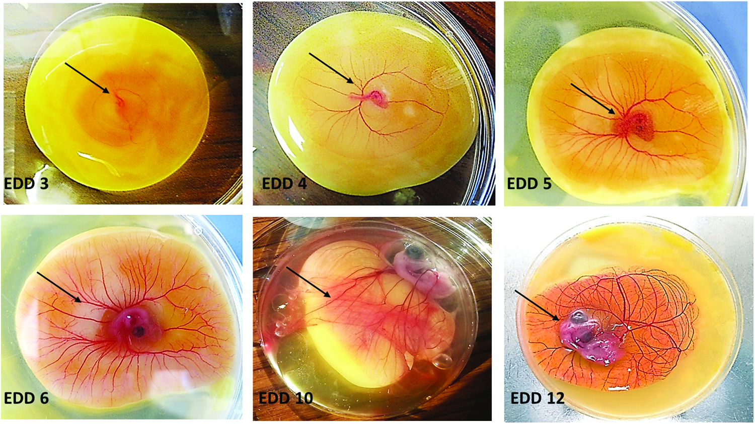
In vitro culture of chick embryos from EDD 3 until EDD 12. Fertilized eggs were incubated until EDD 3 and subsequently the chick embryo was transferred into a Petri dish to resume incubation in vitro. Shell-less (ex ovo) culture was maintained up to EDD 12 and images of the chick embryo (see arrows) were taken at EDD 3, 4, 5, 6, 10, and 12. EDD, embryo developmental day. Color images available online at
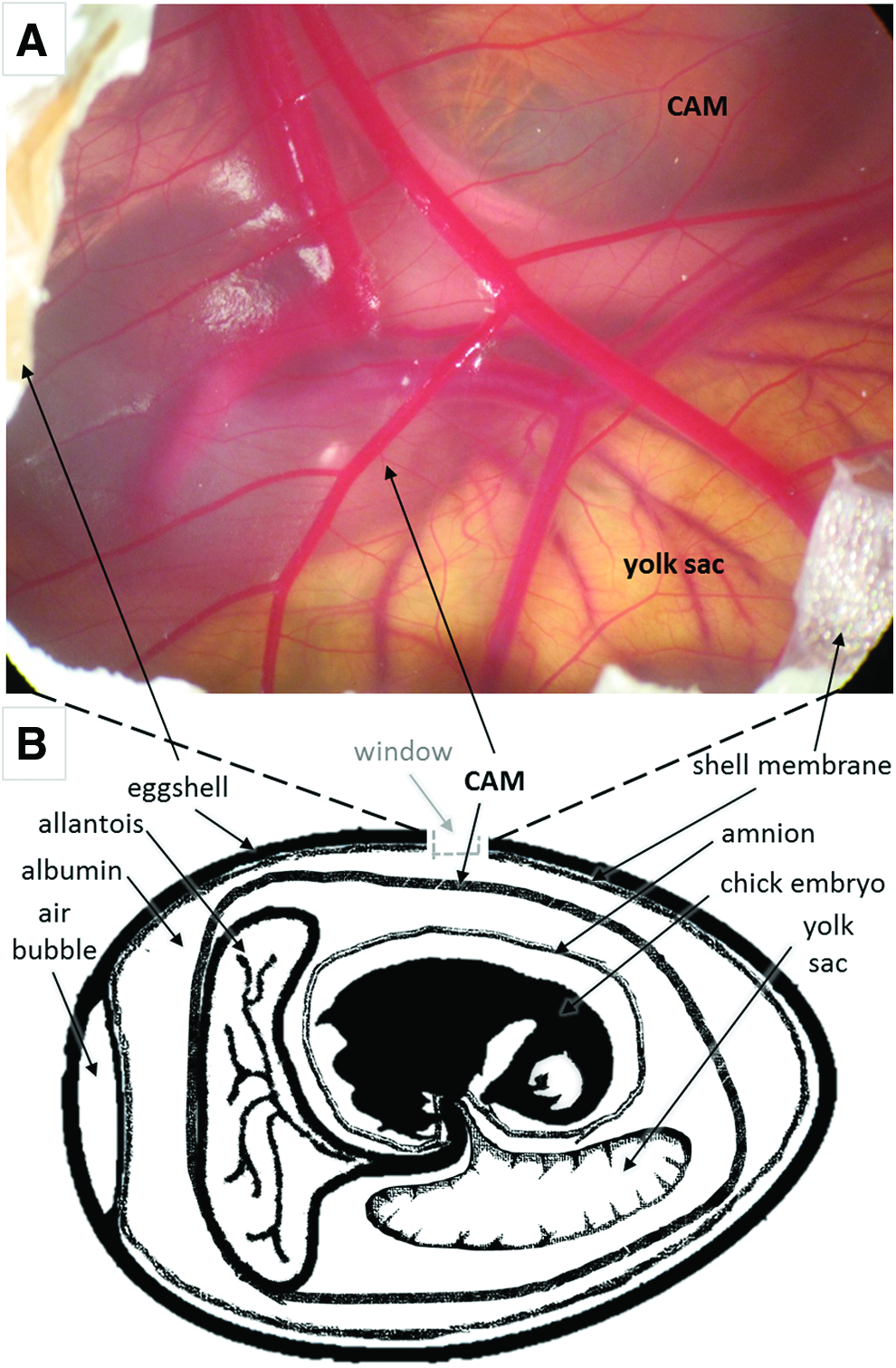
Illustration of the chick embryo and extraembryonic membrane anatomy for CAM assay.
As a consequence of the rapidly developing vascular system present within the CAM, the chick embryo is a commonly used host to perform (anti)angiogenic studies in cancer research.58–60 In essence, the CAM assay enables evaluation of a compound/material/construct that can promote or inhibit the angiogenic response of the extraembryonic membrane (Fig. 3). This is achieved by placing the test component on the CAM surface, approximately between EDD 4 and EDD 10, to observe the angiogenic response. 61 At harvest, the number of developed chick embryos is recorded, as well as the number of integrated samples within the CAM as an indicator of construct biocompatibility and safety. Crucially, graft integration on the CAM is an important parameter to assess the success of the assay (see section “Experimental and Technical Considerations for the CAM Assay” for further details).

Timeline of chick embryo development and CAM assay. Fertile eggs were inspected on arrival (day 0) from the local hatchery before starting incubation in horizontal position at 37.5°C, 60% relative humidity, with rotation scheduled every hour in the incubator
The primitive immune response of the CAM assay
While the chick embryo is conventionally considered an immunodeficient host for graft implantation, a number of studies indicate that the chick embryo is able to elicit a primitive immune response.62–65 It has been shown that as early as EDD 7, the chick thymus starts the process of recruiting lymphocyte precursors. 66 T lymphocytes develop in the chick thymus around EDD 11, whereas lymphocyte B cells develop from the bursa of Fabricius (the equivalent organ to the bone marrow in mammalians) around EDD 12. Both lymphocyte B and T cells start to circulate through the blood stream from EDD 12, together with monocytes and heterophils—the latter acting as a mammalian version of neutrophils. 67 Critically, T and B lymphocytes and monocytes do not become mature until EDD 18, and hence this is why the chick is considered an immunocompromised host. 62 However, Friend et al. showed that no differences in macrophage function were observed between day 14 chick embryos and 16-week-old chickens. 68
A number of studies have reported an immune response following the implantation of (xeno)graft material on the CAM.62–65 One study implanted lymphocytes from various animal species (pigeon, duck, sheep, rat, and guinea pig) onto the CAM and reported swelling of the chick embryo spleen. 62 Sys et al. implanted human osteosarcoma biopsies on the CAM to study their tumorigenic potential and described an inflammatory response together with fibrous deposition and CAM hyperplasia. 63 In a similar manner, the CAM produced significant fibrous deposition after implantation of bacterial endotoxins or cotton threads. 65 In the same study, Valdes et al. reported the presence of leukocytes and macrophages histologically, a possible indication of an acute inflammatory response comparable to that observed in mammalian systems. 65 Other investigations testing the similarities between mammalian and CAM immune systems showed biocompatibility within the CAM to nylon and silicone, commonly used surgical materials in the clinic. 64 The previous studies demonstrated that the chick embryo maintains a primitive immune response, which matures upon the end of the gestational process. Thus, depending on the research question, the potential of an immunogenic reaction from the chick embryo could be considered a positive or a negative feature when performing the CAM assay. Indeed, the culture of tissues from different species (xenograft) requires a suppressed immune response; however, it is the initial inflammatory response which triggers the healing process of the grafted tissue. 69 The second observation becomes particularly important in the context of biomaterial screening, as information around the physiological immune response would be relevant to assess construct efficacy and safety.
The CAM Assay for Tissue Engineering and Biomaterial Applications
The CAM assay in biomedicine
The first use of the CAM was documented in 1911 by Rous and Murphy, who described the culture of chicken sarcomas in the CAM. 70 A few years later, the authors published similar studies with xenotransplants from rat tumors.71–73 From these seminal studies, the use of the CAM has evolved into multiple applications, predominantly related to cancer research60,74–76 and development of viral vaccines.77,78 More recently, the CAM has been primarily used as a highly reactive vascular bed for the study of the angiogenic properties of a variety of compounds such as vascular endothelial growth factor (VEGF), bone morphogenetic protein (BMP), fibroblast growth factor-2, and endothelin.79–81 Other cytokines and growth factors, such as osteogenic protein 1 (BMP7), thrombin peptide, osteocalcin, vitamin D, and human angiotensin, have also been examined.82–85
Additional applications of the CAM include evaluation of the dosage and toxicity of drug delivery systems.54,86 Other studies have examined the effect of applying X-rays on the CAM as a model to determine the side effects in blood vessels after radiotherapy 87 or following hyperglycemia for diabetes research. 88 The CAM has also been used to evaluate novel surgical tools for retina vascularization,89,90 glucose biosensors,64,91 and Doppler tomography measurements of blood flow rate. 92 In summary, the aforementioned list of publications illustrate the wide range of applications of the CAM assay, as well as the large body of data referencing the chick embryo as an in vivo model in biomedical research over the last 30–40 years.
Organ culture on CAM: xenograft model
Given the immature immune system of the chick embryo, as detailed in section “The primitive immune response of the CAM assay”, the CAM has been used to implant tissue (living and decellularized) for xenograft culture, with reports of successful engraftment and vascularization.63,93–97 Kunzi-Rapp et al. described blood vessel formation and infiltration of chick erythrocytes in the preexisting capillaries of a human skin graft and preservation of human specific markers after culture. 93 Furthermore, Carre et al. engrafted healthy mouse fetal skin onto the CAM to then induce a laser injury, generating an in vivo model of wound healing and tissue regeneration. 94 Other publications by Ribatti et al. examined the effect of decellularized brain and aorta tissue from rats on the CAM as extracellular matrix scaffolds and demonstrated graft vascularization.95–97 Additional studies implanting living human tissue on the CAM included cryopreserved ovarian tissue 51 and patient-derived tumors. 63
To date, there has been a limited number of publications in the literature on human bone tissue cultured on the CAM.63,98,99 In 2010 Holzmann et al. studied the effects of the bone banking process on human allograft bone using the CAM assay. 98 To evaluate the angiogenic properties of the various allografts, samples were collected at different banking stages and incubated on an ex ovo CAM for 48 h, with fresh human femoral head bone chips as a control. 98 The vascular reaction of the CAM was significantly higher for control bone chips compared with the allograft samples, however, no attempt was made to measure tissue repair. 98 Recently, our research group has developed a model to culture human bone tissue on the CAM for regenerative medicine applications. 99 Moreno-Jiménez et al. demonstrated avian vascularization of the human bone tissue as well as a significant increase in volume of the bone implants following 7–9 days in vivo implantation. 99 Thus, offering an alternative platform for biomaterial testing as well as a humanized CAM model as a short-term in vivo model. 99
Hence, the aforementioned studies demonstrate the ability of the CAM to culture viable xenograft organs, including human-derived tissue, and thus the possibility of generating humanized in vivo models using the CAM. Moreover, the use of the CAM as an in vivo bioreactor for xenograft culture offers an additional dimension to the standard safety and efficacy applications using the chick assay, offering a more clinically relevant context for biomaterial evaluation.
Biomaterial efficacy using the CAM model: assessment of the angiogenic response
In recent years, the CAM assay has come to the fore as a screening platform for biomaterials. A diversity of constructs with growth factors and/or cells has been implanted onto the CAM over the last 40 years (Table 1). Table 1 provides a detailed summary from a variety of studies implanting biomaterials containing cells/growth factors on the CAM, demonstrating the large diversity in start points, choice of in ovo versus ex ovo approach, output measurements, and incubation time. The present section will review the most common application of the CAM for biomaterial efficacy testing, which is the examination of the angiogenic response of the membrane as an early indicator of construct performance in vivo. Typically, the CAM has been used to assess the angiogenic responses of the biomaterial based on macroscopic evaluation of vessel formation at the implant site (vascular density, vessel branching points/mm2, or blood vessels length) and/or histomorphometric analysis such as immunohistochemistry for CD31, an endothelial cell marker. Alternative methods to quantify angiogenesis include injection of a contrast dye for vessel perfusion and biochemical assays to measure hemoglobin at the implant site (Table 1).
Combination of both avian and mammalian models to test biomaterials.
CAM, chorioallantoic membrane; VEGF, vascular endothelial growth factor; BMP, bone morphogenetic protein; FGF-2, fibroblast growth factor-2; PCL, polycaprolactone; PLGA, poly(lactic-co-glycolic acid); TGF, transforming growth factor; FITC, fluorescein isothiocyanate; n/a, not available data; pTGF-b1, tumor growth factor p1; hrVEGF-A, human recombinant vascular endothelial growth factor-A; PDGF-BB, platelet derived growth factor-BB; hrbFGF, human recombinant basic fibroblast growth factor; hrBMP2, human recombinant bone morphogenic protein-2; PLA, polylactic acid.
In 2001, Valdes et al. used the CAM to test the effect of a variety of biomaterials regularly used in operating theaters, showing comparable results to the mammalian response. 65 Naturally derived materials, such as small intestine submucosa, polymer-derived materials, such as polyglycolic acid (PGA) and PGA modified with poly(lactic-co-glycolic acid) (PLGA) with and without growth factors, have also been tested on the CAM.100,101 Covalent immobilization of angiogenic growth factors (VEGF, Ang-1) on collagen scaffolds for cardiac repair has been examined on the CAM, improving performance over growth factor conjugation. 44 Additional collagen-based scaffolds composed of microporous spheres proved to induce a greater angiogenic response compared with polycaprolactone-based scaffolds. 102
The CAM assay has also been used to evaluate constructs for bone tissue engineering applications. Composite materials, such as bioactive glass nanoparticles with collagen scaffolds, and PLGA combined with amorphous calcium phosphate, have been tested on the CAM to observe an angiogenic response.103,104 In 2009, Vargas et al. examined the biocompatibility and bone mineralization potential of 45S5 Bioglass® using an ex ovo approach, where the authors used the embryo survival rate as an indicator of biocompatibility. 105 Buschmann et al. seeded adipose-derived stem cells in electrospun nanocomposite PLGA–calcium phosphate scaffolds and achieved complete infiltration of blood vessels throughout the scaffold. 104 Yang et al. used the CAM as a vascular bed for the culture of chick femurs containing a bone wedge defect on the diaphysis. 37 The implanted chick femurs were used to examine the effect of BMP-2–PLA scaffolds seeded with patient-derived cells, demonstrating the ability of the chick femur to heal and bridge the defect gap following CAM implantation. 37 Thus, the CAM can serve not only as an angiogenic assay, but also as a bioreactor capable of vascular and nutrient supply for the regeneration of the grafted tissue.
The CAM assay for safety and biocompatibility evaluation
While the CAM has been traditionally used as an in vivo angiogenic assay, there is significant potential in the use of the chick embryo in vivo model to provide additional information such as construct biocompatibility and safety assessment. Indeed, the circulatory system of the embryo and the CAM are connected and, therefore, any compound/construct applied on the CAM can affect the normal development of the chick. Thus, in addition to the vascular response (CAM blood vessel counting), changes in the normal development of the chick (i.e., viability rate) can serve as an indicator of the toxicity of an implanted substance. 86 As a proof of concept, the CAM assay has already been used as a sensitivity assay, replacing the invasive eye irritation test in rabbits since 2005. 53 Following the same approach, regulatory bodies, such as the FDA, are promoting the incorporation of the 3Rs (Reduction, Replacement, and Refinement) in newly submitted research proposals. As an example, the CAM assay was recommended in 2006 as a less sentient alternative for the testing of chronic cutaneous ulcer and burn wound treatments. 35
Documentation of the number of animals employed in each experiment (initial number of CAM eggs, number of viable eggs at the endpoint) and assessment of normal and healthy chick embryo development can serve to aid biomaterial safety evaluation, in addition to the CAM angiogenic response. However, there is a paucity of data available on the starting number of chick embryos used and/or survival rate at harvest, preventing comparison across studies which omit this information.45,99,105 Similar to any in vivo study, reporting of the number of experimental animals (mandatory for in vivo work in a vast majority of peer-reviewed journals (see ARRIVE guidelines, 106 and standard for good scientific practice) would aid the community.
In addition to chick embryo viability rates, a further important parameter to assess in graft biocompatibility is the response of the CAM to the implant or integration of the graft (Fig. 4). Only after a graft becomes integrated within the avian membrane will the graft benefit from the surrogate blood supply of the chick embryo (Fig. 4A). Interestingly, the chick embryo has developed several mechanisms, including material isolation in fat tissue and encapsulation of the material in amniotic fluid, to prevent the interaction with the implant (Fig. 4B).

Example of CAM integration of grafts or rejection at harvest of the CAM assay. Bone tissue grafts were extracted from human femoral heads and CAM implanted in ovo for a period of 9 days. Images taken at harvest of CAM-integrated sample
In summary, the CAM assay can serve as a biocompatibility assessment tool enabling documentation of chick embryo survival rate at harvest as well as the number of CAM-integrated grafts at harvest. Thus, maintenance of chick embryo viability is important to observe differences between actual treatments (i.e., biomaterials). The following section will review the technical aspects that can influence the normal development of the chick embryo and the different types of CAM assay models.
Experimental and Technical Considerations of the CAM Assay
Although the incubation of chick eggs is a relatively simple process, there are various technical aspects to consider to establish and standardize baseline embryo survival rate (ideally ∼90%). Upon fertilization, the chick egg can be stored at a chilled temperature (∼14°C) to prevent the initiation of embryo development. Stasis or developmental arrest can be conducted for 10–14 days postfertilization; however, egg viability reduces in proportion to stasis duration. Before resuming incubation at 37.5°C, eggs should be allowed to gradually increase their temperature over a 5–6 h period (setting eggs), as significant temperature fluctuations can compromise chick embryo viability. Stasis is used to time two different batches of egg incubation, common when implanting organs from donor chick embryos (i.e., EDD 18 chick femurs) into the host CAM (EDD 10–11 chick embryos) to evaluate tissue regeneration and/or repair. 37
The incubation temperature for the chick eggs can be set between 37.7°C and 38.3°C, with a relative humidity established between 52% and 55%, oxygen levels above 20%, and CO2 levels below 0.5%. To prevent the membranes of the chick embryo adhering to the eggshell, incubators need to have a rotation program, which typically involves egg rotation by 90° every hour. Evaluation of fertility and viability of the embryos can be undertaken by candling the eggs. Candling the eggs consist of passing a bright light (i.e., torch) through the eggshell, which helps visualize the air pocket of viable embryos, normally located in the wide portion of the egg. Given the variability in fertility rates (55–95%) from batch to batch of chick embryos, and unforeseeable conditions that can arise during transportation, candling of eggs before commencing an experiment is advisable to enable adjustment of experimental numbers, if required.
Ex ovo versus in ovo
There are two forms of the CAM assay depending on whether the chick embryo continues to develop in the eggshell or, alternatively, in a Petri dish (shell-less culture), respectively, termed in ovo and ex ovo culture. The ex ovo assay permits direct continuous visualization of the implant during the incubation period; however, ex ovo causes an increase in the chick embryo death rate of 50–70% during the initial days of culture, reaching 90% mortality around EDD 14.60,64 The presence of the eggshell is important since the CAM regulates the transport of calcium required for the normal mineralization of the chicken skeleton. 107 As a result, in the context of an ex ovo assay, the CAM can uptake mineral from an implant to compensate for the absence of eggshell. Vargas et al. demonstrated the ability of the CAM to completely resorb bioglass–ceramic scaffold, which resulted in significant mineralization of the chick skeleton compared with control embryos. 105
Since the embryo is allowed to develop normally before graft implantation, lesser number of animals are required when in ovo assays are undertaken, hence adopting the 3Rs policy. 106 For an in ovo approach, the shell of the egg is carefully etched to open a small window, which allows placement of the implant on top of the CAM (Figs. 2B and 3B). Following this, the eggshell window is sealed to preserve sterility and humidity, and the chick egg is placed in the incubator to resume incubation. The start point of the CAM (eggshell windowing) depends on the size/weight of the implant and the desired incubation period. The size of the window created will impact on embryo survival and development. For angiogenic assays, the CAM assay is typically started at EDD 10–11 as this corresponds with a peak in the vascular expansion of the membrane. 79 For tissue engineering applications, the CAM offers a limited period (EDD 10–EDD 18) for in vivo implantation and thus an earlier start point can offer an extended incubation time; however, the size and weight of the implant should be compatible with the maturity of the CAM (EDD) to avoid perforation of the membrane.
The termination date and procedure of the protocol should be determined following the local ethics committee guidelines and approval. In most countries, including the United Kingdom and the United States, the chick embryo becomes a legally protected animal during the last third of the gestational/incubation period (EDD 14), thus requiring a license to conduct any regulated procedures (Animals Scientific Procedures Act [ASPA], UK 1986, amended 2012; Policy on the Humane Care and Use of Laboratory Animals).
Imaging techniques: MRI, positron emission tomography, μCT
A central imaging application for the CAM has been the quantification of vessels using microscopy and histology as the cornerstones for assay assessment. In particular, the number of vessels, including the diameter, density, length of each vessel, and vessel branch points have been the parameters typically evaluated.41,59,108 As fluorescence imaging has advanced, this modality has become a basic tool in the examination of the CAM. Furthermore, fluorescence imaging has been widely used to study cancer metastasis and drug delivery. As an example, the CAM model has been employed to screen for fluorescent tumor markers, where tumor cells or grafts of tumors have been labeled with fluorescent markers and then seeded onto the CAM.109,110 In addition to tumor cell labeling, recently developed quantum dots have also been used in the CAM to visualize blood vessel development and angiogenesis.111,112 Further applications of the CAM model include the development of novel radiotracers for in vivo imaging.113,114 Haller et al. provided strong evidence of shared biodistribution and in situ stability of the radiotracers when comparing the CAM model with a conventional mouse model.113,114 Nevertheless, it is important to consider the radiation dose for the chick egg when employing X-ray-based imaging techniques (positron emission tomography [PET] and CT), as such techniques can compromise the angiogenic readouts of the assay.87,115
MRI has been used as a tool in the CAM model for quantification of the perfusion capacity of scaffolds cultured on the CAM as well as for the evaluation of mineralization using a custom contrast agent.104,116,117 Another emerging tool, which has been implemented in the CAM model, is photoacoustic microscopy (PAM). PAM is an exciting tool that uses generated ultrasound signals, through a laser, to image detailed aspects of tissue. PAM has been used for 3D morphological analysis of vascular networks as well as for analysis of oxygen saturation. 118 PAM has been used in the CAM model to study the dose-dependent effects of angiogenesis inhibitors 119 and for real-time monitoring of vascular changes in the CAM model during tumor destruction. 120 In summary, a wide range of validated imaging techniques (CT, MRI, PAM, PET, etc) are available (and continuously evolving) to analyze outcomes from the CAM assay and to enable the quantification of effects following treatment on the CAM.
Advantages and Disadvantages of the CAM Assay
The main advantage in using the CAM assay is the potential to collect information with regard to safety (biocompatibility and integration) and efficacy (angiogenic response, tissue regeneration/formation) of biomaterials using a minimally invasive, rapid, and cost-effective in vivo model. As detailed in section “Biomaterial efficacy using the CAM model: assessment of the angiogenic response”, the angiogenic response can be quantified using a variety of methods (Table 1); however, distinguishing preexisting vessels from newly formed vessels remains a challenge. A number of authors have proposed methods to overcome this challenge successfully, such as counting vessel numbers at the implant site with respect to vessel numbers in a distal region of the CAM away from the implant, 121 or introducing a nylon mesh or grid between the implant and the CAM to score only the new vessels.58,90,122 In addition to the angiogenic response, assessment of construct biocompatibility is based on (i) the survival rate of the chick embryos at the experimental endpoint, (ii) integration of the implant within the CAM, and (iii) the presence of a primitive inflammatory response. The ability of the chick embryo to reject an implant by nonintegration or development of an inflammatory response is an important parameter to take into consideration in the safety evaluation of a construct. Thus, the CAM assay provides additional information, providing a stepping stone for subsequent in vivo studies and, therefore, a less sentient and high-throughput screening platform for biomaterials.
A common limitation in the use of the CAM assay for xenograft culture (i.e., human tissue) is the inability to differentiate between host and graft tissue due to antibody crossreactivity between species.123,124 To circumvent this issue, genetically modified chick embryos constitutively expressing green florescent protein (GFP) can be used for the CAM assay and thus enable differentiation between host (GFP-CAM) and graft tissue. 99 GFP chick embryos were originally developed by McGrew and colleagues with the central objective to generate therapeutic proteins in chick eggs. 125 The production of transgenic chick embryos was achieved by using a lentiviral vector carrying a reporter transgene (GFP) followed by crossmating to identify homozygous GFP+/GFP+ birds. 125 GFP transgenic chickens have been used to examine gene expression regulation patterns in different tissues 126 as well as to substitute neural tube grafts in wild-type chickens for developmental studies of the nervous and vascular systems. 127
An important constraint of the CAM assay compared with other in vivo models (i.e., mouse subcutaneous implant) is the incubation period, normally limited to 7–10 days in the chick embryo. A potential approach to extend the incubation period on the chick model consists of reimplanting the graft harvested onto a second CAM (double CAM). We have recently explored this concept and implanted human bone tissue on a GFP-CAM for the first incubation period and, at harvest, reimplanted the graft into a wild-type CAM for an additional 7 days culture period (Fig. 5). Chick embryo survival on the second CAM was above 80%, indicative of the excellent integration between graft and host CAM membrane. Histological analysis showed integration of the graft in both CAMs, as well as a close interaction between both GFP+ and GFP− wild type membranes (Fig. 5). Additional evidence of the interaction between the double CAM was evidenced by the fusion of both membranes (GFP staining within wild-type CAM membrane). The implementation of a double-CAM culture approach offers the possibility to extend the culture of the CAM assay and hence increase the in vivo implantation period to allow for tissue regeneration, critical for preclinical testing in regenerative medicine. In summary, while the short-term implantation period available in the CAM can limit the extent of tissue formation compared with commonly used murine models (min. 28 days implantation), the advantages in terms of cost, simplicity of the procedure, minimal invasiveness, access to higher numbers, and rapid angiogenic response, make the CAM an attractive model serving as a unique bridging step between in vitro and in vivo studies in tissue engineering.

Distinction between first and second CAM on human bone tissue following double CAM implantation. Representative images of bone cylinders implanted for 9 days on GFP-CAM, harvested, and reimplanted on a second CAM (wild type) for additional 7 days incubation. Consecutive paraffin sections were stained for AS; Alcian Blue (proteoglycans) and Sirius Red (collagen)
Future Directions
Translatable research relies on the use of in vivo models to examine the safety and efficacy of treatments before the research can reach the clinic. The advances within the regenerative medicine and tissue engineering fields necessitate the development of alternative models to evaluate the new proposed modalities for treatment and the urgent need to refine, reduce, and replace (3Rs) the use of animals in research. In the present review, we have introduced the CAM of the developing chick embryo as a short-term in vivo model together with multiple validated applications in different fields of regenerative medicine research. The CAM assay provides a simple, cost-effective, and high-throughput in vivo model, which can serve as an excellent bridge between in vitro and in vivo models for preclinical research. Numerous studies have compared the outcome from murine and CAM assay models and reported comparable findings when examining similar treatments, demonstrating the potential of the CAM to refine, or even replace, the use of murine hosts in animal experimentation. Future directions will focus on extending the incubation time of the CAM assay by reimplantation on subsequent chick eggs to prolong the in vivo implantation time. Additional research avenues include the use of larger avian species with longer embryo development periods (i.e., ostrich egg), hence extending the incubation time available on the CAM for up to 42 days. Moreover, the implementation of genetically modified chick embryos for the CAM assay offers a significant potential to culture human xenograft tissue for mimicking human physiological conditions and enable differentiation between host and implant tissue. Thus, the humble but complex chick egg and derived CAM model, often used as a poor research surrogate tool, offer a unique research test model to inform the transitional in vivo phase and to evaluate the plethora of technologies and therapeutic strategies proposed of the clinic.
Footnotes
Acknowledgments
Work in the authors' laboratories was supported by grants from the BBSRC (BB/L021072/1 and BB/L00609X/1, European Community Seventh Framework Program Grant, BioDesign [262948] and EU FP7 [FP7/2007–2013 under grant agreement no. 318553] Skelgen) and the UK Regenerative Medicine Platform Hub Acellular Approaches for Therapeutic Delivery (MR/K026682/1) to R.O.C.O. PhD funding from the National Center for the Replacement, Reduction, and Refinement of Animals in Research (NC3Rs) for I.M.-J. is gratefully acknowledged. The work presented here is based on many useful discussions with past and current members of the Bone and Joint Research Group in Southampton, United Kingdom.
Disclosure Statement
No competing financial interests exist.