
Abstract
Cell encapsulation in hydrogels is a technique that offers a variety of applications, ranging from drug delivery to biofabrication of three-dimensional scaffolds. The assembly of cell-laden hydrogel building blocks aims to generate complex biological constructs by manipulating microscale units. An important issue for the clinical implementation of this technique is the long-term storage of a large stock of cell/hydrogel building blocks. In this work, the impact of cryopreservation on the viability and functionality of cells encapsulated in alginate matrices is presented comparing different cryoprotective agents (CPAs). Human osteosarcoma MG63 cells were encapsulated in sodium alginate fiber constructs with wetspinning method and exposed to different formulations of cryopreservation media, containing dimethyl sulfoxide (DMSO), glycerol, and trehalose. The cell-laden fibers were subsequently slow-cooled down to −80°C and stored in liquid nitrogen. After thawing, viability and death pathway of encapsulated cells were investigated, and metabolic activity and proliferative capacity of cells released from the alginate matrix were evaluated. The viability of MG63 cells encapsulated in alginate matrix ranged from 71% ± 4% to 85% ± 2%, depending on the cryoprotective media formulation with no protracted harmful effects from the CPAs. On the other side, cells cryopreserved in encapsulated conditions and released from the hydrogel showed larger metabolic activity and proliferative capacity in tissue culture plate compared to cells cryopreserved in suspension, in particular when DMSO and glycerol were used as CPAs. Results have been correlated with the viscoelastic properties and water content changes of the alginate constructs loaded with the different CPAs.
Introduction
C
Encapsulation of cell offers several potential applications. It can provide an immunoisolation barrier for cells transplantation within a host where they can secrete specific disease treating molecules (e.g., against diabetes, 4 anemia 5 or hemophilia 6 ). Moreover, cell-laden materials can be used as building blocks to biofabricate scaffolds for tissue engineering applications7,8 or for in vitro model to test drug or therapeutic procedures.9,10
Hydrogels are attractive candidates for the incorporation of cells and bioactive compounds, due to their high water content and to the variety of physical properties they can provide depending on their chemistry and cross-linking degree.1,7,11,12 Various methods have been proposed for encapsulating cells; as an example, cells have been encapsulated in microbeads by means of an electrohydrodynamic process 13 or with microfluidic-based techniques, 14 and fiber-shaped structures containing cells have been produced using extrusion-based bioprinting methods.15,16
Sodium alginate is a naturally derived polysaccharide consisting of a linear block copolymer containing β-
An important issue for the practical use of encapsulated cells is their preservation to ensure a steady supply when needed.22–25 Cryopreservation, that is, cooling down cells to very low temperature, is routinely used to slow down or completely stop their biological activity, which is recovered after cells thawing. Cryoprotectants agents (CPAs), added during the cryopreservation process, protect cells from cryodamage by decreasing the freezing point at which intracellular ice forms thus minimizing the damage caused by cooling.26–28 CPAs such as dimethyl sulfoxide (DMSO) and glycerol can penetrate the cell membrane at physiological temperatures, however, their penetration ability rapidly decreases when temperature lowers.22,26,29 Disaccharides like trehalose, mannitol, and sucrose do not cross the cell membrane and act stabilizing the transmembrane proteins. 24 In some cases, multiple types of cryoprotectants are used in association.30,31 Nevertheless, CPAs are generally toxic and must be removed by washing protocols after cells thawing. 27
Several techniques have been proposed for the cryopreservation of cells encapsulated in different materials, tailored for specific applications such as storage of mesenchymal stromal cells, 32 neurospheres, 33 and pancreatic substitutes; 34 preservation of tissue-engineered substitutes;35,36 and assembly of three dimensional constructs containing cells.35,37 A selection of these works is summarized in Table 1.
CPA, cryoprotective agent; DMSO, dimethyl sulfoxide; hMSCs, human mesenchymal stromal cells; hADSCs, human adipose-derived stem cells; mESCs, mouse embryonic stem cells; HepG2, immortalized hepatocyte cell line; SEM, scanning electron microscopy.
The abovementioned studies examined peculiar properties of the proposed method, focusing on evaluating the viability, proliferation, and differentiation potential of encapsulated cells. Moreover, in certain cases the ability of the materials to withstand the cryopreservation in term of structural integrity and mechanical properties was evaluated.31,35,37
However, none of these studies has systematically compared the effect of different cryoprotectants. In this article, we evaluated the effect of cryopreservation and of different cryoprotectants on the biological recovery of MG63 cells encapsulated in alginate filamentous constructs, used as model cells-hydrogel system, made by spinning cells/water alginate solutions onto a gelling bath containing calcium ions. 15 The aim was to optimize the alginate-cryoprotectant formulations and to assess the different ability of DMSO, glycerol, and trehalose to preserve encapsulated cells biological functions after a conventional slow freezing protocol.26,38 Alginate was selected as matrix because cells can be retrieved by dissolving the encapsulating matrix with the use of chelating agents such as EDTA or sodium citrate. 3 Moreover, alginate does not promote cell adhesion and proliferation due to the absence of biorecognition motifs. 18
After thawing, the viability of cells encapsulated in the fibers, and their proliferation and metabolic functions recovery upon dissolution of the alginate matrix were evaluated. All the results were compared with nonencapsulated cells cryopreserved at the same conditions.
The effect of the freezing-thawing on the physical properties of the alginate matrix was evaluated with dynamic rheological tests and by measuring the liquid content of the material before and after freezing.
Materials and Methods
Materials
The following materials were used: sodium alginate powder derived from brown algae alginic acid, calcium chloride dihydrate, DMSO, glycerol, D-(+)-trehalose dihydrate, sodium citrate dihydrate, 0.05% Triton X-100 (Sigma-Aldrich); Calcein-AM, propidium iodide (PI), phosphate buffered saline (PBS), Minimum essential media (MEM), 200 mM
Hydrogel preparation and sterilization
Alginate powder was dissolved in PBS overnight at room temperature to obtain a 2% wt/vol alginate solution. The cross-linking solution consisted of calcium chloride dihydrate dissolved in distilled water at a concentration of 200 mM. For sterilization, the two solutions were filtered through a 0.22-μm filter before use.
Cell culture and encapsulation
MG63 osteosarcoma cells were thawed and expanded using standard protocols. In particular, cells were expanded in tissue culture-treated flasks as monolayer at 37°C under 5% CO2 to 85–90% confluence before encapsulation. Culture medium was composed of MEM containing 10% fetal bovine serum (FBS; GIBCO), 2 mM glutamine, 1 mM sodium pyruvate, 1% nonessential amino acids, and 1% antibiotic antimycotic solution. At sub-confluence cells were detached from the flask with Trypsin solution. Subsequently, cells were centrifuged at 1000 rpm for 10 min, rinsed in PBS to remove any residues of culture medium, and finally dispersed by vortexing inside the buffer. An aliquot of the suspension was used to determine cells concentration using a hemocytometer (Sigma) and Trypan Blue 0.4% as contrasting agent. Cells were centrifuged again and, after removing the supernatant, resuspended in the proper amount of alginate solution to obtain a suspension containing 2× 10⁶ cells/mL.
Four hundred microns diameter alginate fibers about 5 cm long containing cells were formed by wetspinning. 15 The method involved extruding through a 0.5–20 μL filter tip (Corning) 20 μL of cells/alginate solution onto a Petri dish containing the calcium chloride cross-linking solution. Gelation occurred instantaneously upon contact with the solution containing Ca2+ ions. After a few minutes, formed fibers were washed twice with MEM to remove residual calcium.
Cryopreservation and thawing
Before cryopreservation, cell-laden fibers were incubated at 37°C in 5% CO2 atmosphere for 30 min in culture medium containing 20% (v/v) FBS with addition of cryoprotectants (herein referred as cryopreservation media) in the amounts of Table 2 following indications reported in the literature.31,39
The media are prepared by adding the reported compounds to the culture medium.
After incubation, they were moved to cryovials (five fibers per vial) that were inserted in a commercial cooling box (CoolCell Cell Freezing Container, Biocision) and cooled from +37 to −80°C at −1°C/min. The next day, vials were transferred to liquid nitrogen. For in vitro evaluations, vials were fast thawed in a water bath at 37°C and immersed in fresh culture medium that was changed after 2 and 4 h to remove CPAs residues from the alginate matrix. Nonencapsulated cells batches were prepared for comparison by using the same procedure. This method is below referred as standard cryopreservation protocol.
In vitro evaluation
In vitro evaluations were performed on encapsulated cells (live/dead assays and cell death pathway), on cells released from the alginate matrix and cultured on tissue culture plate (TCP) (proliferation and metabolic activity) and on control batches. The complete scheme of the experiments is reported in Figure 1.
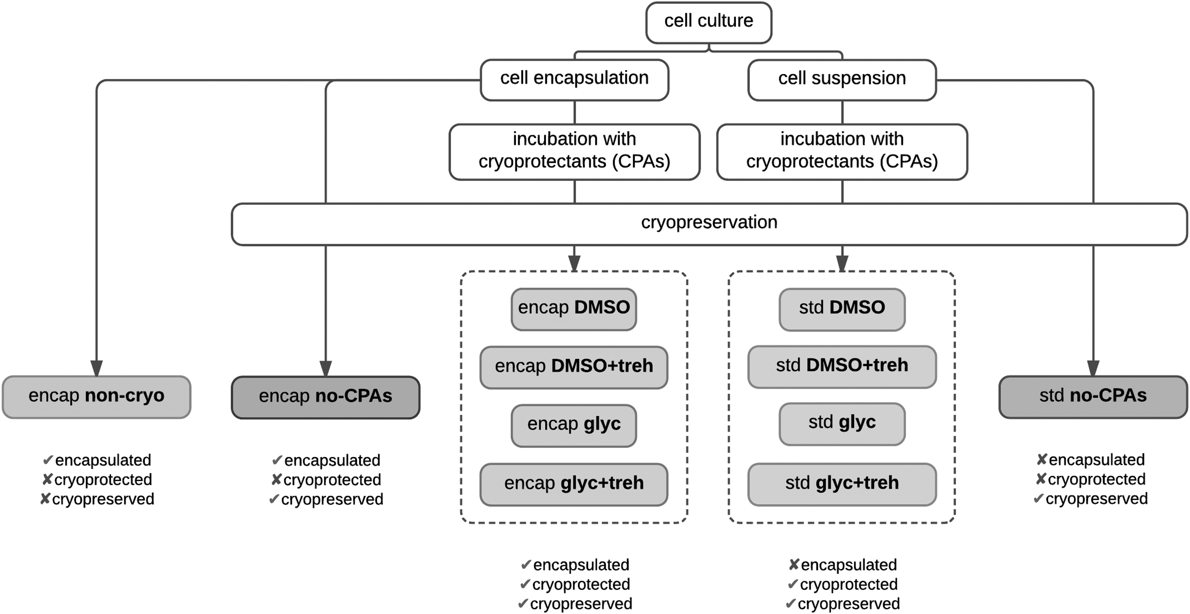
Summary of the samples used for the in vitro analysis associated with their particular fabrication steps.
For cell release, fibers were incubated in a chelating solution (55 mM trisodium citrate, 10 mM HEPES in PBS) at 37°C for 5 min. After centrifugation at 1000 rpm for 5 min the supernatant was removed, and the precipitate with cells was washed in PBS again to remove any residues of chelating solution. An aliquot of suspension was taken to evaluate the cells concentration using the hemocytometer and Trypan Blue as contrasting agent.
Cells viability and distribution in fibers
Confocal microscopy (Nikon A1, Japan) was used to determine cells viability and distribution in the alginate fibers as prepared (no-CPAs control) and in frozen fibers after thawing, at 3, 24, and 72 h. A standard two-color live/dead assay was performed after incubating the fibers in a PBS solution containing 1 μg/mL calcein AM and 20 μg/mL PI for 30 min at 37°C. Confocal images were collected along the Z-axis with 10 μm intervals (488 nm wavelength laser and 500–550 nm detector for calcein; 560 nm wavelength laser and 570–620 nm detector for PI). The viability of encapsulated cells was reported as the ratio of the number of alive cells to the total number of cells in each fluorescent image, automatically counted with the Fiji distribution of image processing software ImageJ. 40 At least five fibers were analyzed for each group.
Flow cytometric apoptosis/necrosis detection
Flow cytometry analysis was used to discriminate in control and released cell samples between apoptosis and necrosis pathways, the two essential processes leading to cell death, after staining the cells with an apoptotic/necrotic detection kit (PK-CA707-30017, PromoKine). Cells were incubated for 15 min in the dark in a buffer solution containing Annexin V-FITC and Ethidium Homodimer III. Subsequently, cells were run at low rates through the FACSCalibur flow cytometer (BD, Singapore) to measure optical transmission, side scattering, and emission fluorescence at 530 ± 30 nm and 585 ± 42 nm of at least 10,000 cells, upon excitation at 488 nm. Low green and low red fluorescence was scored as viable (low left quarter), high green and low red fluorescence was scored as early apoptotic (low right quarter), low green and high red fluorescence was scored as late apoptotic/necrotic (upper right quarter), high green and high red fluorescence was scored as late necrotic (upper left quarter; Fig. 2). Standard error was calculated considering the maximum and minimum number of events occurring in each of the four regions by varying the position of the region boundaries.
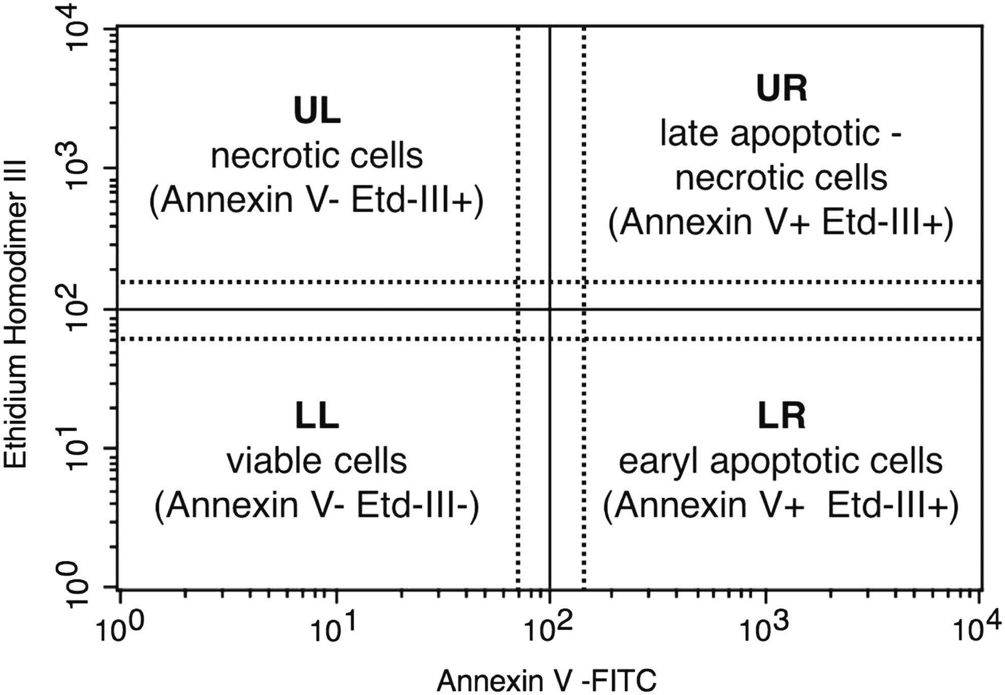
Region classification according to Annexin V and Ethidium Homodimer III intensity after apoptosis/necrosis staining and flow cytometry analysis.
Recovery of cell functionality
Upon thawing, cells were released from the alginate matrix and transferred into a TCP (2000 cells/cm2 manually counted with a hemocytometer). The next day and for 6 days thereafter, the metabolic activity and the proliferation of cells was evaluated with alamarBlue and PicoGreen assays respectively. At 1, 2, 4, and 6 days, cells were incubated for 2 h with culture medium containing 10% alamarBlue and fluorescence intensity was measured on a plate reader (535 ± 25 nm excitation and 590 ± 20 nm emission; Spark 10 M, Tecan, Switzerland). Subsequently, DNA extraction was performed by disrupting the cells membrane with a solution of 0.05% Triton-X in PBS, followed by sonication (UP400S, Hielscher, Germany) for 10 s. PicoGreen was then used for the quantification, measuring the fluorescence intensity of PicoGreen-DNA complex with the plate reader (485 ± 20 nm excitation and 535 ± 25 nm emission). A calibration curve was built up using the DNA standard provided with the assay to correlate fluorescence intensity to DNA concentration. Cells exposed to standard cryopreservation procedure were used as reference. For each test nine replicates were used.
Statistical analysis
Graphpad Prism 7 software was used for statistical analysis. Results are expressed as mean ± standard deviation and significance was tested using two-way analysis of variance with Tukey's post hoc test. A p-value of 0.05 was considered significantly different.
Evaluation of material properties
Rheology
Alginate hydrogels were submitted to rheological test before and after freezing by using a parallel-plate rotational rheometer (Discovery HR-2; TA Instruments) with 40 mm diameter plate. The samples were prepared by casting liquid alginate (as described in par. 3.2) onto gelatin molds containing calcium chloride until complete gelation occurred. Alginate discs (diameter 40 mm, height 2 mm) were then detached from the gelatin substrate and incubated with the different CPAs (as described in par. 3.4) overnight to assure complete diffusion of the CPAs in the alginate matrix. The samples were subsequently cooled to −80°C before transferring into liquid nitrogen and finally thawed at 37°C. After rinsing the sample in DI water to remove the CPA residues, rheological properties were investigated. Frequency sweep experiments were conducted from 0.01 to 10 Hz at a fixed strain and temperature of 2% and 37°C respectively. Storage modulus (G′) and loss modulus (G″) were measured as a function of frequency. At least three samples were evaluated for each condition. As a control, fresh samples were tested after incubation with the CPAs and rinsing in DI water.
Liquid content
The liquid content of the alginate hydrogels before and after freezing was investigated by weighing the samples in the wet and dry state. Alginate discs were fabricated as described in the previous paragraph, incubated in the presence of different CPAs, frozen-thawed, and rinsed in DI water overnight to remove any CPA residues. As a control, fresh samples were tested after incubation with the CPAs and rinsing in DI water. Subsequently, the samples were weighted before and after completely drying in an oven at 65°C overnight. The liquid content of the hydrogels was determined by the following equation, where Mwet and Mdry represent the mass of the samples before and after drying.
At least five samples were evaluated for each condition.
Results
Cells distribution
When injected in the calcium chloride bath, alginate underwent a fast sol-gel transition with the formation of a solid gel fiber. Fibers were left in the cross-linking solution for about 10 min, to allow the diffusion of calcium ions toward the gel core. In Figure 3 a live/dead representation of an alginate fiber containing cells is reported. The mean diameter of the fiber, measured by optical microscopy, is 387 ± 98 μm (n = 9), which is compatible with nutrients diffusion in presence of high cell densities. 13 Moreover, cells appear homogeneously distributed inside the fibers.
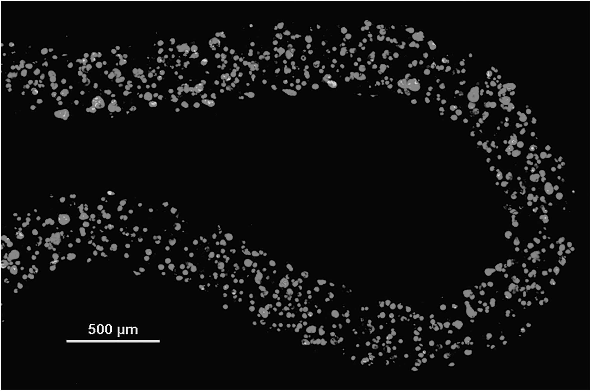
Representative confocal microscopy image of cells encapsulated in a wet-spun alginate fiber stained with live/dead (green/red) fluorescent dyes.
Viability of encapsulated cells
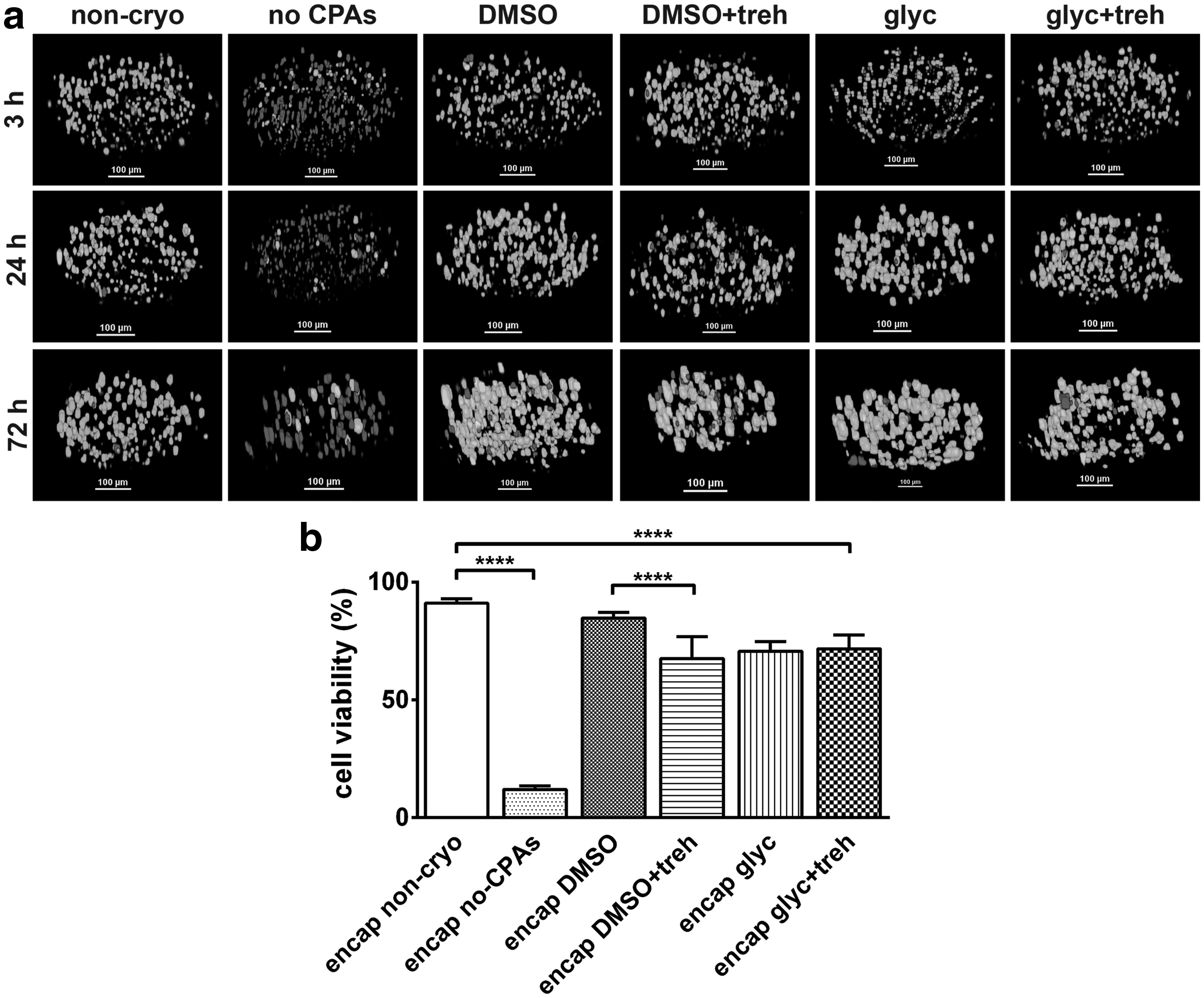
The effect of the different cryoprotective formulations on the viability of encapsulated cells was evaluated on all samples after thawing. Visualization of the alginate fibers containing cells by confocal microscopy, after standard Live/Dead staining, was used to discriminate alive from dead cells (Fig. 4a). Cell viability was evaluated at 3, 24, and 72 h after thawing. Results were compared with two systems: fibers loaded with cells not frozen (non-cryo) and fibers loaded with cells frozen in absence of cryoprotectant (no-CPAs). Images indicated limited cell death in any of the cryopreserved samples, less than or equal to the non-cryo control. For quantitative analysis, live and dead cells were automatically counted and discriminated using the Object Counter plugin of Fiji software immediately after thawing (Fig. 4b). A decrease of viable cell number was observed after cryopreservation compared to non-cryo group (viability 91% ± 2%). A higher retention of cell viability was observed for cells cryopreserved with DMSO (85% ± 2%) compared with glycerol (71% ± 4%), DMSO/trehalose (71% ± 2%), and glycerol/trehalose (72% ± 6%).
Apoptosis/necrosis detection after thawing
The effect of the various cryopreservation protocols on the pathway of cell death during freezing were analyzed by flow cytometry (Fig. 5a). Results were compared with nonfrozen cells and with cells frozen with standard protocol.
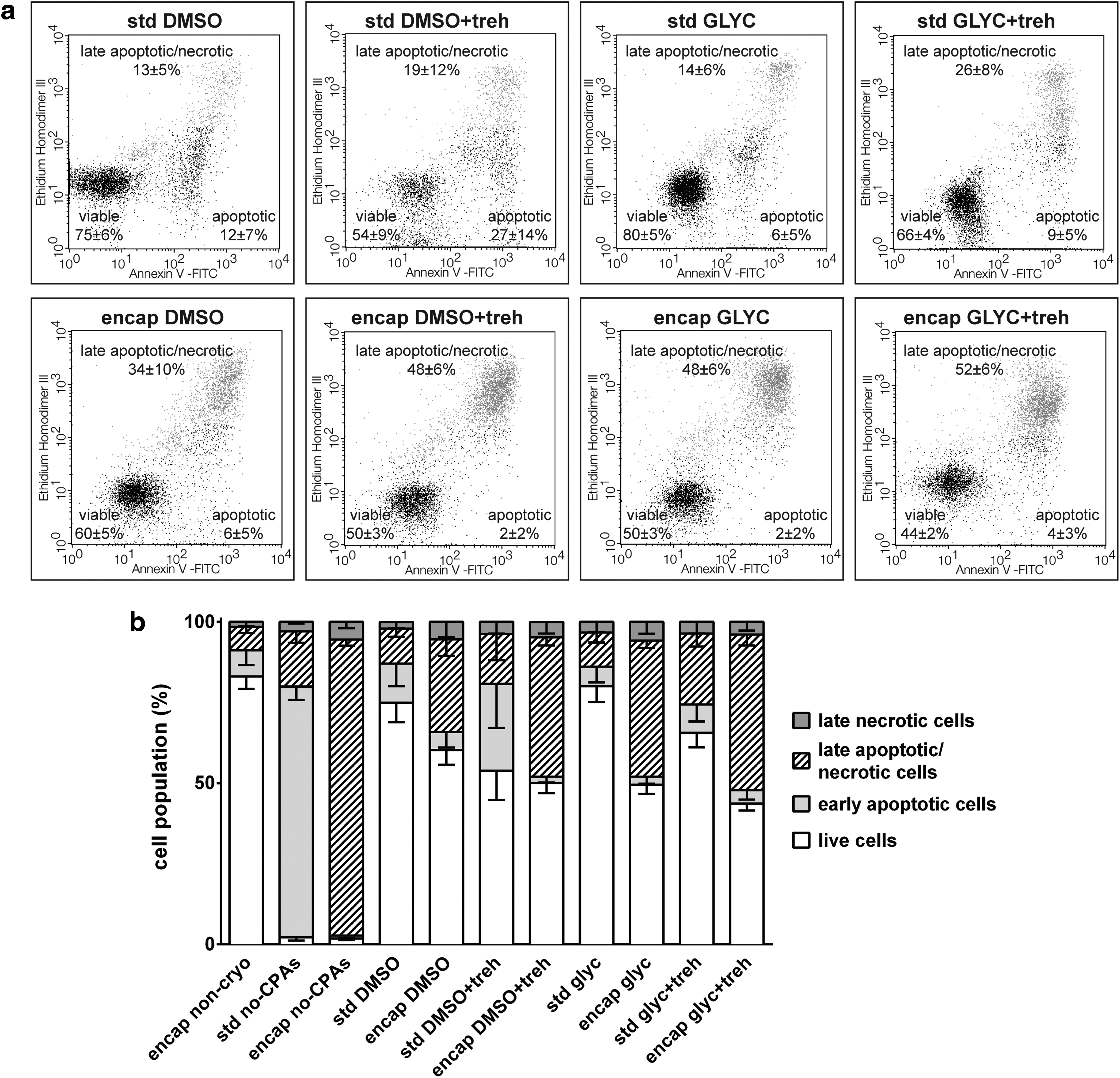
In general, for all cryoprotectants, cell viability was greater than control formulations without any cryoprotectants (no-CPAs controls) (Fig. 5b). We observed a decrease in cell viability for the cryopreserved encapsulated cells compared to the standard cryopreservation protocol both with DMSO (75.0% ± 6.0% vs. 60.3% ± 4.6%) or glycerol (80.1% ± 5.0% vs 49.5% ± 2.8%). Furthermore, we observed a decrease of early apoptosis signal and a shift toward late apoptosis/necrosis signal for encapsulated cells. With trehalose, cell viability decreased both for standard cryopreservation and for cryopreservation after encapsulation (60.3% ± 4.6% vs. 50.1% ± 3.1% for DMSO and 49.5% ± 2.8% vs. 43.7% ± 2.2% for glycerol). This result confirmed the qualitative live/dead confocal analysis evaluations.
Cell proliferation and metabolic activity
The results of cell proliferation and metabolic function are reported in Figures 6 and 7 respectively. Upon thawing, cells were released from the alginate matrix using a calcium ions chelating solution and transferred in TCP. The relative fluoresce intensity was normalized to the value at day one for each group. At day 2, cells frozen with cryoprotectants showed a comparable DNA content. Considering the effect of encapsulation, at days 4 and 6 we observed a significant increase in cell proliferation in presence of DMSO (increment of 12.5 ± 0.9-fold vs. 15.8 ± 1.8-fold at day 6) and glycerol (16.9 ± 1.4 vs. 18.3 ± 1.3 at day 6). The encapsulated group frozen by using DMSO/trehalose exhibited an increase of cell growth with respect to the DMSO alone (15.8 ± 1.8 vs. 18.6 ± 2.4 at day 6). On the contrary, when glycerol was used, the addition of trehalose led to a significant inhibition of cell proliferation (18.3 ± 1.3 vs. 11.5 ± 1.1 at day 6).

Cell number evaluation at each time point from the various frozen groups after thawing. Std: cell frozen with standard protocol (1 M cells/vial in cryopreservation medium. Encap: cell frozen after encapsulation, following dissolution of the gel. Values are normalized to day 1 for each group. Error bars represent mean ± SD (n = 9). **p < 0.01 and ****p < 0.0001.
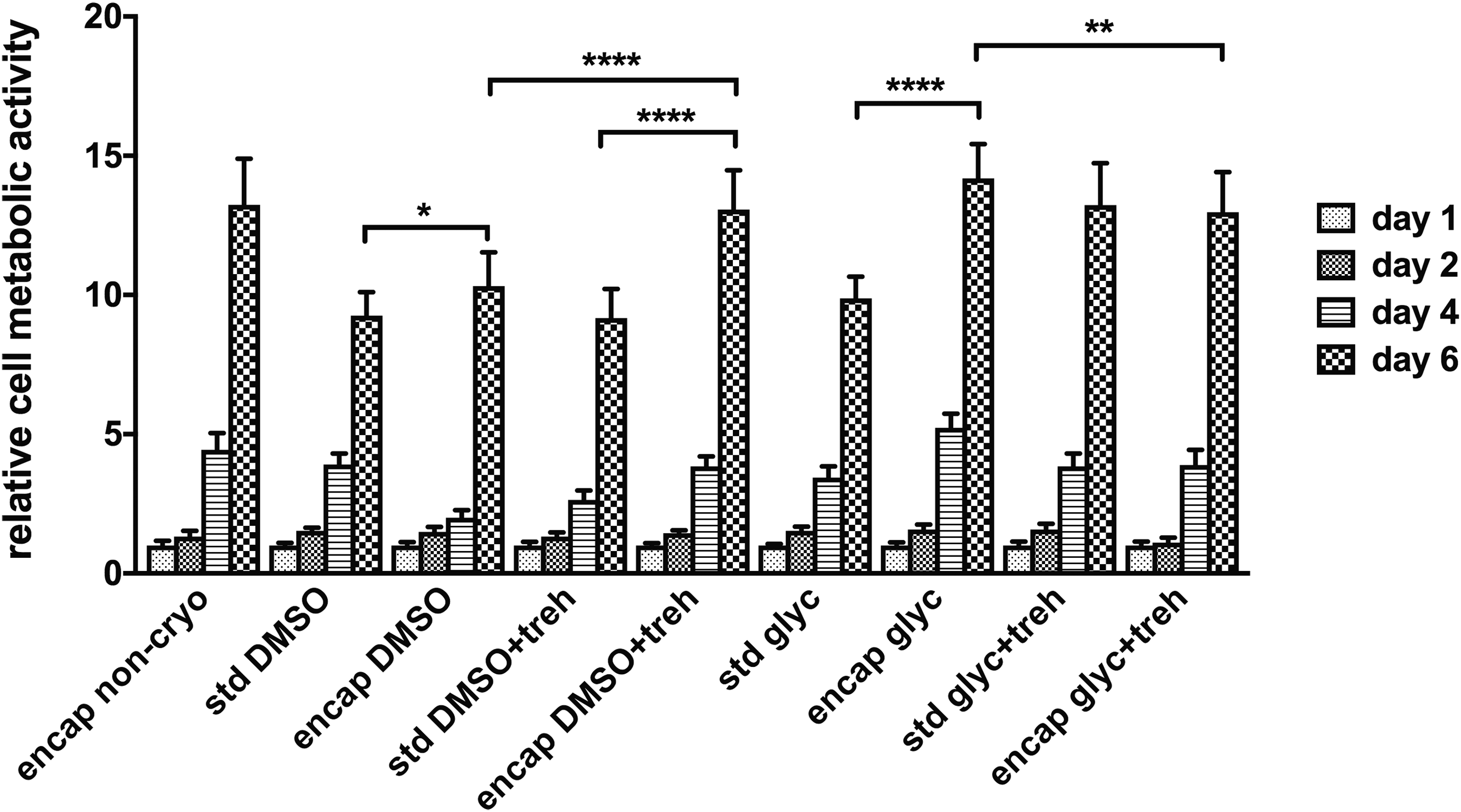
Cell metabolic activity in tissue culture plate at each time point from the various frozen groups. Std: cell frozen with standard protocol (1 M cells/vial in cryopreservation medium). Encap: cell frozen after encapsulation, following dissolution of the gel. Values are normalized to day 1 for each group. Error bars represent mean ± SD (n = 9). *p < 0.05, **p < 0.01 and ****p < 0.0001.
Cells metabolic activity was determined with alamarBlue assay and data were normalized to the value at day 1 for each group. All cryopreserved cells exhibited an increase of metabolic activity from day 1 to 6 and confirmed the trend of cell proliferation signal. Considering the use of a single cryoprotectant, cells frozen after encapsulation exhibited a significantly increased metabolic signal from day 4 onward, both for DMSO (increment of 9.3 ± 0.8-fold vs. 10.3 ± 1.2-fold at day 6) and glycerol (9.9 ± 0.8 vs. 14.2 ± 1.2 at day 6). This result matches the cell proliferation profile, and confirms previous results on fibroblasts-like cells cryopreserved in suspension and in a 3D construct. 41 Regarding the effectiveness of the different CPAs for encapsulated cells, glycerol led to a higher cell recovery 6 days after thawing. The addition of trehalose during cryopreservation was favorable for the metabolism of cells frozen after encapsulation in presence of DMSO (10.3 ± 1.2 vs. 13.1 ± 1.4 at day 6). Overall, encapsulated cell cryopreserved with glycerol led to the best performance in terms of metabolic and proliferative recovery after cryopreservation.
Rheological properties
The rheological behavior of the hydrogel was analyzed to evaluate the influence of a freezing-thawing cycle on the mechanical properties of alginate. Frequency sweep measurements of alginate samples incubated with the different CPAs before freezing and after thawing were compared. In particular, storage and loss modulus were measured in the linear-viscoelastic limit42,43 and are shown in Figure 8. A temperature of 37°C was selected for conducting experiments to mimic in vitro conditions. The results showed that each group is in a relatively stable gel state at 37°C (G′>G″ at any frequency) and the cryopreservation with any CPAs formulation only slightly altered the viscoelastic properties of alginate with some differences when comparing the values of storage and loss modulus before and after freezing-thawing. On the contrary, both the storage (G′) and loss (G″) moduli of samples frozen in absence of any CPAs significantly increased (p < 0.0001 for G′ and p < 0.001 for G″ at 1 Hz) with respect to the values detected for the corresponding fresh gels.

Left: Frequency sweep rheological measurements comparing storage modulus (top) and loss modulus (bottom) of 2% wt alginate samples frozen in presence of the different cryoprotectants. Right: Values of G′ and G″ at 1 Hz. Error bars represent mean ± SD (n = 3). **p < 0.01 and ***p < 0.001. Confidence levels are referred to the no CPAs fresh group.
Liquid content
In this study, we evaluated the influence of cryoprotectants and freezing/thawing on the liquid content of alginate hydrogel. After incubating hydrogel samples with the different CPAs, the samples were either rinsed in water or frozen, thawed, and rinsed in water before measuring the liquid content. As shown in Figure 9, the equilibrium water content for fresh (nonfrozen) hydrogels rinsed in water was not affected by any of the CPAs. On the contrary, we measured an about two points% decrease of the water content when the samples were frozen and thawed after incubation without using any CPA and a small decrease of the absorbed water for gels treated with glycerol or glycerol/trehalose. The water content decrease of the samples frozen without any CPAs agree with the observed increase of G′ and G″ previously reported.
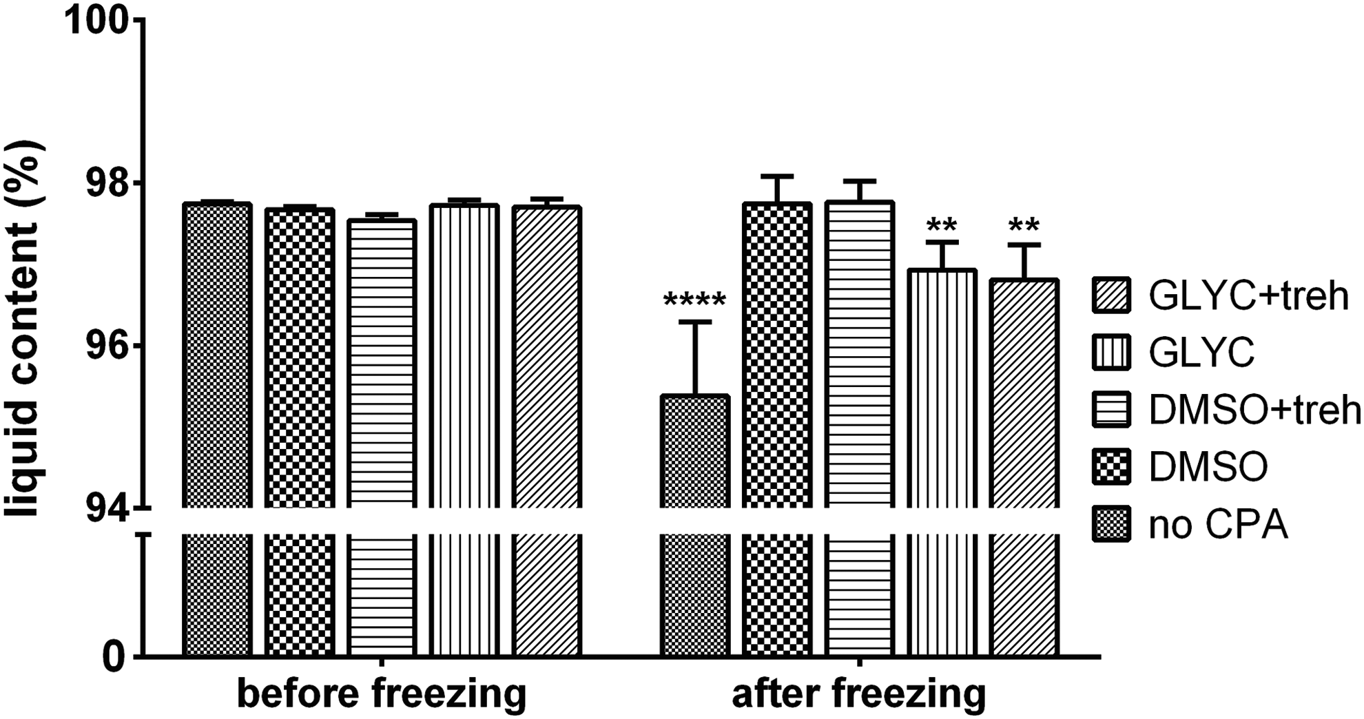
Liquid content study of alginate hydrogels after incubation with the CPAs (before freezing) and after incubation with the CPAs, freezing and thawing (after freezing). Error bars represent mean ± SD (n = 5). **p < 0.01 and ****p < 0.0001. Confidence levels are referred to the relative counterpart before freezing.
Discussion
The issue of preservation and storage of encapsulated cells is an obstacle for translating cell encapsulation to tissue engineering in the form of commercial products.24,27,35 Cryoprotective agents (CPAs) are added during the freezing process to protect cells from cryodamage. 38 DMSO and glycerol, in particular, penetrate cell membrane at physiological temperature and have been extensively used to prevent cell damage during cooling by minimizing intracellular ice formation.22,26 Furthermore, the addition of trehalose and other disaccharides to these cryoprotectants is known to enhance cell survival during cryopreservation of cell suspension 44 and engineered tissues.30,31
Many factors may influence cell viability in a cryopreservation system, including fabrication and freezing process, molecule diffusion kinetics in the hydrogel matrix, cell morphology, and reorganization of the adhesion sites. Therefore, in order to establish a cryopreservation protocol for tissue-engineered products, the interaction between cells, biomaterial, CPAs, and freezing method should be taken into consideration for an extensive investigation of the system. In this study, we propose a systematic approach for assessing the impact of cryopreservation on engineered tissue, considering the analysis of cell recovery and of the material properties.
In this work, we characterized the influence of cryopreservation on encapsulated cells, comparing the effect of different cryoprotectants on cell response after thawing. For this reason, sodium alginate hydrogel was chosen for encapsulating cells to reduce the interaction between cells and their encapsulating matrix. As a model, cells were entrapped in alginate fibers made by wetspinning, which represents a fast and high-throughput encapsulation model. 15 Cell-laden microfibers are easy to prepare, handle, and assemble from an engineering standpoint and are appropriate to reconstruct structures with a hierarchical alignment. 45 To validate the cryopreservation technique, MG-63 cell line was chosen as an established human osteosarcoma cell line for bone tissue engineering models with fully genetic characterization. 46 After cell encapsulation, the hydrogel fibers were frozen in presence of different cryopreservation media containing DMSO, glycerol and trehalose, whose effects on cell functionality were compared after thawing.
All CPA formulations successfully prevented the death of encapsulated cells during cryopreservation compared to cells frozen in absence of any cryoprotectants. The estimated post-thaw viability of cells encapsulated and cryopreserved with DMSO was 7% lower than in the noncryopreserved group and 15% higher than in the group cryopreserved with glycerol, and comparable to the results already published by other groups using the same freezing protocol. 32 The addition of trehalose led to a decrease of cell viability that is larger for the group cryopreserved with DMSO, considering both the encapsulated cells and the control. We also monitored the viability of encapsulated cells up to 3 days after thawing to evaluate possible harmful effects of the CPAs that may remain in contact with the cells. In fact, high concentrations of cryoprotective chemicals can be toxic for cells, and toxicity must be reduced by decreasing the time of cell exposure to the cryoprotectants. 22 The analysis of the Live/Dead data excluded protracted detrimental effect of the CPAs, since appreciable cell death was not detected after the initial post-thawing observation.
To develop methods to mitigate the harmful effect of cryopreservation on encapsulated cells, a detailed study of the possible pathways and mechanism leading to cell death is of primary importance. The exact mechanism leading to apoptosis and necrosis activated during freezing is not well understood and remains to be studied. 47 Moreover, the death pathway activated in one cell type might not be activated in other cell types. Cell cycle after thawing and releasing from the alginate matrix was compared with nonencapsulated protocol. Flow-cytometric analysis highlighted that both encapsulation in alginate and addition of trehalose reduced the viability of cells after cryopreservation. Besides, we observed a difference in cell-death pathway since the encapsulation led to later stage of apoptosis and necrosis of the cell population. A possible explanation is that the hydrogel acts as barrier that could cause a gradient of the cryoprotectant concentration in the fiber, so leading position dependent damage of cells and subsequent necrosis.
The influence of the different cryoprotective formulation on cell metabolism and proliferation were investigated up to 2 weeks, since it has been reported that the effectiveness of cryopreservation cannot be reliably determined immediately after thawing. 48 Moreover, a direct correlation between the viability (Fig. 5) of a certain cell population and the ability of viable cells in that population to proliferate (Fig. 6) and to be metabolically active (Fig. 7) was not detected. Encapsulated cells, thawed and cultured in TCP, showed a larger metabolic activity and proliferative capacity when DMSO and glycerol alone were used as cryoprotectants. Indeed, a previous comparison of fibroblast response to low temperature in suspension and 3D culture indicated a more intense functional expression of stress proteins in 3D constructs. 41 Interestingly, trehalose addition to encapsulated cells had a favorable effect on cell recovery when DMSO was used, and a reverse situation in the case of glycerol. This appears to be contrary to previous studies, which reported how the addition of sugars to cryoprotectants that penetrate cells at physiological temperature leads to a detectable improvement of the results in cryopreservation protocol for both DMSO 31 and glycerol. 49 However, the abovementioned studies used different encapsulation materials and freezing protocols than those used in the current work, and trehalose has been reported to possess inhibitory effects on proliferation of fibroblast-like cells in certain conditions. 50
In our analysis, we detected an increased number of apoptotic cells corresponding to lower proliferation of the reseeded cells. A possible explanation is that Trypan blue method, which was used to counts the alive cells for the reseeding experiments, doesn't discriminate between vital and early apoptotic cells. 51 Thus, the real viable cells that are able to proliferate are less than those counted with the Trypan Blue exclusion method. Moreover, apoptosis has been reported to be responsible for a low cell recovery rate after cryopreservation. 52 The relationship between the addition of trehalose and apoptosis could be attributed to various reasons and its clarification would require complex and laborious gene expression studies. Moreover, the effect of trehalose must be judged considering its complex interactions with the alginate matrix and DMSO/glycerol, which could affect its diffusion capacity from the medium to the cells. Besides, in our study DMSO better preserved the viability of encapsulated cells compared to glycerol; however, its effect (in absence of trehalose) resulted in increased apoptosis after thawing, corresponding to a diminished proliferation and metabolic capacity. This phenomenon can derive from the different ability of glycerol and DMSO to penetrate the alginate layer and interact with the cell membrane. This evidence highlights the importance of analyzing different cryopreservation protocols for different tissue constructs, to find the formulation that best adapt to the chosen cells and materials.
Rheological analysis and liquid content measurement were performed to characterize the influence of cryopreservation on the viscoelastic and swelling properties of alginate hydrogel. In fact, these are critical features of hydrogels used in biomaterials and tissue engineering applications, influencing both tissue morphogenesis and stem cell differentiation. A temperature of 37°C was selected for conducting the experiments to mimic in vitro conditions. Based on our results, the values of tan(δ) (higher than 0.2 at any frequency) reveal a clear viscoelastic behavior of the hydrogels. Moreover, it is clear that all the CPAs successfully preserved the viscoelastic properties of alginate after thawing, at the same time preventing the reduction of water content. We hypothesize that, during the freezing process, the presence of the CPAs can influence the growth of ice crystals from the water present in the matrix, thus affecting the conformational changes of the alginate network after thawing.53,54 In fact, the slow freezing process could generate concentrations fluctuations of the alginate content in solution, and this, thanks to the presence of residues of calcium, could results in a further cross-linking of the gel. 55 Data on the water content (Fig. 9) are consistent with the above assumption, and the increase of G′ in alginate samples after freezing and the stiffness/frequency relationship (Fig. 8). When CPAs are added, ice crystals formation is prevented and concentrations fluctuations are damped. The increase of the matrix rigidity could in theory negatively impact the cells viability. However, the intracellular ice formation remain the main cell death cause.
The collected data demonstrate that cells encapsulated in alginate fibers remained viable after cryopreservation in liquid nitrogen and thawing, hence stocks of cryopreserved cell-laden hydrogel constructs could be thawed when necessary and three-dimensionally assembled. 45
The proposed method can be adopted to compare and select multiple cryopreservation parameters for a given system, including cryopreservation medium formulation, cell density, presence of adhesive motifs in the material and freezing rate. In fact, we evaluated the influence of different parameters on the efficacy of cryopreservation, including the use of different CPAs and the addition of trehalose to the cryoprotective solution. Moreover, this protocol investigates the impact of encapsulation by comparing the results obtained for entrapped cells with those obtained for suspended cells. This work focused on evaluating the recovery of cells in terms of viability and functionality and the impact of freezing on the rheological properties and water content of the encapsulating hydrogel. The outcome of our research thus offers an approach to investigate the effect of cryopreservation on cell-laden hydrogel constructs, which can be adopted as support for different applications, from biofabrication to cell banking to drug releasing devices.
Conclusions
In summary, in the present work the possibility to cryopreserve cell-laden alginate fibers in presence of different cryoprotectants formulations was investigated. Encapsulated cells, when cryopreserved in presence of DMSO and glycerol, maintained a viability degree comparable to cells cryopreserved with standard protocols and no protracted harmful effect of the CPAs were observed after thawing. Furthermore, cryopreserved encapsulated cells expressed a faster recovery of functionality, confirming previous works. 41 Overall, we propose a method to produce and store cell-laden hydrogel constructs that can serve as building blocks for subsequent assembly of tissue constructs according to different biofabrication strategies.9,23,56
Besides, in this article we present an approach for the evaluation of the effect of cryopreservation on the functionality of cell-laden constructs. In fact, the protocol described herein proposes a method for determining the impact of cryopreservation on cell recovery and material properties after freezing in presence of different cryoprotectants, in short- and mid-term (up to 2 weeks after thawing). The proposed approach can be adopted for evaluating the effect of other cryopreservation methods on cells encapsulated in specific hydrogel matrices and designed for specific applications. In fact, integration of cryopreservation techniques with cell micro-scale encapsulation introduces a promising approach to the field of tissue engineering and can be adopted as support for bottom-up engineered tissue assembly and cell banking, expansion and release.18,24
Footnotes
Acknowledgments
The authors acknowledge Dr. Volha Liaudanskaya for helping with DNA quantification measurements and Dr. Mariangela Fedel for helping with flow-cytometry experiment design.
Disclosure Statement
No competing financial interests exist.