
Abstract
Poly-ɛ-caprolactone (PCL) based microspheres have received much attention as drug or growth factor delivery carriers and tissue engineering scaffolds due to their biocompatibility, biodegradability, and tunable biophysical properties. In addition, PCL and polydimethylsiloxane (PDMS) can be fabricated into thermoresponsive shape memory polymers for various biomedical applications (e.g., smart sutures and vascular stents). However, the influence of biophysical properties of PCL-PDMS based microspheres on stem cell lineage commitment has not been well understood. In this study, PDMS was used as soft segments of varying length to tailor the elastic modulus of PCL-based copolymers. It was found that lower elastic modulus (<10 kPa) of the tri-block copolymer PCL-PDMS-PCL promoted vascular differentiation of embryonic stem cells, but the range of 60–100 MPa PCL-PDMS-PCL had little influence on cardiovascular differentiation. Then different sizes (30–140 μm) of PCL-PDMS-PCL microspheres were fabricated and incorporated with embryoid bodies (EBs). Differential expression of KDR, CD31, and VE-cadherin was observed for the EBs containing microspheres of different sizes. Higher expression of KDR was observed for the condition with small size of microspheres (32 μm), while higher CD31 and VE-cadherin expression was observed for the group of medium size of microspheres (94 μm). Little difference in cardiac marker α-actinin was observed for different microspheres. This study indicates that the biophysical properties of PCL-PDMS-PCL microspheres impact vascular lineage commitment and have implications for drug delivery and tissue engineering.
Introduction
C
Efficient scalable differentiation of cardiovascular cells from PSCs usually relies on the microspheres/microparticles to support cell adhesion or to deliver the inducing growth factors in suspension.10–12 Microspheres of different materials such as gelatin, agarose, and poly lactic-co-glycolic acid (PLGA) were shown to induce differential expression of genes and proteins for mesoderm and endoderm lineage commitment from PSCs. 13 In addition, the biophysical properties (e.g., microsphere size and the elastic modulus) of microspheres have profound effects on stem cell lineage commitment. For example, the comparison of different particle sizes showed the accelerated differentiation and more cystic embryoid bodies (EBs) when delivering retinoic acid using small size particles (1–11 μm). 14 PLGA microparticles were also studied in the range of 100–240 μm during chondrogenic differentiation of mesenchymal stem cells (MSCs), where the use of particles of 175 μm resulted in the cartilage-like matrix formation (i.e., collagens I and II and glycosaminoglycan). 15 PLGA microspheres were incorporated into the collagen gels to increase the stiffness of extracellular microenvironment and promoted osteogenic differentiation of MSCs by providing stiff microenvironment. 16 All these studies indicate that biophysical properties of microspheres or microparticles influence stem cell differentiation.
Poly-ɛ-caprolactone (PCL) based microspheres have wide applications in drug delivery and tissue engineering applications.17,18 PCL is biocompatible with a variety of human tissues and its long-term biodegradability allows the drug release up to several months. 19 In addition, PCL-based materials have flexible mechanical properties (Young's modulus, tensile strength, etc.) by tailoring their structures. 18 PCL-based microspheres (about 30–210 μm) can be fabricated by various methods such as emulsion solvent evaporation, spray drying, solution enhanced dispersion, and so on.17,18 Moreover, PCL allows the modification of physical, chemical, and mechanical properties through the formation of copolymers, for example, gelatin, chitosan, polydimethylsiloxane (PDMS), poly(ethylene glycol), poly(vinyl alcohol), and PLGA, or forming composites by blending with other polymers. 18
Among different PCL-based materials, the copolymers of PCL-PDMS-PCL have unique properties and can be used to synthesize and fabricate thermoresponsive shape memory polymer (SMP) based materials.20,21 SMPs have been used in biomedical devices, such as smart sutures, vascular stents, and tissue engineering scaffolds. 22 For example, the cell alignment (i.e., cytoskeleton orientation and nuclear alignment) of MSCs was shown to be modulated by the shape memory-actuated fiber alignment of the scaffolds. 23 In particular, PDMS can serve as soft segments of varying length to tailor the mechanical properties of PCL (serving as hard segments)-based polymers. Varying the segment lengths of PCL and PDMS can tune the mechanical properties of the resulting copolymers to fabricate the copolymers with desired stiffness. 20
The objective of the present study is to fabricate PCL-PDMS-PCL microspheres and evaluate the influence of biophysical properties (i.e., microsphere size and elastic modulus) of PCL-PDMS-PCL copolymers on cardiovascular differentiation of ESCs. This study indicates the importance of biophysical properties of microspheres on cardiovascular lineage commitment and has the significance in biomaterial design for stem cell-based tissue engineering, cellular differentiation, and growth factor delivery. 24
Materials and Methods
Materials
ɛ-caprolactone monomer, poly (dimethylsiloxane)-bis(3-aminopropyl) terminated (molecular weight [Mn] ∼2500 g/mol), hexamethylene diisocyanate (HDI), stannous 2-ethylhexanoate, and solvents were purchased from Sigma-Aldrich (St. Louis, MI). Aminopropyl terminated polydimethylsiloxane DMS A12 (Mn ∼1000 g/mol), DMS A15 (Mn ∼3000 g/mol), DMS A21 (Mn ∼5000 g/mol), and DMS A31 (Mn ∼25,000 g/mol) were purchased from Gelest, Inc. (Morrisville, PA).
Synthesis of PCL-PDMS-PCL copolymers
Macromers of PCLn-block-PDMSm-block-PCLn were synthesized by ring-opening polymerization of ɛ-caprolactone in the presence of
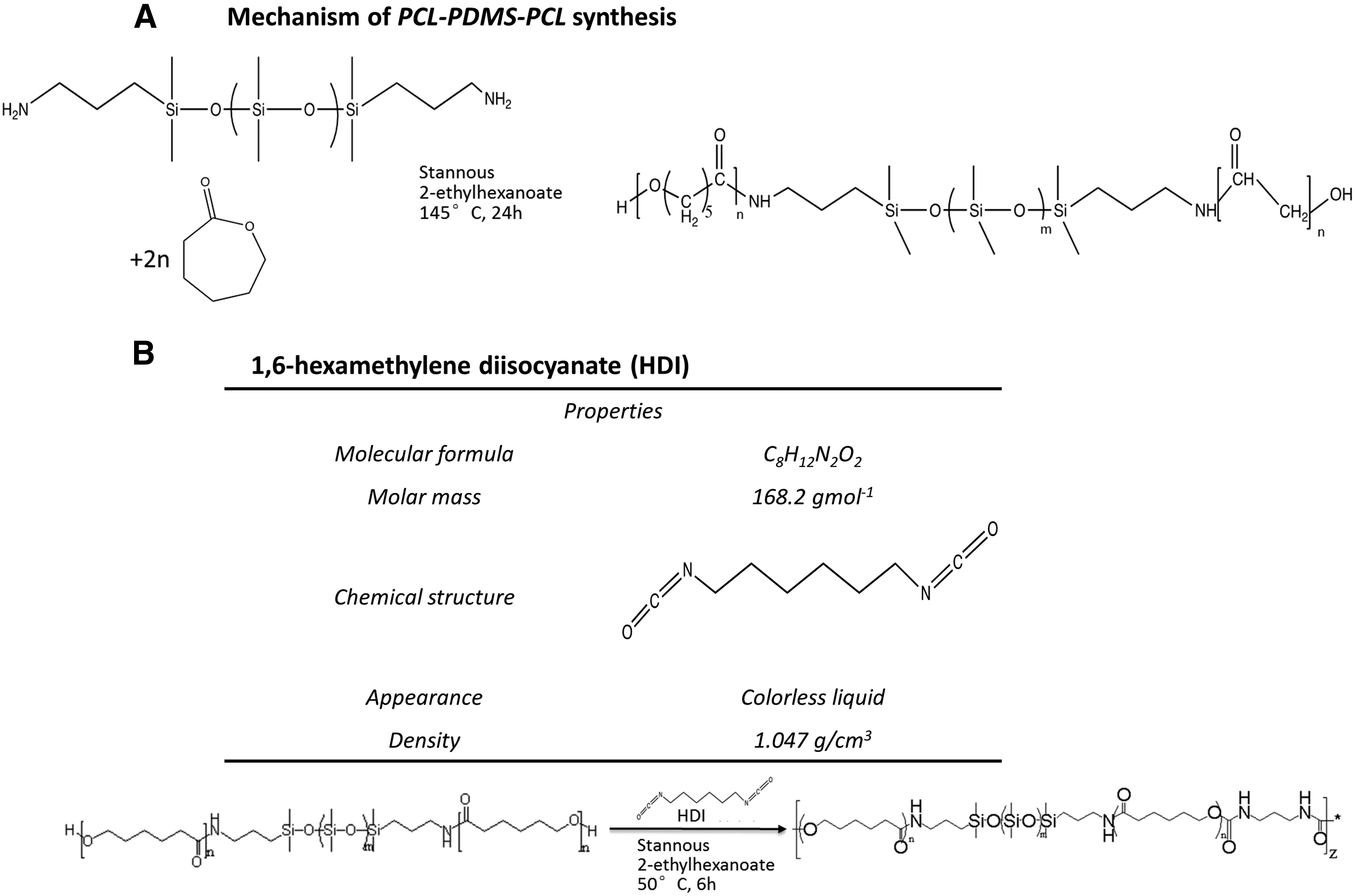
Mechanism of PCL-PDMS-PCL copolymer synthesis.
For the chain propagation between oligomers by adding HDI as a coupling agent (Fig. 1B), the synthesized oligomers were transferred into a 1000 mL flask and cooled down from 145°C to 100°C. Synthesized oligomers were dissolved in 1,2-dichloromethane and water. The 1,2-dichloroethane was then removed at 100°C until no bubbles existed in the solution. When the flask was cooled down from 100°C to 50°C, two drops of stannous octanoate and 0.002 mole HDI were added into the system sequentially and reacted for 5 h.
Dynamic mechanical analysis
Dynamic mechanical analysis (DMA) was used to measure the Young's modulus of different copolymers. The samples were characterized using dynamic mechanical analyzer (Q800; TA Instruments). For tensile modulus, the rectangular samples were exerted under strain at a strain rate of 0.1 min−1 and 30°C. Compressive modulus was measured for samples (low elastic modulus) that cannot be measured for tensile modulus. In a compression model, the cylindrical samples were compressed with a 15-mm compression clamp at a strain rate of 0.1 min−1 and 30°C. The Young's modulus was calculated based on the initial slope of the stress–strain curve.
Differential scanning calorimetry analysis and 1 H-NMR
For differential scanning calorimetry (DSC) analysis, the samples were run on the differential scanning calorimeter (Q100; TA Instruments) from 30°C to 100°C at a heating rate of 10°C/min. The samples were then cooled down from 100°C to 30°C at a cooling rate of 10°C/min. The melting point Tm was calculated from the heat flow-temperature curve. The enthalpy change was determined from the endothermic melting peak. The % crystallinity was calculated using the equation:
where ΔHm is normalized based on the % mass of PCL segments in the PCL-PDMS-PCL macromers. ΔHm0 is 139.5 J/g for 100% crystalline PCL. 26
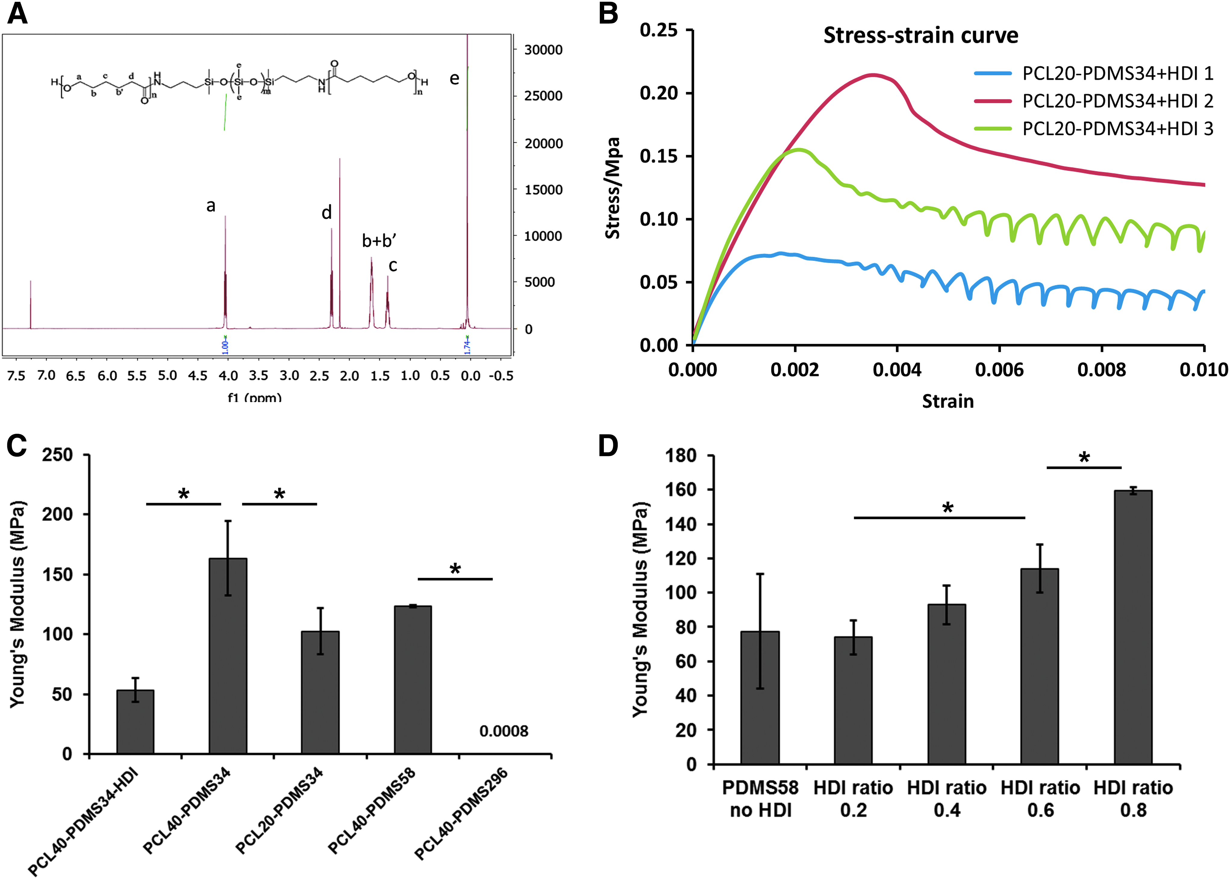
1H-NMR spectra of the copolymers were obtained on a Bruker 500 M spectrometer at 200 MHz. The spectrum was taken in deuterated chloroform at 20°C. The composition of copolymers was calculated from the ratios of absorbance at 0.07 and 4.05 ppm. Average molecular weight (Mn) was calculated according to the following equation:
In this equation, it was assumed that the synthesized copolymers had a constant number of PCL repeating units, n = 40 or 20, and the integration for methylene group in the 1 H-NMR spectrum from PCL repeating units is M and for methyl group in PDMS repeating units is N.
Fabrication of PCL-PDMS-PCL copolymer based microspheres
PCL-PDMS-PCL microspheres were fabricated using a reverse-phase precipitation method. 27 Briefly, after the copolymer synthesis reaction, the solution in 1,2-dichloromethane was slowly poured into a 1000 mL big beaker containing 500 mL methanol. Microspheres were formed and precipitated at the bottom of beaker when stirring the solution at 100 rpm for 2 h. Then the precipitate was filtrated to remove the mixture of 1,2-dichloromethane and methanol. To remove the remaining oligomers and tin (II) 2-ethylhexanoate, the filtrate was washed thrice with methanol. The final products were dried under vacuum for more than 24 h before characterizations or the use in cell culture.
Scanning electron microscopy
The morphology of microparticle samples was examined using field emission scanning electron microscope (SEM) (JEOL 7401F). Samples were sputter-coated with a thin layer of gold before observation. The SEM images were analyzed using ImageJ software to obtain the size distribution of microspheres.
Undifferentiated ESC cultures
Murine ES-D3 line (American Type Culture Collection, Manassas, VA) was maintained on six-well culture plates coated with 0.1% gelatin (Millipore, Temecula, CA) in a standard 5% CO2 incubator.28–31 The culture medium was composed of Dulbecco's modified Eagle's medium (DMEM; Invitrogen, Carlsbad, CA) supplemented with 10% ESC-screened fetal bovine serum (FBS; Hyclone, Logan, UT), 1 mM sodium pyruvate, 0.1 mM β-mercaptoethanol, penicillin (100 U/mL), streptomycin (100 μg/mL) (all from Invitrogen), and 1000 U/mL leukemia inhibitory factor (Millipore). The cells were seeded at 2–4 × 10⁴ cells/cm2 and subcultured every 2–3 days with 0.05% trypsin.
Cell seeding with microspheres and the induction for cardiovascular differentiation
For cell culture, PCL-PDMS-PCL microspheres were sterilized with 70% ethanol and then washed thrice with phosphate-buffered saline (PBS). Different types of microspheres (0.2 mg/mL) were mixed with the cells and seeded in low attachment 24-well plates at 2 × 10⁵ cells in 1 mL of medium. The differentiation medium was composed of DMEM, 10% FBS, 1 mM β-mercaptoethanol, penicillin (100 U/mL), and streptomycin (100 μg/mL) (DMEM-FBS). The medium was supplemented with 10 ng/mL bone morphogenetic protein (BMP)-4 (R&D Systems) for the first 4 days. At day 4, the formed EBs with microspheres were replated into 24-well plates coated with 0.1% gelatin. The cells were fed with differentiation medium without growth factors. At day 2, 5, 11, and 14 after replating, the cells were characterized for cardiovascular markers. For culture medium experiments, the replated cells were maintained in either DMEM-FBS or RPMI-B27 (RPMI + 2% B27) serum-free medium (Invitrogen).
To illustrate the influence of elastic modulus only, the PCL-PDMS-PCL copolymers (9.3 kPa, 58 MPa, and 103 MPa) were processed into 2D disks (diameter: 10 mm, thickness: 1 mm). The day 4 EBs without particles were replated onto different 2D disks coated with 0.1% gelatin. The cells were grown for 11 days and characterized for cell proliferation and cardiovascular marker expression.
Biochemical assays
LIVE/DEAD® Staining Kit (Molecular Probes) was used to assess viability of the cells in EBs with microspheres. 32 After a 5-day culture, the samples were incubated in DMEM containing 1 μM calcein AM (green) and 2 μM ethidium homodimer I (red) for 30 min and imaged under a fluorescent microscope (Olympus IX70, Melville, NY). Cell numbers of microsphere cultures at day 1, 3, and 5 were determined using a hemocytometer after trypsinization. The cell aggregates with microspheres were also incubated with 5 mg/mL 3-(4,5-dimethylthiazol-2-yl)-2,5-diphenyltetrazolium bromide (MTT; Sigma) solution at day 1, 3, and 5 to evaluate metabolic activity. The absorbance of the samples was measured at 500 nm using a microplate reader (Bio-Rad, Richmond, CA).
Reverse transcription polymerase chain reaction
Total RNA was isolated from ESC samples using the RNeasy Mini Kit (QIAGEN, Valencia, CA) according to the manufacturer's protocol followed by the treatment of DNA-Free RNA Kit (Zymo, Irvine, CA).
33
Reverse transcription was carried out using 2 μg of total RNA, anchored oligo-dT primers (Operon, Huntsville, AL), and SuperScript III (Invitrogen, Carlsbad, CA) (according to the protocol of the manufacturer). Primers specific for target genes (Table 1) were designed using the software Oligo Explorer 1.2 (GeneLink, Hawthorne, NY, Table 1). The gene β-actin was used as an endogenous control for normalization of expression levels. Real-time reverse transcription polymerase chain reactions (RT-PCRs) were performed on an ABI7500 instrument (Applied Biosystems, Foster City, CA), using SYBR1 Green PCR Master Mix (Applied Biosystems). The amplification reactions were performed as follows: 2 min at 50°C, 10 min at 95°C, and 40 cycles of 95°C for 15 s and 55°C for 30 s, and 68°C for 30 s. Fold variation in gene expression was quantified by means of the comparative Ct method:
Immunocytochemistry
Briefly, the cells were fixed with 4% paraformaldehyde (PFA) and permeabilized with 0.2–0.5% Triton X-100 (for intracellular markers only). The samples were then blocked and incubated with mouse or goat primary antibody against: KDR (Millipore), CD31 (PECAM-1) (Santa Cruz Biotechnology, Inc., Dallas, TX), VE-cadherin (Santa Cruz Biotechnology, Inc.), and α-actinin (Sigma). After washing, the cells were incubated with the corresponding secondary antibody (Molecular Probes): Alexa Fluor® 594 donkey anti-goat IgG (for CD31 and VE-cadherin) or Alexa Fluor® 488 goat anti-mouse IgG1 (for KDR and α-actinin). For F-actin staining, the cells were incubated with Alexa Fluor 594 Phalloidin (Molecular Probes). For Yes-associated protein (YAP) staining, the cells were incubated with rabbit primary antibody (Santa Cruz Biotechnology, Inc.), followed by Alexa Fluor 594 goat anti-rabbit IgG. The samples were stained with Hoechst 33342 (to reveal cell nuclei) and visualized under a fluorescent microscope.
Flow cytometry
To quantify cardiovascular marker expression, the cells were harvested by trypsinization and analyzed by flow cytometry. 34 Briefly, 1 × 10⁶ cells per sample were fixed with 4% PFA and washed with staining buffer (2% FBS in PBS). The cells were permeabilized with 100% cold methanol (for intracellular markers only), blocked, and then incubated with primary antibodies against KDR, CD31 (PECAM-1), VE-cadherin, Nkx2.5 (Santa Cruz Biotechnology, Inc.), and α-actinin followed by the corresponding secondary antibody: Alexa Fluor 594 donkey anti-goat IgG (for CD31 and VE-cadherin), Alexa Fluor 594 goat anti-rabbit IgG (for Nkx2.5), or Alexa Fluor 488 goat anti-mouse IgG1 (for KDR and α-actinin). The cells were acquired with BD FACSCanto™ II flow cytometer (Becton Dickinson) and analyzed against isotype controls using FlowJo software.
Vascular structure assay
Briefly, 24-well plates were coated with 200 μL/well 1:1 diluted Geltrex (B&D Biosciences) at room temperature for more than 30 min. The cells were plated at 5 × 10⁵ cells on Geltrex-coated plates in 500 μL EGM-2 medium (Lonza, for endothelial cells) and incubated at 37°C in 5% CO2 for 7 days. Cell morphology was photographed by a phase-contrast microscope. The cells were then fixed and assessed for marker expression of CD31, VE-cadherin, and F-actin.
Statistical analysis
Each experiment was carried out at least thrice. The representative experiments were presented, and the results are expressed as mean ± standard deviation. To assess the statistical significance, one-way ANOVA followed by Fisher's LSD post hoc tests was performed. A p-value <0.05 was considered statistically significant.
Results
Fabrication and characterization of PCL-PDMS-PCL copolymers
Macromers of PCLn-block-PDMSm-block-PCLn (n = 20, 40, and 50, m = 10, 34, 58, and 296) were synthesized by ring-opening polymerization of ɛ-caprolactone in the presence of
Characterization of PCL-PDMS-PCL copolymers for chemical and mechanical properties.
p-Value <0.05 in comparison with polymer 1.
p-Value <0.05 in comparison with polymer 2.
p-Value <0.05 in comparison with polymer 3.
Effect of elastic modulus of PCL-PDMS-PCL copolymers
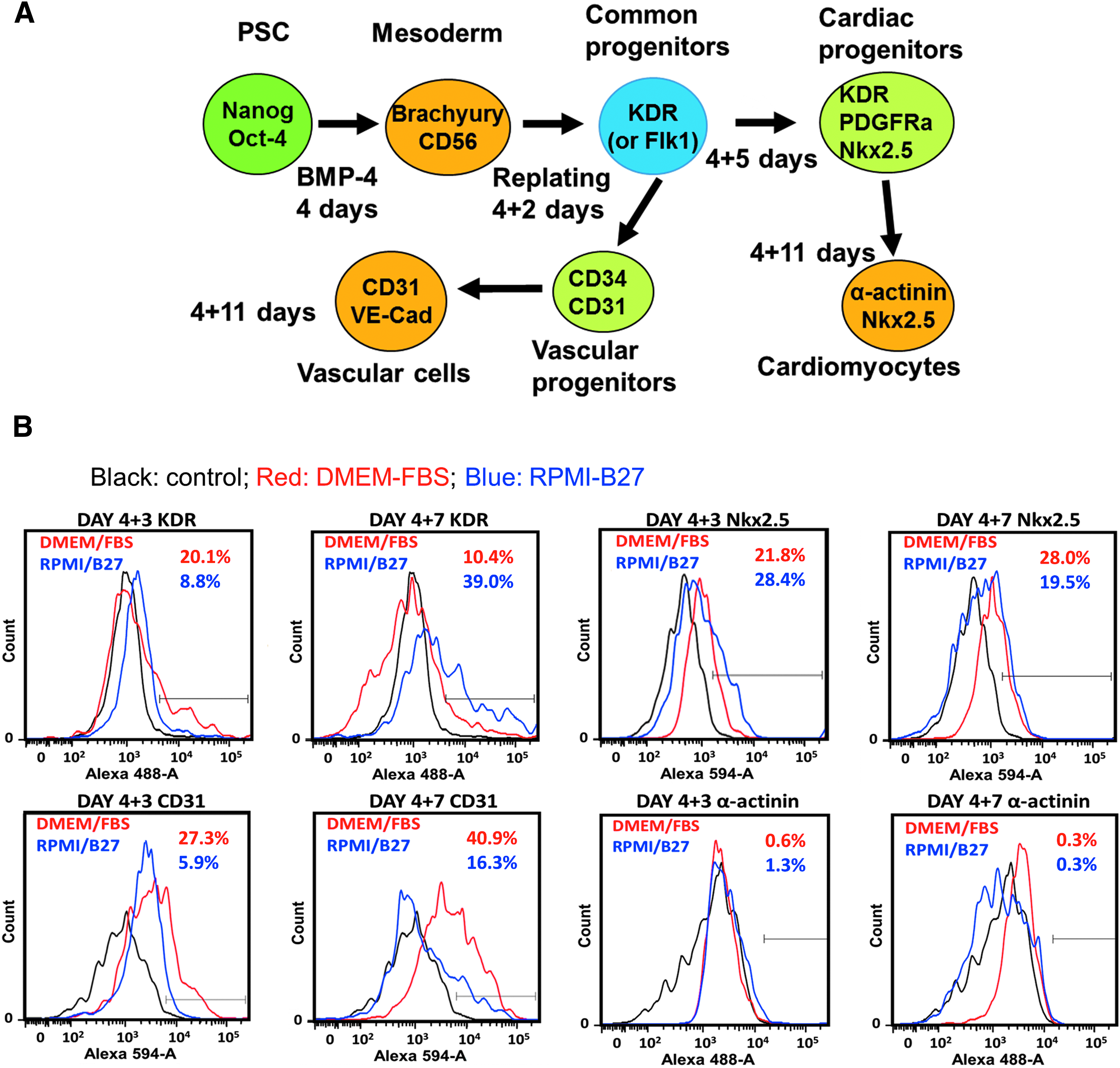
Before the evaluation of the copolymers, the differentiation media for cardiovascular differentiation of ESCs was compared. The differentiation was initiated with BMP-4 induction to obtain KDR+ progenitors which can further differentiate into cardiac cells and vascular cells (Fig. 3A). The day 4 EBs were replated onto the gelatin-coated surface in DMEM-FBS or RPMI-B27 medium. At day 3 after replating, KDR was expressed at a higher level for cells grown in DMEM-FBS compared to RPMI-B27 media (20.1% vs. 8.8% for DMEM-FBS or RPMI-B27, respectively), but decreased quickly at day 7 (10.4% vs. 39.0% for DMEM-FBS or RPMI-B27, respectively) (Fig. 3B). Compared to RPMI-B27 culture, CD31 expression was higher at both day 3 (27.3% vs. 5.9% for DMEM-FBS or RPMI-B27, respectively) and day 7 (40.9% vs. 16.3% for DMEM-FBS or RPMI-B27, respectively) for DMEM-FBS culture. For cardiac markers, Nkx2.5 level was higher for RPMI-B27 condition (28.4% vs. 21.8% for RPMI-B27 or DMEM-FBS, respectively) at day 3 after replating, but became lower (19.5% vs. 28.0% for RPMI-B27 or DMEM-FBS, respectively) than DMEM-FBS culture at day 7 after replating. Both cultures had comparable levels of α-actinin (0.3–1.3%). The temporal expression of KDR and Nkx2.5 (the progenitor markers) suggested that RPMI-B27 may need longer differentiation time than DMEM-FBS. Based on these results, DMEM-FBS medium was used in further study.
Cardiovascular differentiation from ESCs.
2D disks of three types of copolymer (PCL20-PDMS58-PCL20: 9.3 kPa, PCL40-PDMS28-PCL40: 58 MPa, and PCL20-PDMS34-PCL20+HDI: 103 MPa) were evaluated for cardiovascular differentiation (Fig. 4). The highest expression of vascular markers CD31 (63.5%) and VE-cadherin (56.5%) was observed for the 9.3 kPa condition (Fig. 4A). For 58 and 103 MPa conditions, the CD31 expression was 14.3% and 36.4%, respectively, and the VE-cadherin expression was 21.1% and 52.0%, respectively. The lower CD31 and VE-cadherin expression for 58 MPa condition than 103 MPa condition may be due to differentiation variations since vascular differentiation was shown to be promoted at the lower elastic modulus.21,35,36 These data indicate that the modulus range of 58–103 MPa resulted in 14–36% CD31 and 21–52% VE-cadherin for EBs replated on the 2D disks. The expression level of α-actinin was similar (1.6–4.9%) for all three conditions. The fold increase in cell number was the lowest for the 9.3 kPa condition, but was similar for the modulus of 58 and 103 MPa (Fig. 4B), indicating that the substrate with high modulus promotes cell proliferation.

Effect of elastic modulus of PCL-PDMS-PCL copolymer 2D disks on cell proliferation and cardiovascular differentiation.
Biocompatibility of PCL-PDMS-PCL microspheres
PCL-PDMS-PCL (with modulus range of 90–110 MPa) microspheres with three different sizes were then fabricated (Fig. 5). It was noticed that the soft 9.3 kPa copolymer was difficult for microsphere fabrication and the maintenance of sphere size. The morphology of the microspheres was shown in the SEM images, and the size distribution of microspheres was characterized. The average diameter of the three types of microspheres was 32 ± 11 μm (S), 94 ± 39 μm (M), and 140 ± 65 μm (L). After the microspheres (0.2 mg/mL) were mixed with ESCs for EB formation, the microspheres were incorporated into the EBs as shown in the phase contrast images (Fig. 6Ai). The percentages of EBs that incorporated with microspheres (showing as dark particles in the EBs) were assessed based on image analysis. About all the EBs contained the S-spheres, about 40% of EBs contained M-spheres, and about 25% of EBs contained L-spheres. The EBs incorporating the microspheres were viable as shown in the images of LIVE/DEAD assay (Fig. 6Aii). The dead cells were only observed for the single cells that were not able to form the EBs. MTT activity showed the increase with culture time for all three conditions, further confirming the viability of the cells in the EBs (Fig. 6B). M-sphere condition showed the highest level of MTT activity compared to S-sphere and L-sphere conditions. About fourfold increase in cell number was observed over 5-day culture for all the three groups (Fig. 6C). The BrdU expression of the replated cells was also evaluated (Supplementary Fig. S1; Supplementary Data are available online at
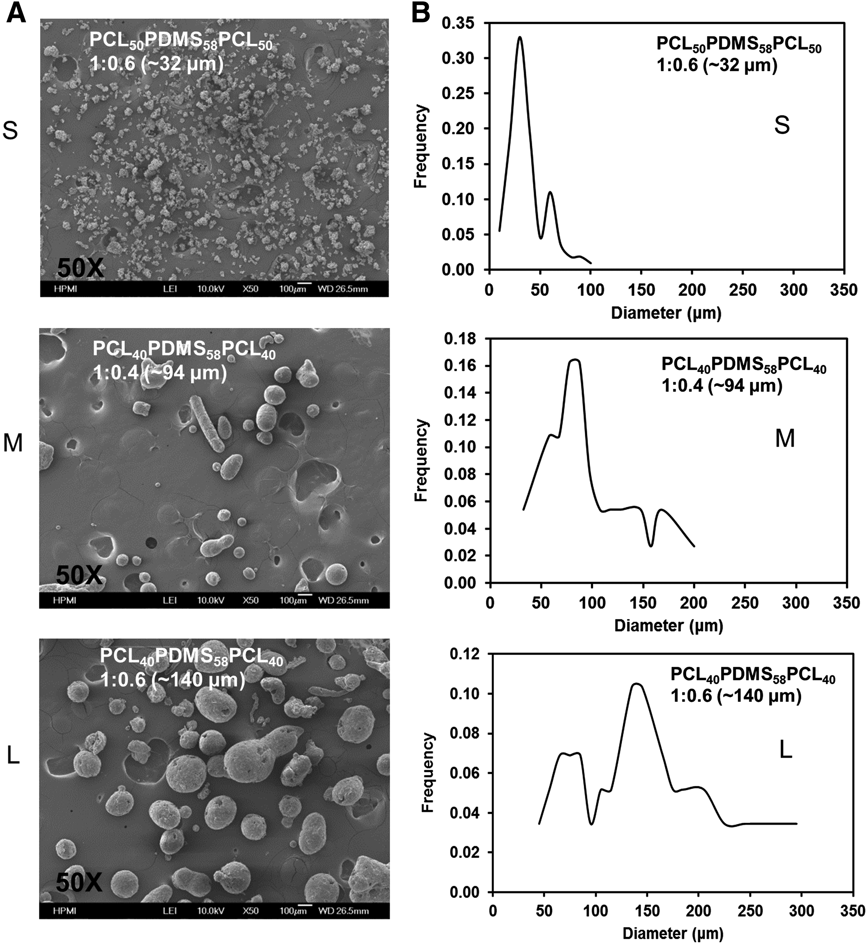
Characterization of PCL-PDMS-PCL copolymer based microspheres.
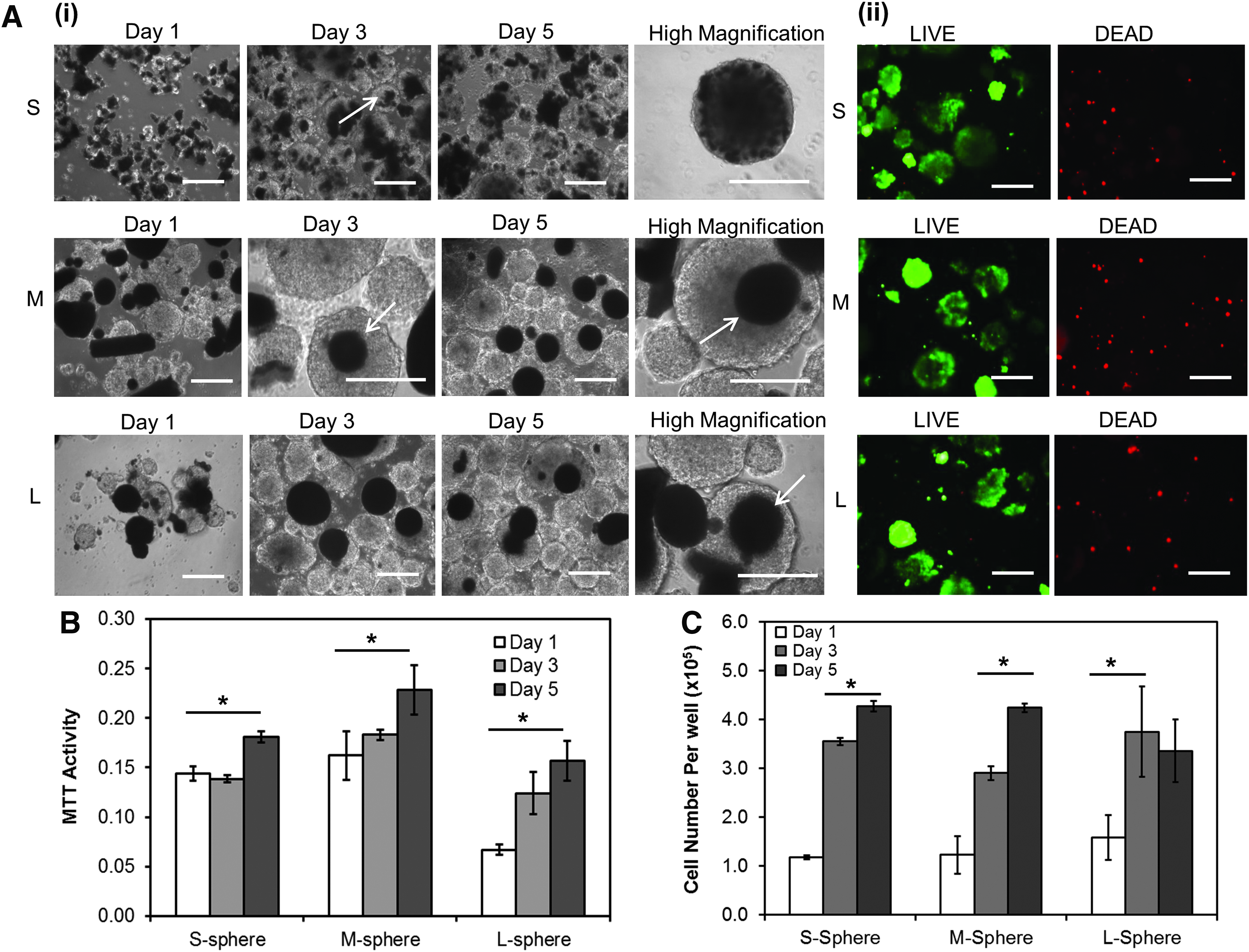
Incorporation of PCL-PDMS-PCL microspheres with EBs.
Cardiovascular differentiation of ESCs grown with microspheres
The EBs incorporating the microspheres were evaluated for the gene expression of Oct-4 and Nanog (Table 3). The decrease of Oct-4 and Nanog indicated the loss of pluripotency for the differentiated cells. The cells were further replated and grown for 11–15 days toward cardiovascular differentiation. At the end of differentiation, the cells expressed CD31 and VE-cadherin but less α-actinin (Fig. 7A). The quantification of progenitor marker showed the high expression of KDR (69.1% ± 3.0% vs. 25.0% ± 9.1% or 26.8% ± 21.3%) at day 3 for S-sphere condition compared to M- and L-sphere conditions. However, the M-sphere condition showed the highest expression of CD31 (67.0% ± 6.0% vs. 29.3% ± 9.3% for S group or 29.9% ± 18.1% for L group) and VE-cadherin (65.5% ± 13.6% vs. 32.8% ± 6.1% for S group or 19.1% ± 9.0% for L group) at day 11 after replating (Fig. 7B, C). For the differentiation in the absence of microspheres, the cells had 53.7% ± 16.1% CD31+ and 17.1% ± 6.6% VE-cadherin+ populations, which were comparable to S and M groups (Supplementary Fig. S2). Since cardiac marker α-actinin expression was low again (3.69% ± 0.04% for EBs without microspheres in Supplementary Fig. S2 and 1.7–4.5% for EBs containing microspheres in Supplementary Fig. S3), the differentiation protocol was more prone to vascular differentiation. The expression of F-actin and YAP was examined for the replated cells of the three groups (Supplementary Fig. S4). The cells expressed the actin stress fibers and nuclear pattern of YAP for all three groups, but no significant difference in expression pattern among different groups was observed.
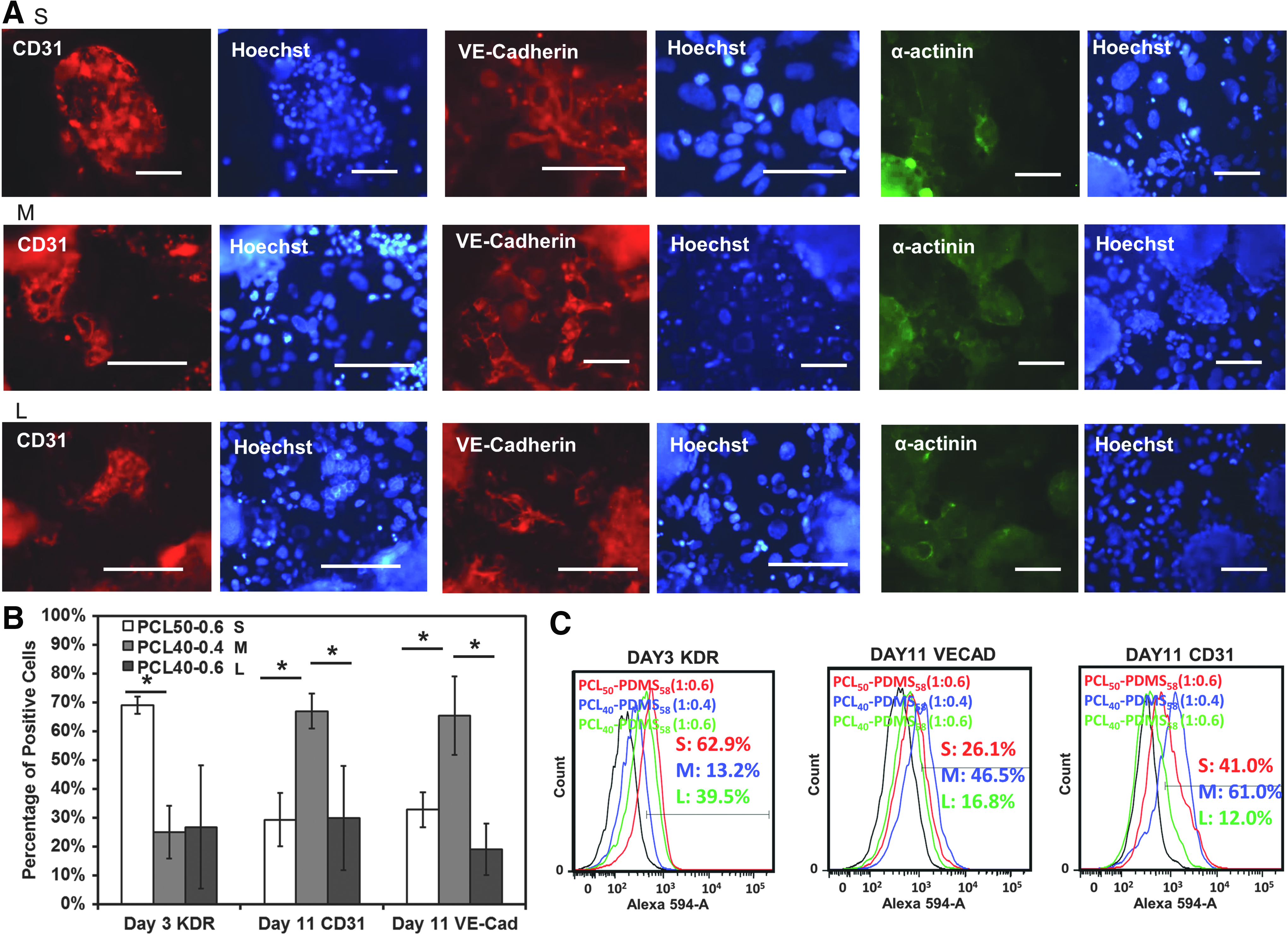
Cardiovascular differentiation from EBs containing PCL-PDMS-PCL microspheres.
p-Value <0.05 in comparison with S-sphere.
The differentiated cells were also harvested and replated onto Geltrex-coated surface in endothelial cell growth medium and growth factors. Under endothelial cell growth condition, CD31 and VE-cadherin positive populations were enriched (Fig. 8). Elongated endothelial cell-like morphology was observed and the M-sphere condition showed more branching points.
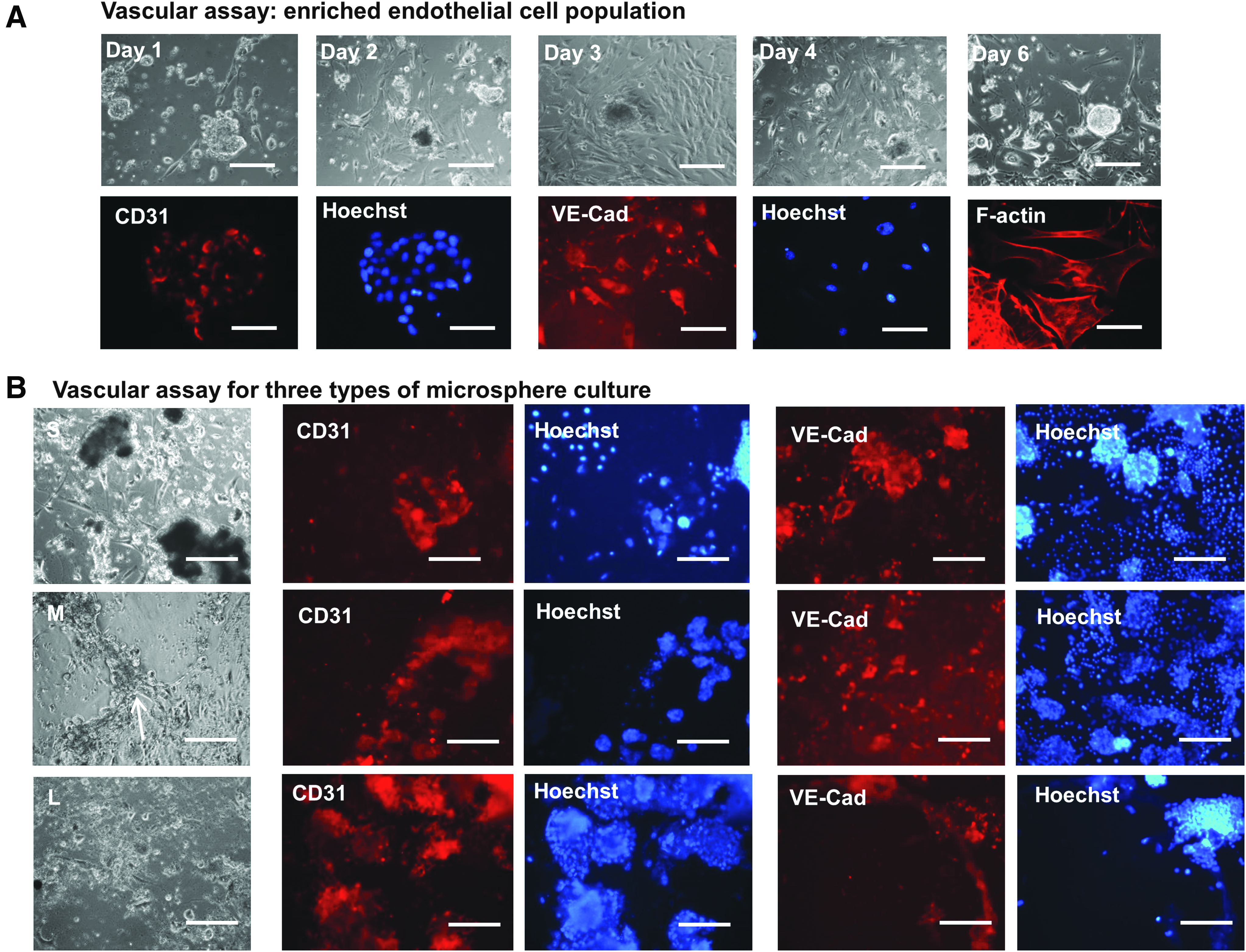
Vascular network assay for the cells derived from EBs incorporating different PCL-PDMS-PCL microspheres.
Discussion
Complex and interactive niche signals, including the mechanical and topographical cues, play a critical role in stem cell lineage commitment. 37 The microstructures and elastic properties of microscale spheres, particles, ribbons, or scaffolds have shown the influence on stem cell fate decisions and drawn attentions in biomaterial design and characterizations.16,38 In this study, PCL-PDMS-PCL copolymers were synthesized, characterized, and used for regulating cardiovascular differentiation from ESCs. In particular, biophysical properties, including microsphere size and elastic modulus of PCL-PDMS-PCL copolymers, were studied.
PCL-PDMS-PCL copolymers are suitable for fabricating thermoresponsive shape memory substrates/scaffolds for various biomedical applications. 39 The use of PCL and PDMS also enables the tuning of elastic properties of the materials by varying the ratio of PCL segments and PDMS segments. This study successfully synthesized PCL-PDMS-PCL copolymers with different PCL and PDMS segments, which were characterized by 1 H-NMR, DSC, and DMA. Increasing PDMS segment length decreased the elastic modulus, while increasing PCL segment length increased the elastic modulus. The elastic properties of PCL-PDMS-PCL copolymers can be further tuned using HDI coupling agent. Potentially, PCL-PDMS-PCL copolymers can be modified with other materials depending on the purpose of the study. For example, tri-block PCL-PDMS-PCL can be copolymerized with DL-lactic acid to control the degradation rate. 40
The influence of PCL-PDMS-PCL copolymers on cardiovascular differentiation in 2D culture was evaluated using 2D disks with different elastic modulus. The effect was mainly observed for vascular differentiation rather than cardiac differentiation. Consistent with the reports from the literature that evaluated other materials,21,36 soft PCL-PDMS-PCL copolymers (<10 kPa) promoted vascular differentiation compared to the hard copolymers (>50 MPa). These results demonstrate the feasibility of using PCL-PDMS-PCL copolymers in a biological system to regulate stem cell differentiation.
The influence of PCL-PDMS-PCL copolymers on cardiovascular differentiation in 3D culture was evaluated using microspheres with different sizes. While PCL-PDMS-PCL copolymers with tunable physical and thermal properties have been recently studied,20,25 the use of such copolymers in stem cell culture has not been well reported. The PCL-PDMS-PCL microspheres were readily incorporated with the EBs and showed the biocompatibility during cardiovascular differentiation of ESCs. The EBs incorporating PCL-PDMS-PCL microspheres displayed high viability, normal MTT activity, and the proliferation activities.
In this study, differential expression of vascular markers was observed for the EBs incorporating the PCL-PDMS-PCL microspheres with different sizes. Higher KDR expression was found for the S-sphere group, but higher CD31 and VE-cadherin were observed for the M-sphere group. It is thought that stiffening EBs with microspheres contribute to the differential expression of vascular markers. Incorporation of gelatin microparticles with stem cell aggregates has shown to increase the stiffness of the aggregates by three to fourfold than those without microparticles. 41 Stiff substrates or scaffolds were reported to favor the early-stage mesoderm commitment from PSCs. For example, elastic modulus of the scaffolds differentially enhanced mesoderm (6 MPa), endoderm (1 MPa), or ectoderm (0.1 MPa) differentiation from human ESCs. 42 Similarly, when increasing the stiffness from 78 kPa to 1.2 MPa, mesoderm differentiation of human ESCs was promoted compared to endoderm differentiation. 43 In this study, the EBs with S-spheres had the highest frequency of microsphere incorporation and almost all the aggregates were stiffened with the microspheres, which may promote mesoderm commitment. M-sphere condition had less frequency of microsphere incorporation followed by the L-sphere condition. The higher CD31 and VE-cadherin expression for M-sphere condition may suggest the balanced mesoderm commitment and further differentiation into vascular lineage.
The effect of microsphere/microparticle size has been observed in the range of 0.24–25 μm for PLGA. The particle size less than 1 μm (e.g., 0.24 μm) was found to be easily taken up by the cells through endocytosis, while microparticles with the size of 6 μm were more universally dispersed in the EBs compared to 25 μm microparticles. 11 The comparison of different particle sizes (1–11 μm) also showed complete cystic EBs when delivering RA using smaller size particle (1 μm). 14 This study revealed the influence of PCL-PDMS-PCL microspheres in the size range of 30–140 μm during ESC mesodermal differentiation.
Both Rho GTPase signaling and Hippo/YAP signaling have been investigated as the mechanotransduction sensors for stem cells.44–46 The soft matrix leads to the cytoplasmic expression of YAP and the increased ability of the cells to form branching endothelial morphology. 21 It was found that stress fibers reduced YAP phosphorylation and promoted nuclear YAP accumulation; thus, disruption of stress fibers using cytochalasin D reduced nuclear YAP. 47 However, in this study, the replated cells with different microspheres all had nuclear expression of YAP, which suggested that the difference in differentiation should result from the suspension stage (e.g., EB culture) of differentiation, where EBs were incorporated with microspheres with different frequencies.
The microspheres are usually used as the carriers (<50 μm) to deliver the drugs or growth factors or as scaffolds or microcarriers (100–200 μm) to influence stem cell differentiation.15,48 Delivery of BMP-4 through gelatin microspheres/microparticles more efficiently increased gene expression of mesoderm and ectoderm lineages because the BMP-4 loaded microparticles were able to use 12-fold less growth factor compared to soluble BMP-4 delivery. 10 Heparin microparticles allowed the delivery of heparin-bounding growth factors BMP-2, vascular endothelial growth factor, and fibroblast growth factor-2, 49 which stimulated the alkaline phosphatase activity of skeletal myoblasts and increased the DNA content mediated by cell particle contact. Similarly, the PCL-PDMS-PCL copolymer-based microspheres can be used as drug carriers and tissue engineering scaffolds as shown for PCL-based microspheres.17,50 Compared with PCL polymer, PCL-PDMS-PCL copolymers have shape memory property that requires further investigation to design smart materials for regulating stem cell fate decisions. As the initial step to apply highly functional materials in stem cell cultures, the present study highlights the importance of biophysical properties of the PCL-PDMS-PCL microspheres during stem cell lineage commitment.
Conclusions
This study synthesized and characterized the biodegradable copolymers that comprised PCL hard segments and PDMS soft segments of variable lengths. The PCL-PDMS-PCL copolymers were evaluated as the substrates and the microspheres that supported cardiovascular differentiation of ESCs. PCL-PDMS-PCL copolymers displayed biocompatibility in ESC differentiation. Small size of microspheres induced higher expression of mesoderm progenitor marker KDR, while medium size of microspheres resulted in higher expression of vascular markers CD31 and VE-cadherin. This study demonstrates the influence of biophysical properties of PCL-PDMS-PCL copolymers on stem cell lineage commitment at the interface of biomaterials and stem cells.
Footnotes
Acknowledgments
The authors thank Ms. Ruth Didier in FSU Department of Biomedical Sciences for her help in flow cytometry analysis and Dr. B.K. Washburn and K. Poduch in FSU Department of Biological Sciences for their help with RT-PCR analysis. This work is supported by FSU start up fund, FSU CRC planning grant, and partially National Science Foundation (grant No. 1652992 to Y.L.).
Disclosure Statement
No competing financial interests exist.
References
Supplementary Material
Please find the following supplemental material available below.
For Open Access articles published under a Creative Commons License, all supplemental material carries the same license as the article it is associated with.
For non-Open Access articles published, all supplemental material carries a non-exclusive license, and permission requests for re-use of supplemental material or any part of supplemental material shall be sent directly to the copyright owner as specified in the copyright notice associated with the article.